. 2025 May 7;15(1):15929.
doi: 10.1038/s41598-025-98846-8.

Targeted metabolomic evaluation of peripheral blood mononucleated cells from patients with PMM2-CDG

Affiliations

Targeted metabolomic evaluation of peripheral blood mononucleated cells from patients with PMM2-CDG

Renata Mangione et al. Sci Rep. .

Abstract

Phosphomannomutase-2 (PMM2) deficiency represents the most common congenital disorder of glycosylation (CDG). Currently, little is known about cell metabolic alterations occurring in these patients. Here, we quantified compounds connected to protein glycosylation (GDP-mannose, UDP-derivatives), energy metabolism (high-energy phosphates, nicotinic coenzymes, oxypurines), oxidative/nitrosative stress (GSH, nitrite, nitrate) and free amino acids in extracts of peripheral blood mononucleated cells (PBMCs), of seven PMM2-CDG patients and ten control healthy donors. Besides marked GDP-mannose decrease, PBMCs of PMM2-CDG patients had higher UDP-glucose (UDP-Glc), UDP-galactose (UDP-Gal) and UDP-Glucuronic levels, lower ATP, GTP and UTP levels, abnormal ATP/ADP, ATP/AMP and NAD+/NADH ratios, increased xanthine, uric acid and nitrite + nitrate levels, and decreased GSH and free amino acids concentrations. These results suggest a new, conceivable metabolic route leading to the increase of specific UDP-derivatives (UDP-Glc, UDP-Gal and UDP-Glucuronic), also potentially explaining the glycogen abnormalities recently found in PMM2-CDG patients. Altogether, this study highlighted various metabolic changes caused by PMM2 deficiency, illustrating the widespread effects of PMM2 mutations (beyond N-glycan biosynthesis) that may significantly vary depending on the cell line considered. Using PBMCs, as a cellular model of lower invasiveness than skin fibroblast, may advantage cell metabolism studies to investigate new therapies specifically targeted to PMM2 deficiency.

Keywords: Energy metabolism; HPLC; Peripheral blood mononucleated cells; Phosphomannomutase2 deficiency; Protein glycosylation; UDP-derivatives.


Conflict of interest statement

Declarations. Competing interests: The authors declare no competing interests. Ethical approval: The study was conducted within a protocol approved by the CT1 Polyclinic Local Ethics Committee entitled: “Clinical evaluation in Phosphomannomutase deficiency” (approval number 221/2020), and written informed consent was obtained from all patients, according to the Declaration of Helsinki.

Figures

Fig. 1
Fig. 1
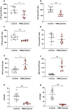
Concentrations of GDP-mannose and UDP-derivatives in controls and PMM2-CDG PBMCs. GDP-mannose (A), UDP-Glucuronic (B), UDP-Gal (C) and UDP-Glc (D) were detected by HPLC in deproteinized extracts of PBMCs obtained from control healthy subjects and PMM2-CDG patients. Mean values and standard deviations are indicated by vertical bars. **Significantly different from controls, p < 0.001. ***Significantly different from controls, p < 0.005.
Fig. 2
Fig. 2
Energy metabolism in PBMCs of controls and PMM2-CDG patients. Concentrations of ATP (A), GTP (B), UTP (C), CTP (D) ADP (E), and AMP (F) were detected by HPLC in deproteinized extracts of PBMCs obtained from control healthy subjects and PMM2-CDG patients. Values of the ATP/ADP (G) and ATP/AMP (H) ratios of PBMCs of controls and PMM2-CDG patients were calculated using the actual concentrations of ATP, ADP and AMP determined in each sample. Mean values and standard deviations are indicated by vertical bars. *Significantly different from controls, p < 0.01, **p < 0.001.
Fig. 3
Fig. 3
Nicotinic coenzyme ratios and acetyl-CoA in PBMCs of controls and PMM2-CDG patients. NAD+/NADH ratio (A), NADP+/NADPH ratio (B) and acetyl-CoA (C) determined in PBMCs obtained from control healthy subjects and PMM2-CDG patients. Values of the nicotinic coenzyme ratios of PBMCs of controls and PMM2-CDG patients were calculated using the actual concentrations of NAD+ , NADH, NADP+ and NADPH determined in each sample. Mean values and standard deviations are indicated by vertical bars. *Significantly different from controls, p < 0.01. **Significantly different from controls, p < 0.02.
Fig. 4
Fig. 4
Correlations of GDP-mannose and UDP-Glc with parameters of energy metabolism. Concentrations of GDP-mannose and GDP, detected in PBMCs of PMM2-CDG patients, showed positive correlation (A), whilst UDP-Glc concentrations were negatively correlated with those of ATP (B) and of the ATP/ADP ratio (C). The Pearson’s correlation coefficients and the corresponding p values are indicated in each panel.
Fig. 5
Fig. 5
Catabolites of ATP, GSH and nitric oxide metabolism in PBMCs of controls and PMM2-CDG patients. Concentrations of xanthine (A) and uric acid (B), GSH (C) and nitrite + nitrate (D) determined in PBMC obtained from control healthy subjects and PMM2-CDG patients. Mean values and standard deviations are indicated by vertical bars. N.D. = Not Detectable. *Significantly different from controls, p < 0.01. **Significantly different from controls, p < 0.001.
Fig. 6
Fig. 6
MetaboAnalyst-based enrichment analysis of the key metabolic alterations in PMM2-deficient PBMCs. Enrichment and pathway analyses were performed using the actual concentrations of the different metabolites detected in control and PMM2-deficient PBMCs, processed through the MetaboAnalyst 6.0 platform. Specifically, the “Pathway Analysis” module within the enrichment analysis section was used to identify significantly impacted metabolic pathways. The analysis was performed using default parameters. Pathways with a p-value of less than 0.05 were considered significantly enriched, indicating potential areas of metabolic perturbation associated with the experimental conditions. Only the top 25 pathways significantly different from controls were considered in the figure.
Fig. 7
Fig. 7
Schematic representation of the metabolic pathways directly affected by PMM2 malfunctioning in PMM2-CDG patients. Increase in mannose-6-phosphate (Man-6-P) causes increased concentrations of fructose-6-phosphate (Fru-6-P) first, and subsequent increased concentrations of glucose-6-phosphate (Glu-6-P), because of the equilibrium reactions catalyzed by mannose phosphate isomerase (MPI) and glucose phosphate isomerase (GPI), respectively. Higher Glu-6-P availability leads to increased formation of UDP-glucose (UDP-Glc). At the same time, the increase of Fru-6-P availability also allows to maintain an adequate synthesis of UDP-GlcNac, thus ensuring a significant level, although partial, of protein glycosylation. Higher levels of UDP-Glc could be responsible for increased rate of glycogen synthesis, thus explaining the abnormal glycogen accumulation observed in the liver of PMM2-CDG patients. Arrows in red indicate the reactions occurring under these pathological conditions. Compounds in the faint yellow boxes are those undergoing significant increases.

References

    1. Schachter, H. & Freeze, H. H. Glycosylation diseases: quo vadis? Biochim. Biophys. Acta. 1792, 925–930. 10.1016/j.bbadis.2008.11.002 (2009). - PMC - PubMed
    1. Peanne, R. et al. Congenital disorders of glycosylation (CDG): quo vadis? Eur. J. Med. Genet.61, 643–663. 10.1016/j.ejmg.2017.10.012 (2018). - PubMed
    1. Freeze, H. H., Jaeken, J. & Matthijs, G. CDG or not CDG. J. Inherit- Metab. Dis.45, 383–385. 10.1002/jimd.12498 (2022). - PMC - PubMed
    1. Ondruskova, N., Cechova, A., Hansikova, H., Honzik, T. & Jaeken, J. Congenital disorders of glycosylation: still hot in 2020. Biochim. Biophys. Acta Gen. Subj.1865, 129751. 10.1016/j.bbagen.2020.129751 (2021). - PubMed
    1. Mohorko, E., Glockshuber, R. & Aebi, M. Oligosaccharyltransferase: the central enzyme of N-linked protein glycosylation. J. Inherit. Metab. Dis.34, 869–878. 10.1007/s10545-011-9337-1 (2011). - PubMed

Substances

LinkOut - more resources