. 2025 Jun;22(6):645-660.
doi: 10.1038/s41423-025-01290-x. Epub 2025 May 8.

Harnessing nutrient scarcity for enhanced CAR-T-cell potency and safety in solid tumors

Affiliations

Harnessing nutrient scarcity for enhanced CAR-T-cell potency and safety in solid tumors

Enzo Manchon et al. Cell Mol Immunol. 2025 Jun.

Abstract

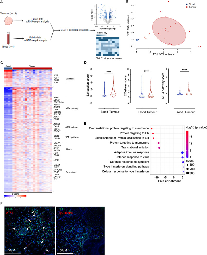
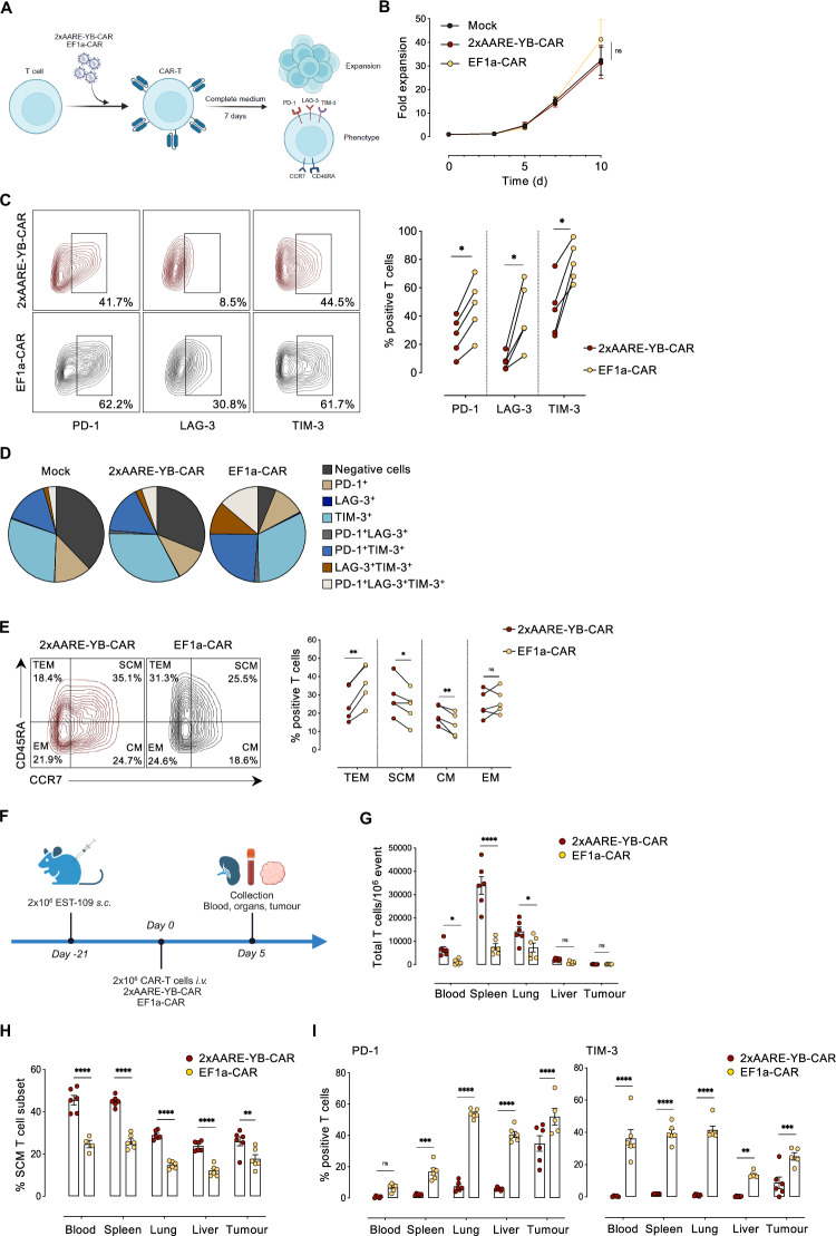
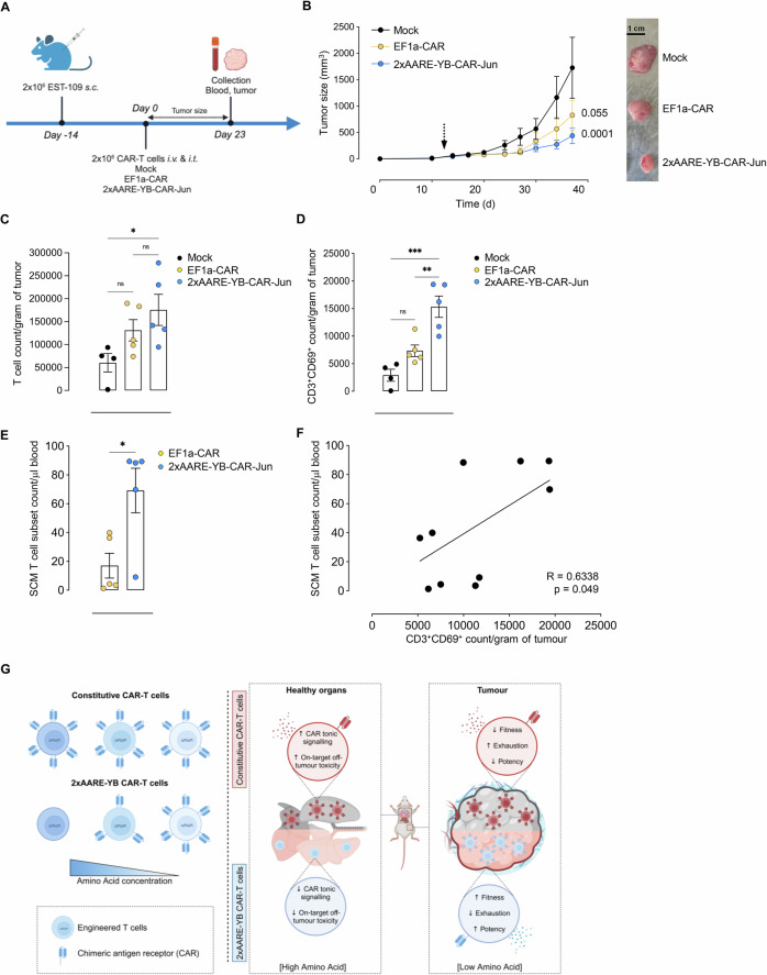
Despite significant advancements, the effectiveness of chimeric antigen receptor (CAR)-T-cell-based therapies in solid tumors remains limited. Key challenges include on-target effects, off-tumor toxicity and reduced CAR-T-cell function within the tumor microenvironment, which is often characterized by metabolic stress triggered by factors such as amino acid scarcity. Activating transcription factor-4 (ATF4) and its upstream regulator GCN2 play crucial roles in the metabolic reprogramming and functionality of CD4+ and CD8+ T cells. ATF4 can be activated by various cellular stress signals, including amino acid deprivation. While ATF4 activation may be associated with T-cell dysfunction, its role in stress adaptation presents an opportunity for therapeutic intervention-particularly in the tumor microenvironment, where T-cell exhaustion is a major challenge. In this study, we developed a strategy to harness the GCN2‒ATF4 axis in CAR-T cells. We employed an amino acid-dependent inducible promoter, which triggers ATF4-dependent gene expression to regulate CAR expression in T cells under conditions of amino acid scarcity within the tumor microenvironment. In vitro and murine xenograft models demonstrate the potential of this system to effectively restrict CAR expression to the tumor site. This targeted strategy not only enhances safety by minimizing off-tumor activity but also CAR-T-cell fitness by reducing exhaustion. By validating this pathophysiologically regulatable CAR expression system for solid tumors, our findings address key limitations of current CAR-T-cell therapies and pave the way for innovative strategies targeting solid malignancies.

Keywords: Amino acid scarcity; CAR-T; Solid tumours; Tumour microenvironment.


Conflict of interest statement

Competing interests: Benjamin Versier, Aravindhan Soundiramourty, Yves Christen, Dominique Charron, Jacques Mallet, and Che Serguera serve Asfalia Biologics. Ludmila Juricek previously served at Asfalia Biologics and currently serves for Coave Therapeutics. The authors declare that a patent application related to this work has been filed. The other authors declare that they have no conflicts of interest.

Figures

Fig. 1
Fig. 1
Amino acid scarcity induces ATF4 in T cells. A Schematic representation of the experimental model used to evaluate the T-cell immunophenotype and subsets under amino acid (AA) and oxygen restriction. B Heatmap illustrating the fold change in the expression of major functional T-cell markers under AA and oxygen restriction conditions compared with the control conditions. C Heatmap illustrating the fold changes in the proportions of stem central memory (SCM), central memory (CM), effector memory (EM), and terminal effector memory (TEM) T-cell subsets under AA and oxygen restriction conditions compared with those under control conditions. D Schematic representation of the ISR pathways that induce ATF4. E Immunoblot showing ATF4 expression in T cells after 24 h of culture under AA restriction and severe hypoxia (0.1% O2). The histograms in the lower panel show the relative expression under different conditions normalized to the expression of the housekeeping control GAPDH. Heatmaps illustrating the mRNA expression of genes associated with the ATF4, ATF6 and XBP1 pathways (F) and other T-cell-associated genes (G) in CD4+ and CD8+ T cells after 24 h of culture under AA restriction and severe hypoxia (0.1% O2). mRNA expression is represented as the fold change in expression normalized to that in the control medium. All the results are presented as the means ± SEMs of three independent healthy donors. Statistical analyses were performed with one-way ANOVA. Asterisks represent significant differences between each group and the control group (*p < 0.05, **p < 0.01, ***p < 0.001)
Fig. 2
Fig. 2
The ATF4 pathway in melanoma-infiltrating T cells. A Schematic representation of the public scRNA-seq data analysis workflow. B Principal component analysis (PCA) based on RNA expression from melanoma tumor-infiltrating T cells (TILs) and healthy donor T cells. C Heatmap illustrating genes differentially expressed between melanoma TILs and healthy donor T cells. D Violin plots of exhaustion, endoplasmic reticulum (ER) stress and ATF4 signature scores on the basis of RNA expression in melanoma TILs compared with healthy donor T cells. E Bubble plot illustrating pathway analysis incorporating genes downregulated and upregulated in melanoma TILs via the GO process. All results are representative of healthy donor blood (n = 4) and melanoma TIL (n = 19) samples and are presented as the mean values ± SEM. Statistical analyses were performed with two-sided Student’s t tests. Asterisks represent significant differences between TILs and blood T cells (****p < 0.0001). F Representative immunofluorescence images of three human primary melanomas. DAPI (nuclei, blue) and an anti-CD3 antibody (green) and either an anti-ATF4 antibody (red) or its IgG isotype control (red) are shown
Fig. 3
Fig. 3
The 2xAARE-YB system induces CAR expression. A Representative immunofluorescence images of melanoma (EST-109) and breast cancer (MDA-MB 231) spheroids after 72 h of coculture with 2xAARE-YB-GFP-T cells in control medium. B GFP intensity of untransduced (mock) or 2xAARE-YB-GFP-T cells infiltrating EST-109 and MDA-MB 231 spheroids over time. C Bar plot illustrating the percentage of (%) GFP+2xAARE-YB-GFP-T cells cultured alone (Ctl) or with spheroids. D Schematic representation of the 2xAARE-YB-regulated CAR expression construct. E Representative histogram and bar plot showing the expression of CAR in 2xAARE-YB-CAR-T cells cultured under different AA restrictions. F Relative surface CAR expression in 2xAARE-YB-CAR-T cells cultured with increasing dilutions of AA. G Surface CAR expression in 2xAARE-YB-CAR-T cells at the indicated times under conditions of AA starvation, rest and re-exposure to AA-restricted conditions. In both F and G, the maximum CAR expression (%) at a 1/1000 dilution for each AA is normalized at 100%. The results are presented as the mean values ± SEM obtained from five independent healthy donors. H Schematic representation of the mouse experimental protocol. Groups (n = 2) of NXG mice (n = 6/group) were s.c. injected with 2×10⁶ CD19 + EST-109 or CD19 + MDA-MB 231 cells. Tumors were developed over 21 days, after which the mice received i.v. and i.t. injections of 2xAARE-YB-CAR or EF1a-CAR-T cells. Blood, spleen, lungs, liver and tumors were collected two days later. I Representative contour plots and bar plots showing the percentages of (%) CAR-positive 2xAARE-YB-CAR- and EF1a-CAR-T cells in the blood, spleen, lungs, liver, CD19 + EST-109 and CD19 + MDA-MB 231 tumors. Not detected (nd). All the results are presented as the means ± SEMs of data obtained from 6 mice/group. Statistical analyses were performed with one- and two-way ANOVA. Asterisks represent significant differences between groups (*p < 0.05, **p < 0.01, ***p < 0.001, ****p < 0.0001)
Fig. 4
Fig. 4
Expanded 2xAARE-YB-CAR-T cells exhibit a memory phenotype and reduced exhaustion. A Experimental procedure and readout of expanded CAR-T cells. B Fold expansion of untransduced (mock), 2xAARE-YB-CAR, or EF1A-CAR-T cells. C Representative contour plots and connected dot plots representing the percentages of (%) PD-1-, LAG-3- and TIM-3-positive 2xAARE-YB- and EF1a-CAR-T cells after their expansion. D Pie chart illustrating the percentage of (%) PD-1-, LAG-3- and TIM-3-positive 2xAARE-YB- and EF1a- CAR-T cells after their expansion as well as the percentage of (%) double- or triple-positive cells as indicated. E Representative contour plots and connected dot plots representing the percentages of (%) SCM, CM, EM, and TEM 2xAARE-YB-CAR or EF1a-CAR-T-cell subsets after expansion. The results are presented as the mean values ± SEM obtained from five independent healthy donors. F Schematic representation of the mouse experimental protocol. Groups (n = 2) of NXG mice (n = 6/group) were s.c. injected with 2×10⁶ CD19 + EST-109 cells. Tumors were developed over 21 days, after which the mice received i.v. injections of 2xAARE-YB-CAR or EF1a-CAR-T cells. Blood, spleen, lungs, liver and tumors were collected 5 days later. G Bar plot illustrating the number of 2xAARE-YB-CAR and EF1a-CAR-T cells/106 cells that were isolated from the blood, spleen, lungs, liver and tumors. H Bar plot illustrating the percentage (%) of the 2xAARE-YB-CAR and EF1a-CAR-T-cell SCM subsets. I Bar plot illustrating the percentage of (%) PD-1- and TIM-3-positive 2xAARE-YB-CAR-T or EF1a-CAR-T cells in the blood, spleen, lungs, liver, and tumor. All the results are presented as the means ± SEMs of data obtained from 6 mice/group. Statistical analyses were performed with one- and two-way ANOVA. Asterisks represent significant differences between groups (*p < 0.05, **p < 0.01, ***p < 0.001, ****p < 0.0001)
Fig. 5
Fig. 5
Conditional expression of c-Jun enhances the cytotoxicity of 2xAARE-YB-CAR-T cells. A Schematic representation of the 2xAARE-YB-CAR and 2xAARE-YB-CAR-Jun constructs. B Representative histograms and connected dot plots illustrating c-Jun expression in 2xAARE-YB-CAR (red) and 2xAARE-YB-CAR-Jun (blue) T cells after 48 h of amino acid restriction. C Schematic representation of the 2D cytotoxicity assay. D Representative contour plots and bar plot representing the percentage (%) of specific lysed CD19+ EST-109 tumor cells after 30 h of coculture with 2xAARE-YB-CAR and 2xAARE-YB-CAR-Jun T cells at an E:T ratio of 2:1 under AA-restricted conditions. E Connected dot plots illustrating the percentage of (%) PD-1-, LAG-3- and TIM-3-positive 2xAARE-YB-CAR or 2xAARE-YB-CAR-Jun T cells after 48 h of AA restriction. All results are presented as the mean values ± SEM obtained from four independent healthy donors. Statistical analyses were performed with one- and two-way ANOVA. Asterisks represent significant differences between groups (*p < 0.05, **p < 0.01)
Fig. 6
Fig. 6
The 2xAARE-YB system generates efficient and less exhausted CAR-T cells within a 3D spheroid model. A Cytotoxic activity of untransduced (mock), 2xAARE-YB-CAR-Jun, 2xAARE-YB-CAR-Jun and EF1a-CAR-T cells against CD19 + EST-109 and CD19 + MDA-MB 231 spheroids over time under AA restriction. Asterisks represent significant differences between the CAR groups and mock-treated T cells (*p < 0.05, **p < 0.01, ****p < 0.0001). B Density plot representing the distribution of 2xAARE-YB-CAR, 2xAARE-YB-CAR-Jun and EF1a-CAR spheroid-infiltrating T cells in t-SNE after 96 h of coculture with CD19 + EST-109 spheroids and T cells under AA-restricted conditions. The color varies according to the cell abundance. C Scatterplots of t-SNEs and bar plots representing the intensity and percentage (%) of PD-1-, LAG-3-, and TIM-3-positive 2xAARE-YB-CAR, 2xAARE-YB-CAR-Jun and EF1a-CAR spheroid-infiltrating T cells after 96 h of coculture with CD19 + EST-109 spheroids and T cells under AA-restricted conditions. The color varies according to the surface marker abundance. All results are presented as the mean values ± SEM obtained from four independent healthy donors. Statistical analyses were performed with one- and two-way ANOVA. Asterisks represent significant differences between groups (*p < 0.05, **p < 0.01)
Fig. 7
Fig. 7
The 2xAARE-YB system generates potent antitumour CAR-T cells. A Schematic representation of the mouse experimental protocol. Groups (n = 3) of NXG mice (n = 5/group) were s.c. injected with 2 × 10⁶ CD19+ EST-109 cells. Tumors were developed over 14 days, after which the mice received i.v. and i.t. injections of mock-, 2xAARE-YB-CAR-Jun- or EF1a-CAR-T cells. Tumor size was monitored every 3–4 days. Blood and tumors were collected 23 days after injection. B CD19+ EST-109 tumor growth curves, with an arrow indicating the point at which the CAR-T cells were injected (left panel). Representative image of tumors collected on day 23 (right panel). The numeric values represent the significant differences between the CAR groups and mock-treated T cells. C Bar plot illustrating the absolute counts of mock-, 2xAARE-YB-CAR-Jun- and EF1a-CAR-T cells/gram of tumor cells. D Bar plot illustrating the absolute counts of CD69-positive mock-, 2xAARE-YB-CAR-Jun- and EF1a-CAR-T cells/gram of tumor. E Bar plot illustrating the absolute counts of the SCM mock-, 2xAARE-YB-CAR-Jun- and EF1a-CAR-T-cell subsets/μl of blood. F Scatter plot illustrating the relationship between absolute counts of the SCM T-cell subset in the blood and CD69-positive CAR-T cells in tumors. All the results are presented as the means ± SEMs of 5 mice/group. Statistical analyses were performed with two-sided Student’s t tests and one- and two-way ANOVA. Asterisks represent significant differences between groups (*p < 0.05, **p < 0.01, ***p < 0.001, ****p < 0.0001). G Graphical summary of the 2xAARE-YB system as an effective strategy for generating CAR-T cells targeting solid tumors

References

    1. Maude SL, Frey N, Shaw PA, Aplenc R, Barrett DM, Bunin NJ, et al. Chimeric antigen receptor T cells for sustained remissions in leukemia. N Engl J Med. 2014;371:1507–17. - PMC - PubMed
    1. Cappell KM, Sherry RM, Yang JC, Goff SL, Vanasse DA, McIntyre L, et al. Long-term follow-up of anti-CD19 chimeric antigen receptor T-cell therapy. J Clin Oncol. 2020;38:3805–15. - PMC - PubMed
    1. Holstein SA, Lunning MA. CAR T-Cell Therapy in hematologic malignancies: a voyage in progress. Clin Pharmacol Ther. 2020;107:112–22. - PubMed
    1. Zhang Y, Qin D, Shou AC, Liu Y, Wang Y, Zhou L. Exploring CAR-T-Cell therapy side effects: mechanisms and management strategies. J Clin Med. 2023;12:6124. - PMC - PubMed
    1. Saleki K, Mohamadi MH, Alijanizadeh P, Rezaei N. Neurological adverse effects of chimeric antigen receptor T-cell therapy. Expert Rev Clin Immunol. 2023;19:1361–83. - PubMed

Substances