. 2025 May 7;41(1):82.
doi: 10.1007/s10565-025-10029-3.

ELF4 improves sepsis-induced myocardial injury by regulating STING signaling-mediated T cells differentiation

Affiliations

ELF4 improves sepsis-induced myocardial injury by regulating STING signaling-mediated T cells differentiation

Yawen Zheng et al. Cell Biol Toxicol. .

Abstract

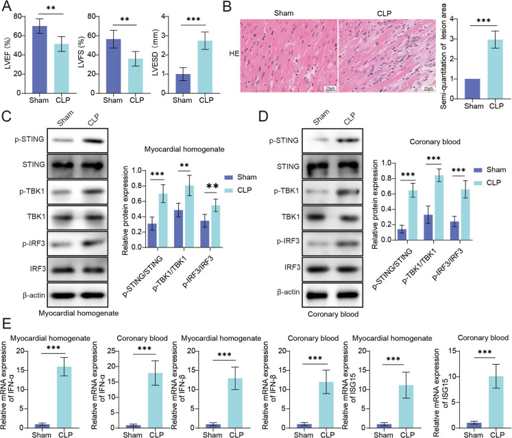
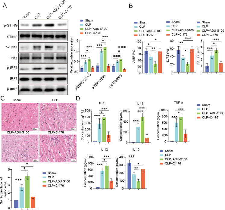
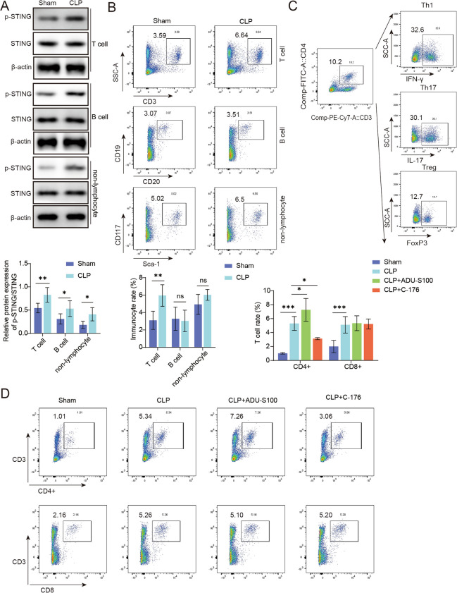
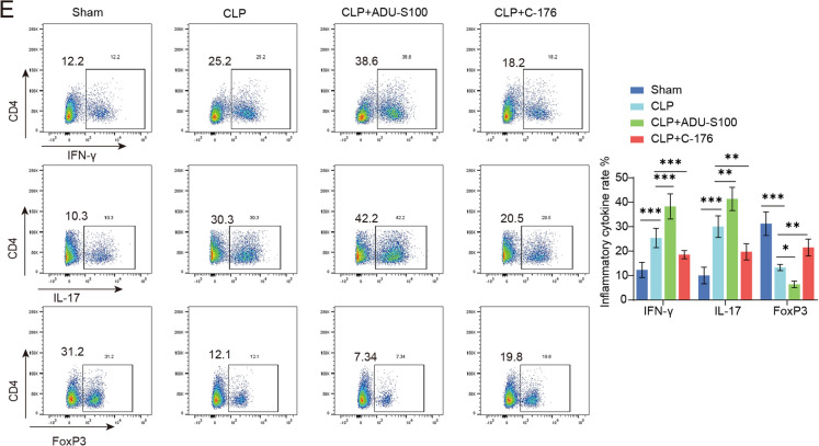
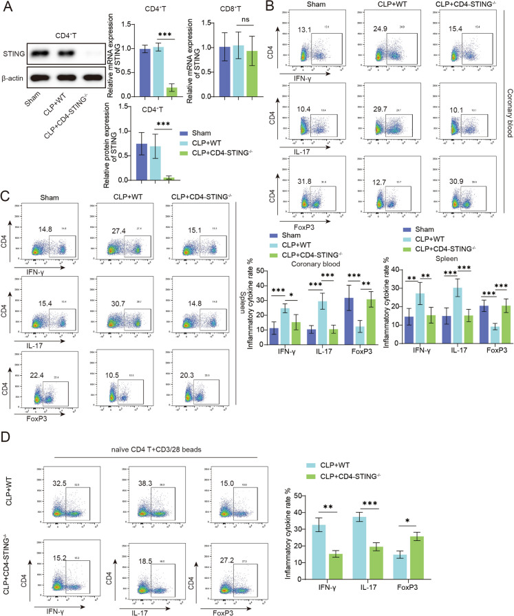
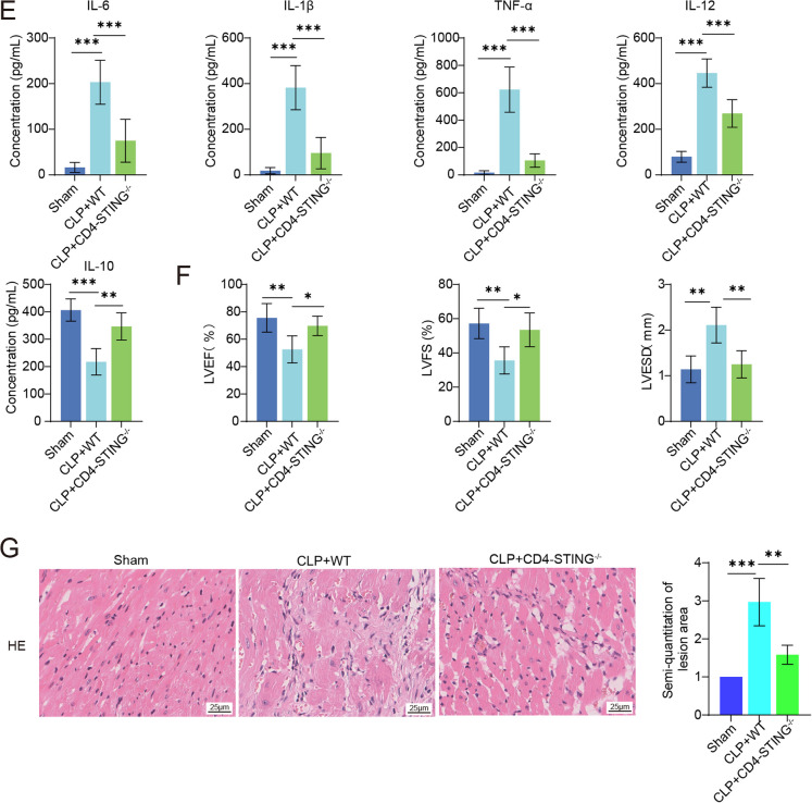
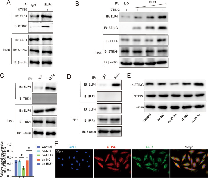
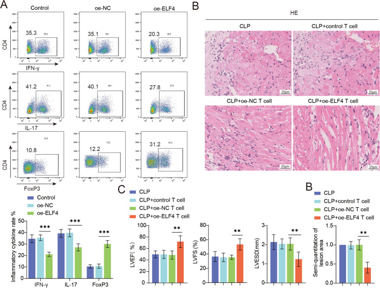
Septic cardiomyopathy (SCM) is a common complication caused by sepsis. T cells differentiation is involved in SCM progression. However, the role and underlying mechanisms of T cells-mediated immunity in SCM remain unclear. This study aimed to investigate the role of STING-mediated T cells differentiation in SCM. Cecal ligation and puncture (CLP) surgery was conducted in mice to establish SCM model. The mice were injected intraperitoneally with STING agonist ADU-S100 and C-176 after modeling. Wild type (WT) mice and CD4-STING-/- mice were employed. Besides, overexpressing vectors of ELF4 (oe-ELF4), short hairpin RNA targeting ELF4 (sh-ELF4) were transfected into 293T cells. STING signaling was found to be activated in sepsis-induced myocardial immune injury in mice. The administration of ADU-S100 exacerbated myocardial injury and inflammation, while C-176 alleviated these effects. Additionally, STING activation influenced T cells differentiation, with an increase in Th1 and Th17 cells and a decrease in Treg cells. Conditional knockout of STING in CD4+ T cells reduced Th1 and Th17 populations and improved myocardial function and histology. Furthermore, ELF4 was found to inhibit STING activation, reducing T cells differentiation into pro-inflammatory subsets. Overexpression of ELF4 in CD4+ T cells ameliorated myocardial damage and improved cardiac function in CLP mice, suggesting that the ELF4-STING signaling axis plays a protective role in sepsis-induced myocardial injury by regulating T cells differentiation.

Keywords: ELF4; STING; Septic cardiomyopathy; T cells differentiation.


Conflict of interest statement

Declarations. Ethical approval: The animal use protocol listed has been reviewed and approved by the Institutional Animal Care and Use Committee (IACUC) of The Second Xiangya Hospital, Central South University (No. 2021229). Conflict of interest: The authors declare no competing interests.

Figures

Fig. 1
Fig. 1
CLP mice exhibit augmented STING activation in SCM. C57BL/6 mice was subjected to CLP surgery to conduct a septic mouse model. A. Echocardiography was used to evaluate cardiac function parameters (LVEF, LVFS, LVESD). B. Representative images stained by HE for assessing the pathologic changes. C-D. Western blot analysis evaluated the protein levels of STING, p-STING, TBK1, p-TBK1, IRF3 and p-IRF3. E. RT-qPCR analysis determined the mRNA levels of IFN-α, IFN-β and ISG15. Data was shown as mean ± SD. N = 5. **p < 0.01, ***p < 0.001
Fig. 2
Fig. 2
Activation of STING aggravated sepsis-induced myocardial damage. C57BL/6 mice was subjected to CLP surgery to conduct a septic mouse model. The STING agonist (ADU-S100) and STING antagonist (C-176) were administrated at 1 h and 12 h after CLP operation. Mice in each group were sacrificed 24 h after CLP. The coronary blood samples were acquired from each group mice. A. The protein levels of STING, p-STING, TBK1, p-TBK1, IRF3 and p-IRF3 were evaluated by western blot analysis. B. Cardiac function parameters (LVEF, LVFS, LVESD) were examined by Echocardiography. C. Representative images stained by HE for assessing the pathologic changes. D. ELISA analysis was performed for quantifying the levels of IL-6, IL-12, IL-1β, TNF-α and IL-10. N = 5. Data was shown as mean ± SD. *p < 0.05, **p < 0.01, ***p < 0.001
Fig. 3
Fig. 3
STING activation promoted T cells enrichment in SCM. C57BL/6 mice was subjected to CLP surgery to conduct a septic mouse model. A. The expressions of STING in T cells, B cells and non-lymphocytes in coronary blood were evaluated western blot assay. B. The frequency of T cells, B cells and non-lymphocyte in the coronary blood of mice were assessed using flow cytometry. C. The differentiation rate of CD4+ T cells toward to Th1, Th17 and Treg cells were analyzed by flow cytometry. D. The STING agonist (ADU-S100) and STING antagonist (C-176) were administrated at 1 h and 12 h after CLP operation. The coronary blood samples were acquired from each group mice. D. The overall percentage of CD4+ T cells and CD8+ T cells were quantified by flow cytometry. E. The differentiation rate of CD4+ T cells toward to Th1, Th17 and Treg cells were analyzed by flow cytometry. Data was shown as mean ± SD. N = 5. *p < 0.05, **p < 0.01, ***p < 0.001
Fig. 3
Fig. 3
STING activation promoted T cells enrichment in SCM. C57BL/6 mice was subjected to CLP surgery to conduct a septic mouse model. A. The expressions of STING in T cells, B cells and non-lymphocytes in coronary blood were evaluated western blot assay. B. The frequency of T cells, B cells and non-lymphocyte in the coronary blood of mice were assessed using flow cytometry. C. The differentiation rate of CD4+ T cells toward to Th1, Th17 and Treg cells were analyzed by flow cytometry. D. The STING agonist (ADU-S100) and STING antagonist (C-176) were administrated at 1 h and 12 h after CLP operation. The coronary blood samples were acquired from each group mice. D. The overall percentage of CD4+ T cells and CD8+ T cells were quantified by flow cytometry. E. The differentiation rate of CD4+ T cells toward to Th1, Th17 and Treg cells were analyzed by flow cytometry. Data was shown as mean ± SD. N = 5. *p < 0.05, **p < 0.01, ***p < 0.001
Fig. 4
Fig. 4
STING promoted T cells differentiating into Th1/17. CD4+ T cells-specific STING conditional knockout (CD4-STING−/−) mice were subjected to CLP surgery, and WT mice as control. Post surgery 24 h, the spleen tissues and coronary blood samples were obtained for next analysis. A. Western blot and RT-qPCR assays were conducted to evaluated the expression of STING in CD4+ T cells and CD8+ T cells. B. The differentiation rate of CD4+ T cells toward to Th1, Th17 and Treg cells in coronary blood samples were analyzed by flow cytometry. C. The differentiation rate of CD4+ T cells toward to Th1, Th17 and Treg cells in spleen tissues were analyzed by flow cytometry. D. CD4 + T from WT or CD4-STING−/− mice were isolated for T cells differentiation test under indicating cytokine panel. E. ELISA was employed to detect the levels of inflammatory factors including IL-6, IL-1β, IL-12, TNF-α and IL- 10. F. Cardiac function parameters (LVEF, LVFS, LVESD) were evaluated by echocardiography. G. Representative images stained by HE for assessing the pathologic changes. Data was shown as mean ± SD. N = 5. *p < 0.05, **p < 0.01, ***p < 0.001
Fig. 4
Fig. 4
STING promoted T cells differentiating into Th1/17. CD4+ T cells-specific STING conditional knockout (CD4-STING−/−) mice were subjected to CLP surgery, and WT mice as control. Post surgery 24 h, the spleen tissues and coronary blood samples were obtained for next analysis. A. Western blot and RT-qPCR assays were conducted to evaluated the expression of STING in CD4+ T cells and CD8+ T cells. B. The differentiation rate of CD4+ T cells toward to Th1, Th17 and Treg cells in coronary blood samples were analyzed by flow cytometry. C. The differentiation rate of CD4+ T cells toward to Th1, Th17 and Treg cells in spleen tissues were analyzed by flow cytometry. D. CD4 + T from WT or CD4-STING−/− mice were isolated for T cells differentiation test under indicating cytokine panel. E. ELISA was employed to detect the levels of inflammatory factors including IL-6, IL-1β, IL-12, TNF-α and IL- 10. F. Cardiac function parameters (LVEF, LVFS, LVESD) were evaluated by echocardiography. G. Representative images stained by HE for assessing the pathologic changes. Data was shown as mean ± SD. N = 5. *p < 0.05, **p < 0.01, ***p < 0.001
Fig. 5
Fig. 5
ELK4 interacted with STING to inhibit the activation of STING in CD4+ T cells. A-E. 293T was transfected by STING plasmid. A-D. Co-IP experiment verified the direct interaction between ELF4 and STING in 293T. The interaction between ELF4 and TBK1 or IRF3 was also evaluated by Co-IP experiment. E. 293T were transfected with oe-ELF4, sh-ELF4 or corresponding NCs. The protein levels of STING and p-STING were detected by western blot assay. F. The confocal co-localization images of STING and ELF4 in 293T. Data was shown as mean ± SD. N = 3. *p < 0.05
Fig. 6
Fig. 6
ELF4 overexpression alleviated cardiomyocyte injury by suppressing STING signaling-mediated Th1/17 differentiation. A. Mouse primary CD4T cells were isolated and transfected with overexpressing ELF4 plasmids for differentiation test ex vivo. The proportion of Th1, Th17 and Treg were detected by flow cytometry. B. Undifferentiated control T cells, OE-NC T cells, and OE-ELF4 T cells were adoptively transferred into WT mice, followed by CLP surgery. Representative images stained by HE for assessing the pathologic changes. C. Echocardiography was used to evaluate cardiac function parameters (LVEF, LVFS, LVESD). Data was shown as mean ± SD. N = 5. **p < 0.01, ***p < 0.001

Similar articles

References

    1. Anyalebechi JC, et al. CD8(+) T cells are necessary for improved sepsis survival induced by CD28 agonism in immunologically experienced mice. Front Immunol. 2024;15:1346097. - PMC - PubMed
    1. Benoit-Lizon I, et al. CD4 T cell-intrinsic STING signaling controls the differentiation and effector functions of T(H)1 and T(H)9 cells. J Immunother Cancer. 2022;10(1):e003459. - PMC - PubMed
    1. Berger G, et al. STING activation promotes robust immune response and NK cell-mediated tumor regression in glioblastoma models. Proc Natl Acad Sci U S A. 2022;119(28):e2111003119. - PMC - PubMed
    1. Busch K, et al. Inhibition of the NLRP3/IL-1beta axis protects against sepsis-induced cardiomyopathy. J Cachexia Sarcopenia Muscle. 2021;12(6):1653–68. - PMC - PubMed
    1. Cao M, et al. The transcription factor ELF4 alleviates inflammatory bowel disease by activating IL1RN transcription, suppressing inflammatory TH17 cell activity, and inducing macrophage M2 polarization. Front Immunol. 2023;14:1270411. - PMC - PubMed

MeSH terms