. 2025 May 7;16(1):95.
doi: 10.1186/s13244-025-01955-1.

CT of acute abdomen in the elderly

Affiliations

CT of acute abdomen in the elderly

Juliette Coutureau et al. Insights Imaging. .

Abstract

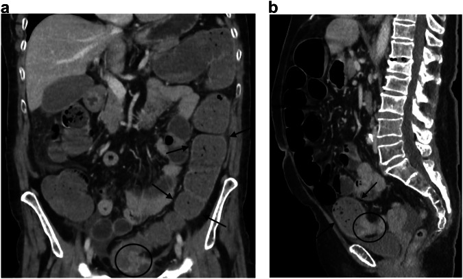
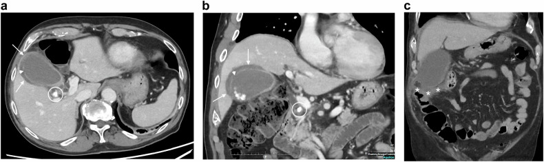
Abdominal disorders represent 10 to 15% of all Emergency Department visits in elderly patients. This educational review focuses on acute abdomen pathologies specific to the elderly and on their imaging patterns and proposes a strategy for performing CT scans in this population. Bowel obstruction is the most common cause of emergency surgery in the elderly with a higher proportion of colonic obstructions, in particular obstructive colorectal cancer and sigmoid volvulus. Concerning abdominal inflammatory processes, such as cholecystitis, appendicitis, and diverticulitis, gangrenous cholecystitis and complicated appendicitis are relatively frequently encountered due to delayed diagnoses. Bowel ischemia, which includes acute mesenteric ischemia (AMI) and ischemic colitis (IC), is also much more common after the age of 80. Although ischemic colitis is mainly related to cardiovascular risk factors, it can also result from a persistent distension above a colonic cancer or from fecal impaction. Finally, extra-abdominal pathologies responsible for acute abdominal pain, such as inferior myocardial infarction, should not be overlooked. In clinical practice, when possible thanks to sufficient and appropriate radiological resources, we recommend a scan without injection of contrast and an injection depending on the results of the unenhanced scan, decided by the radiologist present at the CT scan room during the examination. CRITICAL RELEVANCE STATEMENT: CT is critical in the diagnosis and management of patients over 75 years old with an acute abdomen, given the difficulty of clinico-biological diagnosis, the frequency of complicated forms, and the morbidity induced by delayed diagnosis. KEY POINTS: The most common site and cause of bowel obstruction in the elderly is large bowel obstruction due to colon cancer. Discrepancy between a poor clinical examination and complicated forms on imaging, particularly for inflammation and infections, is responsible for late diagnosis and increased morbidity. Ischemia, including of the small bowel, colon, and gallbladder are common cause of acute abdomen in elderly. In patients with upper quadrant pain, consider extra-abdominal causes such as pneumonia or myocardial infarction.

Keywords: Acute abdomen; Cholecystitis; Elderly patients; Intestinal obstruction; Mesenteric ischemia.


Conflict of interest statement

Declarations. Ethics approval and consent to participate: Not applicable. Consent for publication: Not applicable. Competing interests: The authors declare that they have no competing interests.

Figures

Fig. 1
Fig. 1
Acute mesenteric ischemia (AMI) due to mesenteric thrombosis and non-occlusive mesenteric ischemia in two different patients. AMI due to thrombosis: axial portal phase CT slice (a) in an 88-year-old woman shows decreased enhancement of the small bowel wall (plain circle) compared to another segment (dotted circle). Some of the concerned segments show a thin, “virtual” wall (asterisk). Axial portal phase CT slice at an upper level (b) shows a proximal thrombus in the superior mesenteric artery (arrow). Sagittal arterial phase CT (c) shows an occlusion of an extensively calcified mesenteric artery (arrows). AMI due to NOMI: axial and coronal portal phases (d, e) in a 78-year-old man shows dilatation of the small bowel lumen associated with a decreased enhancement of the small bowel wall and parietal pneumatosis (arrowheads). The mesenteric vessels are permeable (dotted circle)
Fig. 2
Fig. 2
Ischemic colitis. Axial and coronal portal phase CT (a, b) in a 90-year-old man shows a segmental thickening of the left colonic angle with a stratified enhanced wall (arrows) and peripheric fat stranding (asterisk), suggesting a reperfusion form of ischemic colitis
Fig. 3
Fig. 3
Ischemic colitis complicating a right colonic cancer. Axial and coronal portal phase CT (a, b) in an 82-year-old woman shows a stratified enhanced thickening (arrows) upstream a short and irregular unstratified thickening (dotted circle) of the right colonic wall
Fig. 4
Fig. 4
Volvulus of the sigmoid. Axial and sagittal portal phase CT (a, b) in an 86-year-old woman shows distension of the large bowel (arrows) above a unique “beak-sign” (dotted circle) corresponding to the transition point of an organo-axial sigmoid volvulus. c Volvulus of the sigmoid. Axial portal phase CT (c) in a 91-year-old man shows a mesenteric-axial sigmoid volvulus as suggested by the presence of two adjacent “beak-signs” (arrows)
Fig. 5
Fig. 5
Ischemic colitis complicating fecal impaction. Axial unenhanced and portal phase CT (a, b) in an 80-year-old woman shows compacted feces in the right and transverse colon (asterisk), associated with a smooth annular wall thickening (arrows), suggesting ischemic colitis due to the distension. Axial subtraction sequence (c) confirms a lack of enhancement of the right colonic wall (arrowheads)
Fig. 6
Fig. 6
Large bowel obstruction caused by sigmoid cancer. Coronal and sagittal portal phase CT (a, b) in a 78-year-old woman shows a distension of the large bowel (arrows) above an asymmetric and short-segment thickening of the sigmoid wall (circle) narrowing the colonic lumen
Fig. 7
Fig. 7
Small bowel obstructions due to causes specifically encountered in the elderly. Obturator hernia in an 85-year-old woman. Axial portal phase CT (a) shows an intestinal segment between the obturator externus and pectineus muscles (dotted circle). Coronal portal phase CT (b) shows distension of the small bowel (asterisk) up to a transition point corresponding to the collar of the hernia (arrow), suggesting a small bowel strangulation due to an obturator hernia. Biliary ileus in a 90-year-old woman. Coronal portal phase CT (c) shows distension of the small bowel (arrows) above a radio-opaque gallstone (circle)
Fig. 8
Fig. 8
Gangrenous cholecystitis. Axial and coronal portal phase CT (a, b) in an 80-year-old man shows an irregular thickening of the gallbladder wall (arrows) with interrupted contrast enhancement (arrowhead). The cystic duct is obstructed by a gallstone (circle). Coronal portal phase CT (c) in an 81-year-old man shows increased density of fatty tissue around the gallbladder (asterisk) and interruption of the inferior part of the gallbladder wall
Fig. 9
Fig. 9
Volvulus of the gallbladder. Axial and coronal portal phase CT (a, b) in an 84-year-old woman shows dilatation of the gallbladder and horizontal rotation of its axis with poor enhancement of its walls (arrows)
Fig. 10
Fig. 10
Complicated appendicitis. Axial portal phase CT (a, b) in a 76-year-old man shows a collection in the right psoas muscle (arrows), in which an appendicolith can be distinguished (circle), suggesting a complicated appendicitis with a psoas abscess
Fig. 11
Fig. 11
Stercoral peritonitis due to colonic cancer with upstream perforation. Coronal and axial portal phase CT (ac) in a 78-year-old man shows free intra-peritoneal gas (arrowheads) and feces associated with fat stranding (dotted circle). The site of the perforation is located above a short-segment thickening of the right colonic wall (arrows), showing a perforation due to distension above a tumoral stenosis
Fig. 12
Fig. 12
Acute inferior myocardial infarction as a differential diagnosis of acute abdominal pain. Axial and coronal portal phase CT (a, b) in a 90-year-old man shows a segmental perfusion defect of the inferior myocardia (arrows) corresponding to the territory of the circumflex artery, suggestive of an acute infarction

References

    1. Eurostat (2024) Population structure and ageing. Statistics explained. Retrieved January 09, 2025, from https://ec.europa.eu/eurostat/statistics-explained/index.php?title=Popul...
    1. Neilsberg Research (2024) United States population by age. Retrieved January 09, 2025, from https://www.neilsberg.com/insights/united-states-population-by-age/
    1. Natesan S, Lee J, Volkamer H, Thoureen T (2016) Evidence-based medicine approach to abdominal pain. Emerg Med Clin North Am 34:165–190. 10.1016/j.emc.2015.12.008 - PubMed
    1. Baz Ü, Satar S, Kozacı N, Açıkalın A, Gülen M, Karakurt Ü (2014) Geriatric patient admissions to emergency service. J Acad Emerg Med 13:53–57. 10.5152/jaem.2013.007
    1. Ukkonen M, Jämsen E, Zeitlin R, Pauniaho SL (2019) Emergency department visits in older patients: a population-based survey. BMC Emerg Med 19:20. 10.1186/s12873-019-0236-3 - PMC - PubMed

LinkOut - more resources