. 2025 May 7;31(1):176.
doi: 10.1186/s10020-025-01228-z.

Melatonin alleviates sepsis-induced acute lung injury by inhibiting necroptosis via reducing circulating mtDNA release

Affiliations

Melatonin alleviates sepsis-induced acute lung injury by inhibiting necroptosis via reducing circulating mtDNA release

Yuce Peng et al. Mol Med. .

Abstract

Background: Sepsis is a life-threatening condition that often leads to severe complications, including acute lung injury (ALI), which carries high morbidity and mortality in critically ill patients. Melatonin (Mel) has shown significant protective effects against sepsis-induced ALI, but its precise mechanism remains unclear.

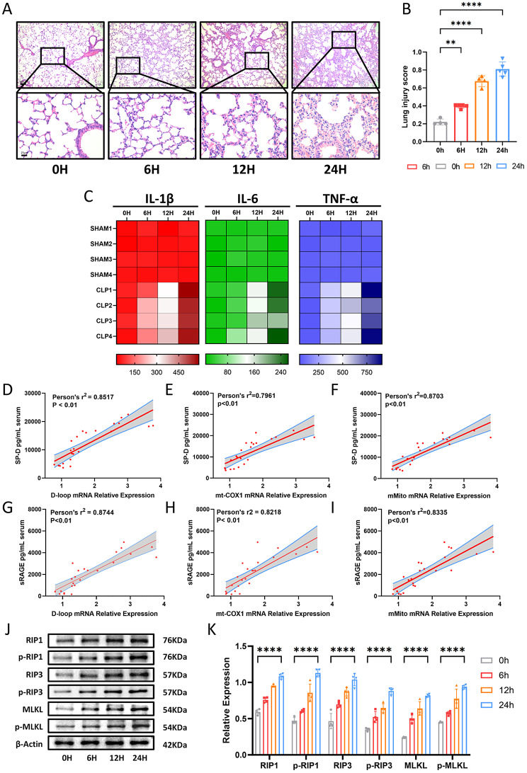
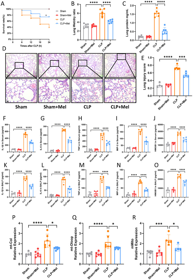
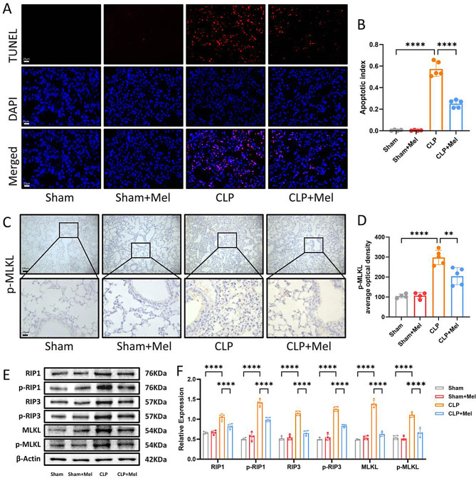
Methods: A cecal ligation and puncture (CLP) model was used to induce sepsis in male C57BL/6 mice, which were divided into four groups: Control, Sham, CLP, and CLP + Mel. ALI severity was evaluated via hematoxylin and eosin (H&E) staining, lung wet/dry ratio, and serum biomarkers (SP-D, sRAGE). Inflammatory cytokines (IL-1β, IL-6, TNF-α) were measured in serum and bronchoalveolar lavage fluid using ELISA. Circulating mitochondrial DNA (mtDNA) subtypes (D-loop, mt-CO1, mMito) were quantified by real-time PCR. TUNEL staining was performed to assess lung cell apoptosis. Necroptosis and STING pathway activation were analyzed via Western blot and immunofluorescence.

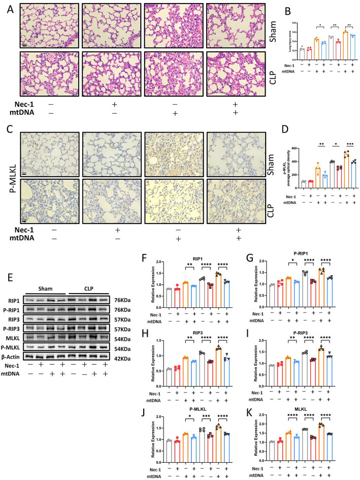
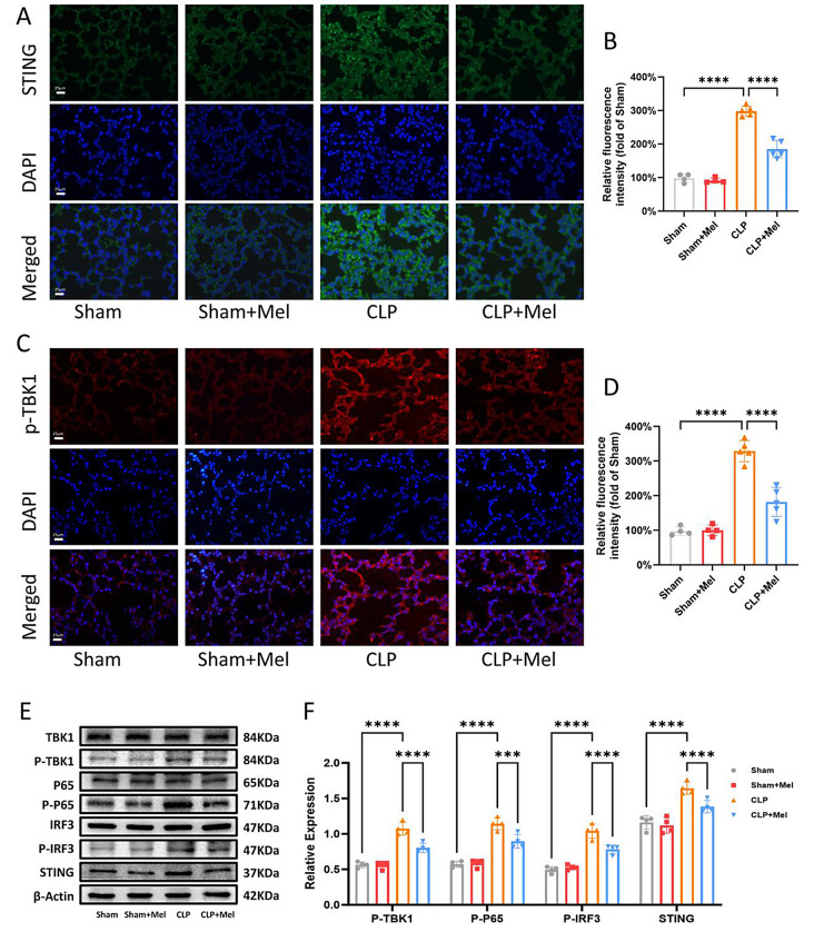
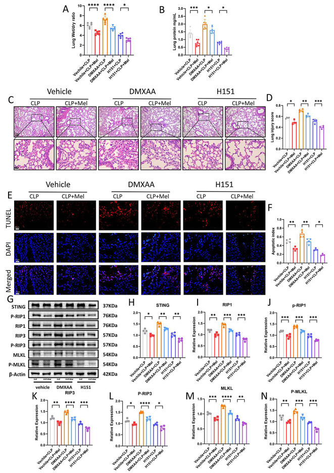
Results: Sepsis led to increased circulating mtDNA levels and activation of necroptosis signaling pathways. Melatonin treatment alleviated sepsis-induced ALI, improving survival, reducing inflammatory cytokines and mtDNA release, and suppressing necroptosis. Intraperitoneal injection of mtDNA in mice activated necroptosis, while RIP1 inhibitor Nec-1 counteracted mtDNA-induced lung damage and necroptosis in sepsis-induced ALI. Additionally, melatonin significantly inhibited STING pathway activation. Further experiments revealed that STING modulation influenced necroptosis protein expression and mediated melatonin's protective effects in sepsis-induced ALI.

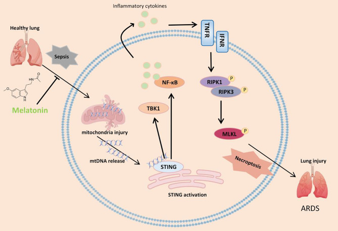
Conclusion: Melatonin mitigates sepsis-induced ALI by suppressing necroptosis through inhibition of STING activation and reduction of mtDNA release. These findings suggest melatonin as a potential therapeutic strategy for sepsis-induced ALI.

Keywords: ALI; Melatonin; Necroptosis; Sepsis; mtDNA-STING.


Conflict of interest statement

Declarations. Ethics approval and consent to participate: All animal experimental procedures were conducted in accordance with the National Institutes of Health (NIH) guidelines, and ethical approval was obtained from the Committee of Guangdong Provincial People’s Hospital. Consent for publication: The author confirms that the work described has not been published previously and is not under consideration for publication elsewhere. Competing interests: The authors declare no competing interests.

Figures

Fig. 1
Fig. 1
An Increase in Circulating mtDNA Is Associated with Inflammation and Necroptosis in Septic ALI. (A) Representative H&E-stained photomicrographs of murine lung tissues from the control (0 h), 6 h, 12 h, and 24 h groups. Bar = 25 μm (original magnification ×100, ×400). (B) Lung injury scores for the control (0 h), 6 h, 12 h, and 24 h groups. (C) Plasma levels of inflammatory cytokines (IL-1β, IL-6, and TNF-α) measured by ELISA in mice after CLP at 0, 6, 12, and 24 h. (D-I) Correlations between serum mtDNA levels and lung injury biomarkers (SP-D and sRAGE) assessed using the Spearman correlation test. mtDNA levels were analyzed via quantitative real-time PCR using three mtDNA primers (D-loop, mt-COX1, mMito). (J) Western blot analysis of necroptosis signaling in murine lung tissues at 0, 6, 12, and 24 h after CLP. (K) Quantification of Western blot results. Data are presented as mean ± SD from at least four independent experiments. *p < 0.05; **p < 0.01; ***p < 0.001; ****p < 0.0001; ns, not significant. One-way ANOVA followed by Tukey’s multiple comparisons test
Fig. 2
Fig. 2
Melatonin Mitigates Septic ALI by Reducing Circulating mtDNA Release and Inflammatory Injury. (A) Survival rates of mice with or without melatonin therapy 24 h after CLP (log-rank [Mantel-Cox] test, n = 20). (B) Lung wet/dry ratio in mice with or without melatonin therapy at 24 h after CLP. (C) BALF protein levels in mice with or without melatonin therapy at 24 h after CLP. (D) Representative H&E-stained photomicrographs of lung tissues from Sham, Sham + Mel, CLP, and CLP + Mel groups. Bar = 25 μm (original magnification ×100, ×400). (E) Lung injury scores from the Sham, Sham + Mel, CLP, and CLP + Mel groups. (F-J) Plasma levels of inflammatory cytokines (IL-1β, IL-6, TNF-α, MIP-2, and HMGB1) measured by ELISA in Sham, Sham + Mel, CLP, and CLP + Mel groups. (K-O) BALF levels of inflammatory cytokines (IL-1β, IL-6, TNF-α, MIP-2, and HMGB1) measured by ELISA in the same groups. (P-R) qPCR analysis of mt-Col, mt-Dloop, and mMito mRNA levels in indicated tissues. Data are presented as mean ± SD. *p < 0.05; **p < 0.01; ***p < 0.001; ****p < 0.0001; ns, not significant. One-way ANOVA followed by Tukey’s multiple comparisons test
Fig. 3
Fig. 3
Melatonin Alleviates Sepsis-Induced ALI by Inhibiting RIP1/RIP3/MLKL-Mediated Necroptosis. (A) TUNEL staining analysis of cell death in Sham, Sham + Mel, CLP, and CLP + Mel groups. Bar = 25 μm (original magnification ×400). (B) Apoptotic index represented by the ratio of positive cells to DAPI-stained cells. (C) Representative images of IHC analysis for p-MLKL expression in lung tissues. (D) Quantification of p-MLKL optical density in immunohistochemistry using ImageJ. Bar = 25 μm (original magnification ×100, ×400). (E) Western blot analysis of necroptosis signaling in the lungs of Sham, Sham + Mel, CLP, and CLP + Mel groups. (F) Quantification of Western blot results. Data are presented as mean ± SD from at least four independent experiments. *p < 0.05; **p < 0.01; ***p < 0.001; ****p < 0.0001; ns, not significant. One-way ANOVA followed by Tukey’s multiple comparisons test
Fig. 4
Fig. 4
Circulating mtDNA Induces ALI in CLP Mice by Activating RIP1/RIP3/MLKL-Mediated Necroptosis. (A) Representative H&E-stained photomicrographs of lung tissues from Sham and CLP groups, with or without Nec-1 and mtDNA injection. Bar = 25 μm (original magnification ×400). (B) Lung injury scores in Sham and CLP groups, with or without Nec-1 and mtDNA injection. (C) Representative IHC images for p-MLKL expression in lung tissues. (D) Quantification of p-MLKL optical density in immunohistochemistry using ImageJ. Bar = 25 μm (original magnification ×400). (E) Western blot analysis of necroptosis signaling in the lungs of Sham and CLP groups, with or without Nec-1 and mtDNA injection. (F-G) Quantification of Western blot results. Data are presented as mean ± SD from at least four independent experiments. *p < 0.05; **p < 0.01; ***p < 0.001; ****p < 0.0001; ns, not significant. One-way ANOVA followed by Tukey’s multiple comparisons test
Fig. 5
Fig. 5
Melatonin Alleviates STING Pathway Activation After Sepsis. (A) Immunofluorescence staining of STING in lung tissues of mice. Green, STING immunostaining; blue, DAPI-stained DNA. Scale bar = 25 μm (original magnification ×400). (B) Quantification of relative fluorescence intensity for STING. (C) Immunofluorescence staining of p-TBK1 in lung tissues of mice. Red, p-TBK1 immunostaining; blue, DAPI-stained DNA. Scale bar = 25 μm (original magnification ×400). (D) Quantification of relative fluorescence intensity for p-TBK1. (E) Western blot analysis of STING signaling in the lungs of Sham, Sham + Mel, CLP, and CLP + Mel groups. (F) Quantification of Western blot results. Data are presented as mean ± SD from at least four independent experiments. *p < 0.05; **p < 0.01; ***p < 0.001; ****p < 0.0001; ns, not significant. One-way ANOVA followed by Tukey’s multiple comparisons test
Fig. 6
Fig. 6
The mtDNA-STING Signaling Pathway Promotes ALI Development by Activating Necroptosis. (A) Lung wet/dry ratio in mice with or without melatonin therapy at 24 h after CLP. (B) BALF protein levels in mice with or without melatonin therapy at 24 h after CLP. (C) Representative H&E-stained photomicrographs of lung tissues from Vehicle, DMXAA, and H151 groups, with or without melatonin therapy. Bar = 25 μm (original magnification ×100, ×400). (D) Lung injury scores in the Vehicle, DMXAA, and H151 groups, with or without melatonin therapy. (E) TUNEL staining analysis of cell death in Vehicle, DMXAA, and H151 groups, with or without melatonin therapy. Bar = 25 μm (original magnification ×400). (F) Apoptotic index represented by the ratio of positive cells to DAPI-stained cells. (G) Western blot analysis of STING signaling in lung tissues of Vehicle, DMXAA, and H151 groups, with or without melatonin therapy. (H-N) Quantification of Western blot results. Data are presented as mean ± SD from at least four independent experiments. *p < 0.05; **p < 0.01; ***p < 0.001; ****p < 0.0001; ns, not significant. One-way ANOVA followed by Tukey’s multiple comparisons test
Fig. 7
Fig. 7
Schematic illustration of this study. Melatonin reduces mtDNA leakage, thereby inhibiting STING activation, suppressing necroptosis, and ultimately mitigating lung injury and improving lung function

Similar articles

Cited by

References

    1. Alsabani M, et al. Reduction of NETosis by targeting CXCR1/2 reduces thrombosis, lung injury, and mortality in experimental human and murine sepsis. Br J Anaesth. 2022;128(2):283–93. - PMC - PubMed
    1. Barber GN. STING: infection, inflammation and cancer. Nat Rev Immunol. 2015;15(12):760–70. - PMC - PubMed
    1. Bos LDJ, Ware LB. Acute respiratory distress syndrome: causes, pathophysiology, and phenotypes. Lancet. 2022;400(10358):1145–56. - PubMed
    1. Bowdish ME, et al. A randomized trial of mesenchymal stromal cells for moderate to severe acute respiratory distress syndrome from COVID-19. Am J Respir Crit Care Med. 2023;207(3):261–70. - PMC - PubMed
    1. Brault M, et al. Intracellular nucleic acid sensing triggers necroptosis through synergistic type I IFN and TNF signaling. J Immunol. 2018;200(8):2748–56. - PMC - PubMed

MeSH terms