Review
. 2025 May 7;24(1):138.
doi: 10.1186/s12943-025-02309-7.

Protein lipidation in the tumor microenvironment: enzymology, signaling pathways, and therapeutics

Affiliations
Review

Protein lipidation in the tumor microenvironment: enzymology, signaling pathways, and therapeutics

Mengke Xu et al. Mol Cancer. .

Abstract

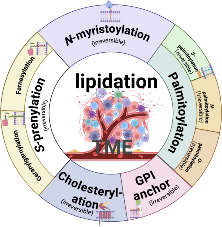
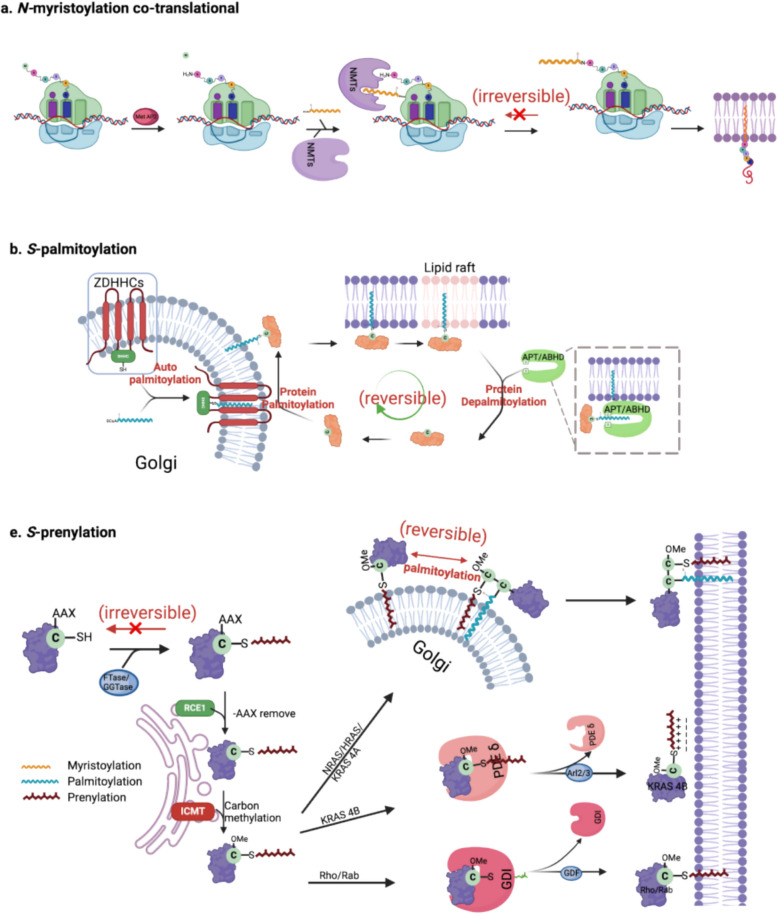
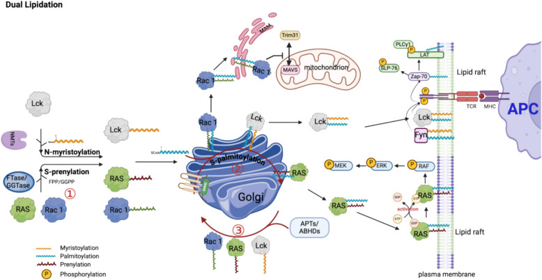
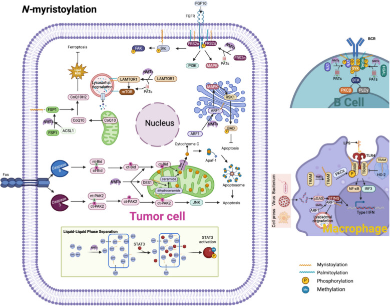
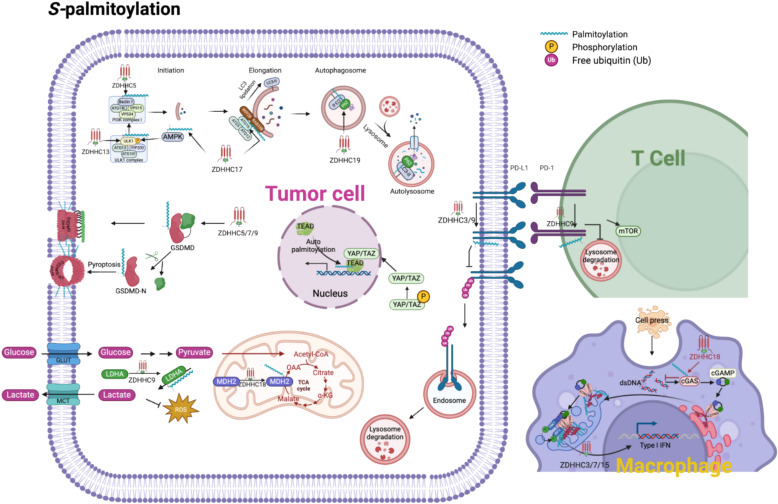
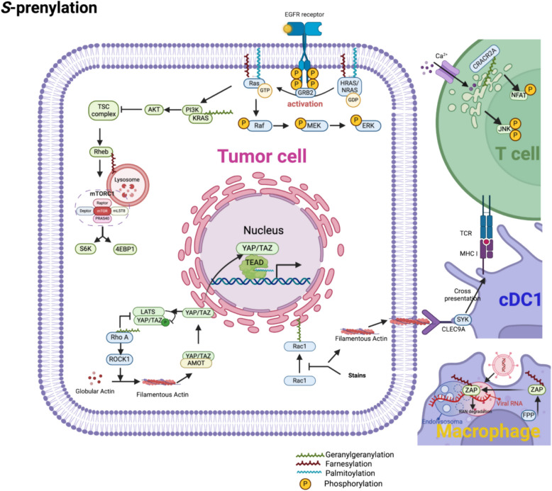
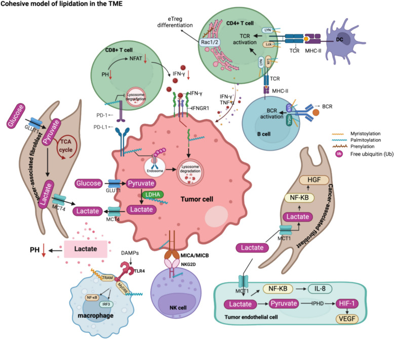
Protein lipidation is a pivotal post-translational modification that increases protein hydrophobicity and influences their function, localization, and interaction network. Emerging evidence has shown significant roles of lipidation in the tumor microenvironment (TME). However, a comprehensive review of this topic is lacking. In this review, we present an integrated and in-depth literature review of protein lipidation in the context of the TME. Specifically, we focus on three major lipidation modifications: S-prenylation, S-palmitoylation, and N-myristoylation. We emphasize how these modifications affect oncogenic signaling pathways and the complex interplay between tumor cells and the surrounding stromal and immune cells. Furthermore, we explore the therapeutic potential of targeting lipidation mechanisms in cancer treatment and discuss prospects for developing novel anticancer strategies that disrupt lipidation-dependent signaling pathways. By bridging protein lipidation with the dynamics of the TME, our review provides novel insights into the complex relationship between them that drives tumor initiation and progression.

Keywords: N-myristoylation; S-palmitoylation; S-prenylation; Lipidation; Tumor microenvironment.


Conflict of interest statement

Declarations. Ethics approval and consent to participate: Not applicable. Consent for publication: Not applicable. Competing interests: The authors declare no competing interests.

Figures

Fig. 1
Fig. 1
Overview of protein lipidation. Shown are five major types of lipidation modification, including N-myristoylation, S-palmitoylation, S-prenylation, Cholesterylation, GPI anchor, as well as their subtypes. GPI anchor: Glycosylphosphatidylinositol anchor; TME: Tumor microenvironment
Fig. 2
Fig. 2
Processes of the three major classes for protein lipidation. a N-myristoylation is a co-translational lipid modification involving the irreversible covalent attachment of a myristoyl group (a 14-carbon saturated fatty acid derived from myristoyl-CoA) onto the N-terminal glycine residue of target proteins. During translation, the initiator methionine is typically removed by Methionine aminopeptidase 2 (MetAP2), exposing a glycine suitable for lipidation. This reaction is catalyzed by two closely related enzymes, N-myristoyltransferase 1 and 2 (NMT1/2), which display high catalytic efficiency (k_cat/K_M ~ 1.33 × 10^4 M⁻1 s⁻1), depending on the substrate peptide sequence context. Mechanistically, upon substrate binding, NMT1/2 enzymes undergo conformational transitions from an open, ligand-free state to a closed, catalytically competent state, precisely aligning peptide substrates and myristoyl-CoA for effective acyl transfer. The resultant irreversible lipid modification ensures stable membrane anchoring, thereby critically regulating protein trafficking, localization, and signaling pathways. b S-palmitoylation is a reversible, post-translational modification that typically occurs at the Golgi apparatus through a two-step enzymatic process. Initially, zinc finger DHHC-type palmitoyltransferases (ZDHHCs) undergo auto-palmitoylation, covalently attaching a 16-carbon palmitate derived from palmitoyl-CoA to their own cysteine residue. Subsequently, this palmitate moiety is transferred onto specific cysteine residues of substrate proteins. ZDHHC family members, such as ZDHHC20, exhibit high catalytic efficiency (k_cat/K_M ~ 1.13 × 10^5 M⁻1 s⁻1), facilitating rapid and selective protein S-palmitoylation. S-palmitoylated proteins typically display an enhanced affinity for plasma membrane localization, particularly within lipid rafts, influencing their trafficking, membrane partitioning, and downstream signaling dynamics. The reversible nature of this modification is mediated by depalmitoylation enzymes, predominantly acyl-protein thioesterases (APTs) and α/β hydrolase domain-containing proteins (ABHDs). These enzymes remove the palmitoyl group, enabling dynamic and reversible cycling of substrate proteins between intracellular compartments and the plasma membrane, thus fine-tuning protein function in response to cellular cues. c S-prenylation is a form of irreversible lipid modification initiated by the enzymatic attachment of either a farnesyl (15-carbon) or geranylgeranyl (20-carbon) isoprenoid lipid group to cysteine residues located within the conserved C-terminal CAAX motif (where C is cysteine, A an aliphatic amino acid, X determines prenylation specificity). Farnesyltransferase (FTase, catalytic efficiency k_cat/K_M ~ 1.8 × 10^5 M⁻1 s⁻1) and geranylgeranyltransferase (GGTase) catalyze these reactions, utilizing prenyl donors derived from intermediates of the mevalonate (MVA) pathway, farnesyl pyrophosphate (FPP) and geranylgeranyl pyrophosphate (GGPP), respectively. Prenylation markedly increases protein hydrophobicity, facilitating initial association with the cytosolic leaflet of the endoplasmic reticulum (ER). Subsequent post-prenylation modifications occur at the ER membrane, involving two sequential enzymatic steps to further enhance membrane affinity: Ras-converting enzyme 1 (Rce1) cleaves off the terminal -AAX amino acid residues. Isoprenylcysteine carboxyl methyltransferase (ICMT) then carboxymethylates the newly exposed C-terminal prenylcysteine residue. Different prenylated proteins exhibit distinct membrane-targeting strategies. NRAS and HRAS undergo additional S-palmitoylation by ZDHHC, which stabilizes their plasma membrane association within lipid rafts. In contrast, KRAS4B membrane localization primarily depends on electrostatic interactions facilitated by Phosphodiesterase δ (PDEδ) shuttle proteins. Meanwhile, prenylated small GTPases of the Rho and Rab families initially bind to guanine nucleotide dissociation inhibitors (GDIs) in the cytosol, before their transfer to the plasma membrane, regulating membrane cycling and signaling dynamics. ABHD: α/β-Hydrolase domain-containing protein; APT: Acyl-protein thioesterase; ARL2/3: ADP-ribosylation factor-like protein 2/3; FTase: Farnesyltransferase; GDI: Guanine nucleotide dissociation inhibitor; GDF: Guanine nucleotide dissociation factor; GGTase: Geranylgeranyltransferase; ICMT: Isoprenylcysteine carboxyl methyltransferase; NMT: N-myristoyltransferase; PDEδ:Phosphodiesterase δ; RAB: Ras-related in brain protein; RCE1: Ras-converting enzyme 1; RAS: Rat sarcoma viral oncogene homolog; RHO: Ras homolog family member; ZDHHC: Zinc finger DHHC-type containing protein
Fig. 3
Fig. 3
The comprehensive kinetic model illustrates dual lipidation interplay within the TME signaling pathways. A detailed representation of dual lipidation dynamics in T-cell receptor (TCR) signal transduction, mitochondrial antiviral signaling and Ras-dependent proliferative signaling within the TME. Initially, the proto-oncogene tyrosine-protein kinase Lck undergoes N-myristoylation. Subsequently, Lck undergoes reversible S-palmitoylation at cysteine residues, primarily catalyzed by ZDHHC within the Golgi apparatus. Palmitoylation significantly enhances Lck membrane affinity, promoting localization into lipid raft microdomains. Upon antigen presentation by antigen-presenting cells (APCs), dual lipidation anchors Lck at the plasma membrane, where it phosphorylates ITAM motifs on CD3 chains, initiating the T-cell activation cascade. Downstream signaling involves adaptor proteins (LAT, SLP- 76) whose S-palmitoylation stabilizes signaling assemblies within lipid rafts, enhancing the activity of PLCγ1, triggering secondary messenger production and signal amplification. A parallel dual lipidation mechanism governs small GTPases, notably HRAS and NRAS, involving initial irreversible S-prenylation catalyzed by FTase. This prenylation event primes Ras proteins for subsequent reversible S-palmitoylation via ZDHHC enzymes at the Golgi apparatus, enhancing their membrane microdomains: affinity and facilitating efficient trafficking to the lipid raft of plasma membrane (PM). At the PM, palmitoylated Ras isoforms upload GTP and tranlocated to the disordered (non-raft) regions of the PM then activate downstream effectors (RAF/MEK/ERK pathway), which promote oncogenic signaling cascades critical for proliferation and survival in the TME. S-palmitoylation dynamically modulates Ras localization between distinct cellular compartments. Thioesterase enzymes (APT1/2) reversibly remove the palmitoyl groups, enabling Ras isoforms to cycle between the PM and Golgi. This continuous palmitoylation–depalmitoylation cycling tightly regulates Ras signaling activity and membrane occupancy. The small GTPase Rac1 similarly undergoes dual lipidation cycles. Following viral infection, Rac1 translocates into cholesterol-enriched microdomains within mitochondria-associated membranes (MAMs), where it inhibits the interaction between MAVS and the E3 ligase Trim31. This prevents Trim31-mediated ubiquitination of MAVS, thereby blocking MAVS aggregation and downstream antiviral activation. ABHD: α/β-Hydrolase domain-containing protein; APC: Antigen-presenting cell; APT: Acyl-protein thioesterase; ERK: Extracellular signal-regulated kinase; Fyn: Proto-oncogene tyrosine-protein kinase Fyn; LAT: Linker for activation of T cells; Lck: Lymphocyte-specific protein tyrosine kinase; MAVS: Mitochondrial antiviral-signaling protein; MEK: Mitogen-activated protein kinase kinase; MHC: Major histocompatibility complex; PLCγ1: Phospholipase C gamma 1; Rac1: Ras-related C3 botulinum toxin substrate 1; RAF: Rapidly accelerated fibrosarcoma kinase; RAS: Rat sarcoma viral oncogene homolog; SLP- 76: SH2 domain-containing leukocyte protein of 76 kDa; TCR: T-cell receptor; Trim31: Tripartite motif-containing protein 31
Fig. 4
Fig. 4
The role of N-myristoylation in the TME. This figure depicts comprehensive signaling networks modulated by protein N-myristoylation within the tumor microenvironment (TME). Detailed mechanisms include: 1. ARF1-mediated apoptosis suppression: N-myristoylation-dependent membrane localization of ADP-ribosylation factor 1 (ARF1) activates ribosomal protein S6 kinase 1 (RSK1), leading to phosphorylation and inhibition of the pro-apoptotic protein Bcl- 2-associated death promoter (BAD). This cascade negatively regulates apoptosis, enhancing tumor cell survival. 2. FGF10/FGFR/Src pathway: Myristoylation of Src kinase facilitates its stable anchoring to lipid raft microdomains, where it promotes fibroblast growth factor receptor (FGFR)-mediated phosphorylation of focal adhesion kinase (FAK). This signaling enhances cell adhesion, migration, and prostate tumor progression driven by paracrine signaling from fibroblast growth factor 10 (FGF10). 3. ARF1/STING-mediated autophagy: Myristoylation-dependent membrane recruitment of ARF1 potentiates stimulator of interferon genes (STING)-mediated autophagy flux, modulating innate immunity and tumor-immune cell interactions. This regulatory axis represents a novel immune evasion strategy exploited by tumors. 4. Apoptosis via PAK2 and Bid: Upon apoptosis induction, N-myristoylated caspase-cleaved C-terminal fragments of p21-activated kinase 2 (PAK2)and the pro-apoptotic protein BH3-interacting domain death agonist (Bid) efficiently translocate to mitochondrial membranes. This translocation accelerates cytochrome C release, activating the apoptosome and driving apoptosis through the c-Jun N-terminal kinase (JNK) pathway. 5. Autophagy via LAMTOR1 and mTORC1: N-myristoylation and S-palmitoylation of late endosomal/lysosomal adaptor, MAPK, and MTOR activator 1 (LAMTOR1) facilitate its lysosomal targeting, essential for the recruitment and activation of mechanistic target of rapamycin complex 1 (mTORC1). This regulatory pathway critically controls autophagy initiation, influencing cellular metabolism and survival within the nutrient-stressed TME. 6. Ferroptosis regulation via ACSL1-FSP1: Acyl-CoA synthetase long-chain family member 1 (ACSL1)-dependent N-myristoylation of ferroptosis suppressor protein 1 (FSP1) targets it to the plasma membrane, enhancing coenzyme Q10 (CoQ₁₀) reduction activity. This mechanism reduces lipid peroxide accumulation and lipophilic radical formation, protecting cancer cells from ferroptotic cell death. 7. Toll-like receptor 4 (TLR4) signaling activation via TRAM: Following stimulation with lipopolysaccharide (LPS) or pathogen-associated molecular patterns (PAMPs), myristoylated TRIF-related adaptor molecule (TRAM) rapidly relocates to plasma membrane microdomains. Protein kinase C epsilon (PKCε) phosphorylates TRAM, enhancing its binding affinity for the adaptor protein TIR-domain-containing adapter-inducing interferon-β (Trif). This complex subsequently activates interferon regulatory factor 3 (IRF3)-mediated type I interferon (IFN-I) production and nuclear factor-κB (NF-κB)-dependent proinflammatory cytokine secretion, orchestrating macrophage-driven inflammation within the TME. 8. B-cell receptor (BCR) signaling enhancement via Lyn kinase: Myristoylation and subsequent -palmitoylation of the Src-family kinase Lck/Yes-related novel protein tyrosine kinase (Lyn) stabilize B-cell receptor (BCR) complexes within lipid raft domains on the plasma membrane. This dual lipidation enhances BCR clustering, antigen recognition, and downstream signaling cascades mediated by phospholipase C gamma (PLCγ), augmenting adaptive immune responses within the TME. 9. ARF1-STING axis (extended detail): N-myristoylation of ARF1 enhances membrane association and subsequent interaction with STING, promoting STING aggregation and downstream activation of autophagy, shaping the tumor-immune interface. 10. Liquid–liquid phase separation (LLPS) via EZH2 and STAT3: Enhancer of zeste homolog 2 (EZH2)-mediated liquid–liquid phase separation (LLPS) is enhanced by N-myristoylation-driven hydrophobic interactions, facilitating formation of membraneless condensates. These condensates efficiently concentrate activators of signal transducer and activator of transcription 3 (STAT3), promoting STAT3 phosphorylation and subsequent transcriptional activation of oncogenic genes critical for tumor proliferation, immune evasion, and epigenetic reprogramming. ACSL1: Acyl-CoA synthetase long-chain family member 1; ARF1: ADP-ribosylation factor 1; BAD: Bcl- 2-associated death promoter; BCR: B-cell receptor; BTK: Bruton’s tyrosine kinase; CoQ₁₀: Coenzyme Q10; ct-Bid: Cleaved C-terminal fragment of BH3-interacting domain death agonist; ct-PAK2: Cleaved C-terminal fragment of p21-activated kinase 2; EZH2: Enhancer of zeste homolog 2; FAK: Focal adhesion kinase; Fas: Fas cell surface death receptor; FGF10: Fibroblast growth factor 10; FGFR: Fibroblast growth factor receptor; FRS2α: Fibroblast growth factor receptor substrate 2-alpha; FSP1: Ferroptosis suppressor protein 1; IFN: Interferon; IRF3: Interferon regulatory factor 3; JNK: c-Jun N-terminal kinase; LAMTOR1: Late endosomal/lysosomal adaptor, MAPK and MTOR activator 1; LPS: Lipopolysaccharide; Lyn: Lck/Yes-related novel protein tyrosine kinase; mTOR: Mechanistic target of rapamycin; NFKB: Nuclear factor-κB; NMT: N-myristoyltransferase; nt-Bid: N-terminal fragment of BH3-interacting domain death agonist; nt-PAK2: N-terminal fragment of p21-activated kinase 2; PAT: Protein acyltransferase; PAK2: p21-activated kinase 2; PKC-β: Protein kinase C beta; PKCε: Protein kinase C epsilon; PLCγ: Phospholipase C gamma; STAT3: Signal transducer and activator of transcription 3; STING: Stimulator of interferon genes; TLR4: Toll-like receptor 4; TRAM: TRIF-related adaptor molecule
Fig. 5
Fig. 5
The role of S-palmitoylation within the TME. S-palmitoylation influences several critical cellular processes across different cell types in the TME. 1.Hippo Pathway: Upon dephosphorylation, Yes-associated protein (YAP) and transcriptional coactivator with PDZ-binding motif (TAZ) translocate into the nucleus, where they selectively interact with S-palmitoylated TEA domain transcription factor (TEAD) proteins. This YAP/TAZ-TEAD transcriptional complex activates oncogenic gene expression, promoting tumor cell proliferation, survival, and therapeutic resistance. 2. Autophagy: S-palmitoylation controls multiple steps of the autophagy cascade. Under nutrient deprivation or mechanistic target of rapamycin (mTOR) inhibition, AMP-activated protein kinase (AMPK) phosphorylates unc- 51-like kinase 1 (ULK1) at Ser317 and Ser777, enhancing ULK1 activation. Simultaneously, Zinc finger DHHC-type containing 13 (ZDHHC13)-mediated S-palmitoylation facilitates ULK1 recruitment to autophagosome formation sites, amplifying its ability to phosphorylate autophagy-related 14-like protein (ATG14L) in the class III phosphatidylinositol 3-kinase (PI3 K) complex. Additionally, ZDHHC5-mediated Beclin 1S-palmitoylation enhances its interaction with ATG14L and vacuolar protein sorting 15 (VPS15), synergistically activating PI3 K lipid kinase activity to initiate autophagosome formation. ZDHHC17 primes AMPK activation, forming a feedforward loop essential for sustained autophagic flux. During autophagosome elongation, ZDHHC17-mediated ATG16L1S-palmitoylation stabilizes its interactions with WD repeat domain phosphoinositide-interacting protein 2B (WIPI2B) and Rab33B, ensuring efficient LC3 lipidation (LC3-II) and autophagosome maturation. Finally, the selective autophagy receptor p62/SQSTM1 is palmitoylated by ZDHHC19, enhancing its LC3-II affinity, thereby facilitating the degradation of ubiquitinated cargo via lysosomal fusion. 3. Pyroptosis: S-palmitoylation of gasdermin D (GSDMD) at cysteine 191 (C191), catalyzed by ZDHHC5, ZDHHC7, and ZDHHC9, facilitates its membrane localization and pore formation in response to NLR family pyrin domain containing 3 (NLRP3) inflammasome activation. While gasdermin D N-terminal domain (GSDMD-N) was previously thought to require cleavage for activation, new evidence suggests that S-palmitoylation alone can prime uncleaved GSDMD for membrane pore formation, establishing a novel checkpoint for pyroptosis regulation. 4. Energy Metabolism Regulation: ZDHHC9-mediated S-palmitoylation of lactate dehydrogenase A (LDHA) enhances its catalytic activity, driving aerobic glycolysis (Warburg effect), increasing lactate secretion, and reducing reactive oxygen species (ROS) accumulation by shifting metabolism away from mitochondrial oxidative phosphorylation (OXPHOS). This metabolic adaptation supports tumor growth, invasion, and acidifies the TME. Additionally, S-palmitoylation of malate dehydrogenase 2 (MDH2) at C138 enhances its enzymatic activity, boosting ATP production and facilitating tumor cell proliferation under metabolic stress conditions. 5. Immune Checkpoint: S-palmitoylation of programmed death-ligand 1 (PD-L1) prevents its monoubiquitination, protecting it from lysosomal degradation via the endosomal sorting complex required for transport (ESCRT) pathway. This stabilization prolongs PD-L1 surface expression, enabling persistent T-cell suppression by engaging programmed death- 1 (PD- 1) on tumor-infiltrating lymphocytes (TILs). Similarly, S-palmitoylation of PD- 1 prevents its degradation, stabilizing PD- 1 inhibitory signaling, which enhances mechanistic target of rapamycin (mTOR) activation, ultimately promoting tumor survival, proliferation, and immune evasion. 6. cGAS-STING Pathway: S-palmitoylation of cyclic GMP-AMP synthase (cGAS) inhibits DNA binding, leading to reduced 2′3'-cyclic GMP-AMP (cGAMP) synthesis and weakening stimulator of interferon genes (STING) activation, thereby suppressing innate immune responses. However, S-palmitoylation of STING promotes its Golgi aggregation, a prerequisite for TANK-binding kinase 1 (TBK1) activation and interferon regulatory factor 3 (IRF3) phosphorylation, ultimately enhancing the type I interferon (IFN-I) response. AMPK: AMP-activated protein kinase; ATG14L: Autophagy-related 14-like protein; ATG16L1: Autophagy-related protein 16-like 1; Beclin 1: Bcl- 2-interacting coiled-coil protein; cGAMP: 2′3'-cyclic GMP-AMP; cGAS: Cyclic GMP-AMP synthase; GSDMD: Gasdermin D; LDHA: Lactate dehydrogenase A; LC3-II: Microtubule-associated protein 1 light chain 3-II; mTOR: Mechanistic target of rapamycin; MDH2: Malate dehydrogenase 2; p62/SQSTM1: Sequestosome- 1; PD- 1: Programmed death- 1; PD-L1: Programmed death-ligand 1; ROS: Reactive oxygen species; STING: Stimulator of interferon genes; TEAD: TEA domain transcription factor; ULK1: Unc- 51-like kinase 1; VPS15: Vacuolar protein sorting 15; WIPI2B: WD repeat domain phosphoinositide-interacting protein 2B; YAP: Yes-associated protein; ZDHHC: Zinc finger DHHC-type containing protein
Fig. 6
Fig. 6
The role of S-prenylation within the TME. Key pathways and molecules influenced by S-prenylation are summarized. 1. RAS pathway: S-prenylation of Ras (rat sarcoma viral oncogene homolog) proteins initiates the Ras signaling cascade by promoting GDP–GTP exchange, enabling Ras translocation to the membrane, activation of the Ras–Raf–MEK–ERK (rapidly accelerated fibrosarcoma-mitogen-activated protein kinase kinase-extracellular signal-regulated kinase) pathway, and facilitating the interaction between KRAS (Kirsten rat sarcoma viral oncogene homolog) and PI3 K (phosphatidylinositol 3-kinase), which activates the PI3 K pathway, regulating modulating excessive inflammatory responses. 2. Rheb-mTORC1: S-prenylation of Rheb (Ras homolog enriched in brain) localizes it to the lysosomal surface, where it activates mTORC1 (mechanistic target of rapamycin complex 1). Upstream, AKT (protein kinase B) inhibits TSC1/TSC2 (tuberous sclerosis complex 1/2), preventing Rheb suppression and sustaining mTORC1 signaling, which activates S6 K (ribosomal protein S6 kinase) and 4EBP1 (eukaryotic translation initiation factor 4E-binding protein 1), promoting protein synthesis and tumor metabolism. 3. Hippo Pathway: S-prenylation of RhoA (Ras homolog family member A) activates YAP/TAZ (yes-associated protein/transcriptional coactivator with PDZ-binding motif) by inhibiting LATS1/2 (large tumor suppressor kinase 1/2), driving oncogenic TEAD/YAP (TEA domain transcription factor-YAP) transcription. Moreover, RhoA indirectly hyperactivates RhoA/ROCK1 (Rho-associated coiled-coil containing protein kinase 1)/actomyosin mechano-signaling to promote oncogenic TEAD/YAP transcription further. 4. Rac1: Inhibition of Rac1 (Ras-related C3 botulinum toxin substrate 1) geranylgeranylation triggers exposure of damaged filaments, recognized by CLEC9 A (C-type lectin domain family 9 A) on cDC1 s (conventional dendritic cells type 1), and cross-presentation to activate CD8⁺ T cells. 5. TCR (T-cell receptor): S-prenylation localizes CRACR2 A (calcium release-activated channel regulator 2 A) to the Golgi, regulating calcium influx, JNK (c-Jun N-terminal kinase) signaling, and vesicle transport, enhancing TCR activation and immune synapse formation. 6. ZAP (zinc-finger antiviral protein): Farnesylation of ZAP long isoform targets endolysosomes, facilitating viral RNA degradation and innate immunity. 4EBP1: Eukaryotic translation initiation factor 4E-binding protein 1; AKT: Protein kinase B; AMOT: Angiomotin; cDC1: Conventional dendritic cell type 1; CLEC9 A: C-type lectin domain family 9 A; CRACR2 A: Calcium release-activated channel regulator 2 A; ERK: Extracellular signal-regulated kinase; FPP: Farnesyl pyrophosphate; JNK: c-Jun N-terminal kinase; KRAS: Kirsten rat sarcoma viral oncogene homolog; LATS: Large tumor suppressor kinase; MEK: Mitogen-activated protein kinase kinase; mTORC1: Mechanistic target of rapamycin complex 1; PI3 K: Phosphatidylinositol 3-kinase; Rac1: Ras-related C3 botulinum toxin substrate 1; Rheb: Ras homolog enriched in brain; RhoA: Ras homolog family member A; ROCK1: Rho-associated coiled-coil containing protein kinase 1; S6 K: Ribosomal protein S6 kinase; TEAD: TEA domain transcription factor; TSC: Tuberous sclerosis complex; YAP/TAZ: Yes-associated protein/Transcriptional coactivator with PDZ-binding motif; ZAP: Zinc-finger antiviral protein; ZDHHC: Zinc finger DHHC-type containing protein
Fig. 7
Fig. 7
Cohesive model of lipidation in the TME. 1. TCR Signaling: N-myristoylation and S-palmitoylation regulate T-cell receptor (TCR) signaling by ensuring membrane localization and activation of Lck and Fyn in CD4⁺ T cells. Upon TCR activation, CD4⁺ T cells release cytokines such as IFN-γ and TNF-α, modulating immune responses and shaping the TME. 2. BCR Signaling: In B cells, N-myristoylation and S-palmitoylation ensuring the proper membrane anchoring of Lyn and HGAL. Following BCR activation, B cells facilitate antigen presentation via major histocompatibility complex class II (MHC II) through human leukocyte antigen HLA-DM/HLA-DO, enhancing CD4⁺ T-cell activation and adaptive immunity. 3. PD- 1/PD-L1: S-palmitoylation stabilizes PD-L1 and PD- 1, preventing their lysosomal degradation. This prolongs PD- 1/PD-L1 interactions, thereby inhibiting TCR activation, reducing T-cell cytotoxicity, and allowing tumors to evade immune surveillance. 4. IFN-γ Signaling: S-palmitoylation of interferon gamma receptor 1 (IFNGR1)serves as a lysosomal sorting signal, reducing IFN-γ responsiveness and impairing MHC-I upregulation, which dampens CD8⁺ T-cell activation. Additionally, excessive lactate production by tumor cells acidifies the TME, lowering pH, suppressing NFAT, and reducing IFN-γ secretion, further contributing to immune evasion. 5. Lactate Metabolism: S-palmitoylation of lactate dehydrogenase A (LDHA) enhances glycolytic flux, increasing lactate accumulation in the TME. Lactate acidifies the TME, suppressing NFAT,thereby impairing T-cell function. Cancer-associated fibroblasts (CAFs) and tumor endothelial cells uptake lactate via monocarboxylate transporter 1 (MCT1) and utilize it as an energy source, and activate the NF-κB/HGF signaling and NF-κB/IL- 8 pathway, respectively. while glycolytic tumor cells and CAFs export lactate via MCT4, forming a metabolic symbiosis. Lactate also stabilizes HIF- 1α, inducing VEGF production, further supporting tumor growth and vascularization. 6. TLR4 in Macrophages: Upon recognition of damage-associated molecular patterns (DAMPs), Toll-like receptor 4 (TLR4) in macrophages is activated. S-palmitoylation of MyD88 by ZDHHC6 is required for the recruitment of TLR signaling complexes, driving NF-κB activation and proinflammatory cytokine release. N-myristoylation of TRAM directs its localization to plasma membrane microdomains, where PKCε phosphorylation facilitates TRIF-mediated IFN-I signaling in response to TLR4 activation, shaping macrophage polarization within the TME. 7. NK Cell: Natural killer (NK) cells rely on S-palmitoylation to enhance receptor-ligand clustering within lipid rafts, such as NKG2D-MICA/MICB interactions, thereby optimizing tumor cell recognitionand cytotoxicity. TCR: T-cell receptor; BCR: B-cell receptor; Lck: Lymphocyte-specific protein tyrosine kinase; Fyn: Proto-oncogene tyrosine-protein kinase Fyn; Lyn: Lck/Yes-related novel protein tyrosine kinase; CD4⁺ T cells: Cluster of differentiation 4-positive T cells; NFAT: Nuclear factor of activated T cells; TNF-α: Tumor necrosis factor-alpha; IFN-γ: Interferon gamma; PD- 1: Programmed cell death protein 1; PD-L1: Programmed death-ligand 1; MHC II: Major histocompatibility complex class II; LDHA: Lactate dehydrogenase A; MCT1: Monocarboxylate transporter 1; MCT4: Monocarboxylate transporter 4; HIF- 1α: Hypoxia-inducible factor 1-alpha; VEGF: Vascular endothelial growth factor; NF-κB: Nuclear factor kappa-light-chain-enhancer of activated B cells; HGF: Hepatocyte growth factor; TLR4: Toll-like receptor 4; TRAM: TRIF-related adaptor molecule; MyD88: Myeloid differentiation primary response 88; IRF3: Interferon regulatory factor 3; DAMPs: Damage-associated molecular patterns; NK: Natural killer cells; NKG2D: Natural killer group 2D receptor; MICA/MICB: MHC class I polypeptide-related sequence A/B; PHD: Prolyl hydroxylases

Similar articles

References

    1. Anderson NM, Simon MC. The tumor microenvironment. Curr Biol. 2020;30:R921-r925. - PMC - PubMed
    1. Hinshaw DC, Shevde LA. The Tumor Microenvironment Innately Modulates Cancer Progression. Cancer Res. 2019;79:4557–66. - PMC - PubMed
    1. Li W, Li F, Zhang X, Lin H-K, Xu C. Insights into the post-translational modification and its emerging role in shaping the tumor microenvironment. Signal Transduct Targeted Ther. 2021;6:422. - PMC - PubMed
    1. Tate EW, Soday L, de la Lastra AL, Wang M, Lin H. Protein lipidation in cancer: mechanisms, dysregulation and emerging drug targets. Nat Rev Cancer. 2024;24:240–60. - PubMed
    1. Jiang H, Zhang X, Chen X, Aramsangtienchai P, Tong Z, Lin H. Protein Lipidation: Occurrence, Mechanisms, Biological Functions, and Enabling Technologies. Chem Rev. 2018;118:919–88. - PMC - PubMed

LinkOut - more resources