. 2025 May 7;23(1):337.
doi: 10.1186/s12951-025-03402-1.

Cyclic cell-penetrating peptide-engineered ceria nanoparticles for non-invasive alleviation of ultraviolet radiation-induced cataract

Affiliations

Cyclic cell-penetrating peptide-engineered ceria nanoparticles for non-invasive alleviation of ultraviolet radiation-induced cataract

Luyang Jiang et al. J Nanobiotechnology. .

Abstract

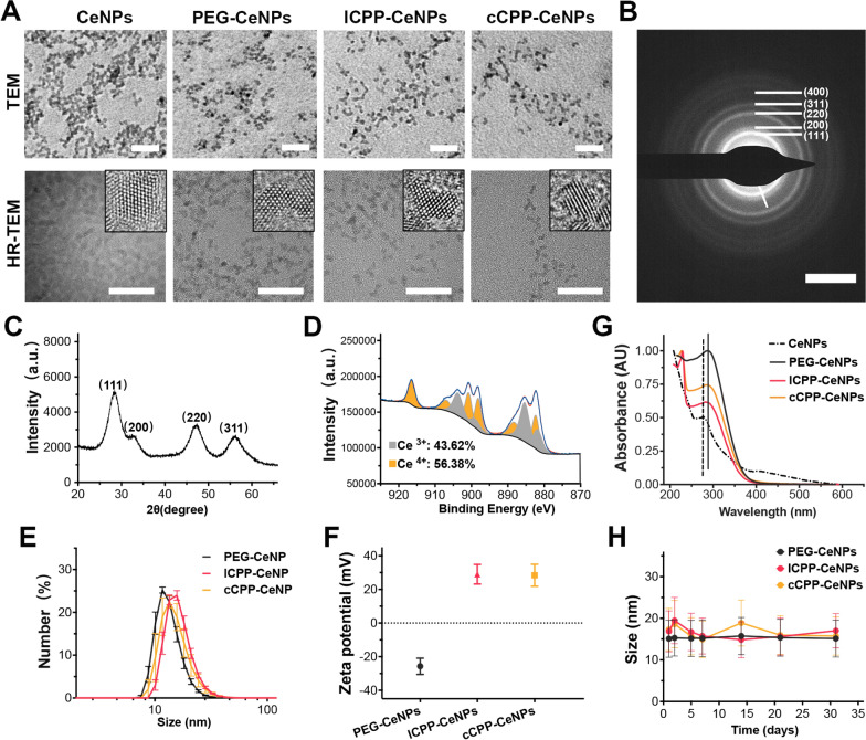
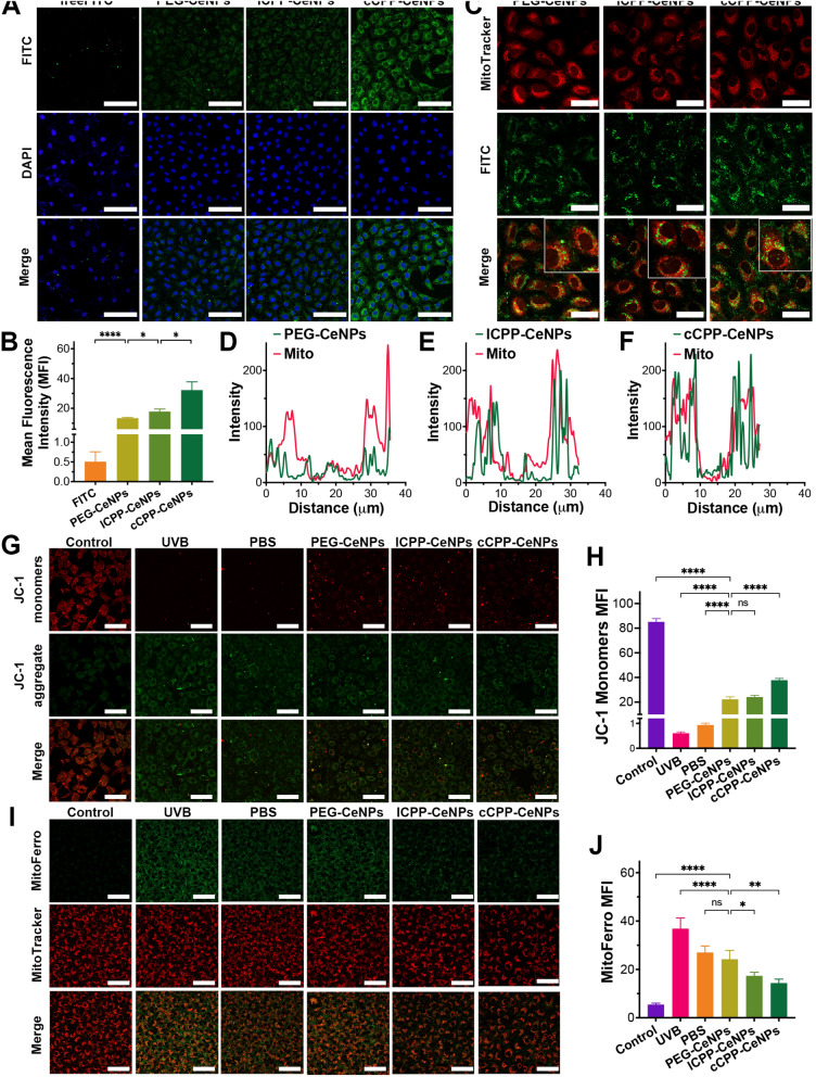
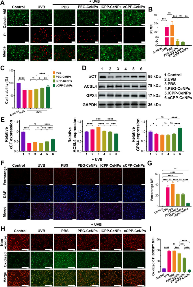
Oxidative stress, which results from the accumulation of free radicals, plays a substantial role in cataract formation. Antioxidants have shown promise in mitigating or even preventing this process. However, delivering antioxidants noninvasively to the anterior segment of the eye has been a significant challenge. In this study, we developed ceria nanoparticles modified with cyclic cell-penetrating peptides to overcome the obstruction of the dense corneal barrier on topical drug delivery. Our results demonstrated that modified ceria nanoparticles with cell-penetrating peptides (CPPs) facilitate the opening of tight junctions in human corneal epithelial cells. This characteristic considerably enhances the trans-corneal transport of nanoparticles and improves cellular uptake efficiency, while also contributing to their intracellular enrichment toward mitochondria. Further experiments confirmed that the modified ceria nanoparticles effectively counteracted ferroptosis induced by oxidative stress in lens epithelial cells both in vitro and in vivo, substantially reducing cataract formation. The successful development of ceria nanoparticles modified with cyclic cell-penetrating peptides (cCPPs) opens new avenues for research in cataract prevention and treatment. Additionally, the modified ceria nanoparticles could serve as a noninvasive drug delivery system, which holds remarkable potential for advancing drug delivery in diseases affecting the anterior segment of the eye.

Keywords: Antioxidants; Cataracts; Ceria nanoparticles; Cyclic cell-penetrating peptides; Ferroptosis; Oxidative stress.


Conflict of interest statement

Declarations. Ethics approval and consent to participate: All animal experiments have complied with the Association for Research in Vision and Ophthalmology Statement and the guidelines for the Animal Care and Use Committee, Zhejiang University. All animal experimental protocols were approved by the Animal Ethics Committee, the Second Affiliated Hospital, School of Medicine, Zhejiang University (Approval number: 2023–084). Consent for publication: Not applicable. Competing interests: The authors declare no competing interests.

Figures

Fig. 1
Fig. 1
Schematic diagram showing the synthesis process and mechanism of cCPP-CeNPs for alleviating cataracts
Fig. 2
Fig. 2
Illustration of ceria nanoparticles and their morphology, structure, size, surface charges, and stability. A Representative TEM and HR-TEM images of CeNPs dispersed in chloroform, PEG-CeNPs, lCPP-CeNPs and cCPP-CeNPs dispersed in water (scale bar: 20 nm). B The SAED pattern (right panel) of CeNPs dispersed in chloroform (scale bar: 5 nm). C XRD analysis of CeNPs. D XPS spectra of CeNPs. E Hydrodynamic diameters of PEG-CeNPs, lCPP-CeNPs and cCPP-CeNPs. F ζ-potentials of PEG-CeNPs, lCPP-CeNPs and cCPP-CeNPs. G UV–Vis spectrum of ceria nanoparticles before (dotted line) and after (solid line) surface modification. H Size changes of PEG-CeNPs, lCPP-CeNPs and cCPP-CeNPs in artificial tears over 30 days
Fig. 3
Fig. 3
ROS scavenging performance and auto-regeneration feature of ceria nanoparticles. A Color changes of CeNPs upon adding H2O2 and subsequent aging. B The UV–Vis spectrum depicting H2O2 quenching of the water-soluble PEG-CeNPs, lCPP-CeNPs, and cCPP-CeNPs. The band exhibited a red shift from the control (dotted line) to the as-reacted solution (solid line) upon H2O2 adding. C SOD-mimetic activities of PEG-CeNPs, lCPP-CeNPs, and cCPP-CeNPs. D Experimental X-band (9.867 GHz) ESR spectra of aqueous solutions of Control, 0.5 mM cCPP-CeNPs, 1.0 mM cCPP-CeNPs, and 5.0 mM cCPP-CeNPs. E and F Representative ROS staining images (E) of lens epithelial cells in the presence of Rosup and ceria nanoparticles and statistics of relative mean fluorescence intensity (F) (scale bar: 100 µm). In all histograms, data is presented as the mean ± SD (Standard Deviation) (n = 3). *p < 0.05, **p < 0.01, ***p < 0.001, ****p < 0.0001; ns, no significance
Fig. 4
Fig. 4
Ophthalmic penetrating mechanism of CeNPs. A Confocal images of the cornea and anterior capsule after administering eyedrops containing free-FITC, PEG-CeNPs, lCPP-CeNPs, and cCPP-CeNPs (scale bar: 500 µm). White arrows point to the anterior capsule of the lens. B Immunofluorescence imaging of ZO-1 in the corneal epithelium of mice (scale bar: 40 µm). C Immunofluorescence imaging of ZO-1 in HCECs (scale bar: 50 µm). D TEM images of mice corneal tissue following treatment with PBS, PEG-CeNPs, lCPP-CeNPs, and cCPP-CeNPs. Red frames indicate corneal epithelial tight junctions (scale bars: 2 µm)
Fig. 5
Fig. 5
Cellular uptake and intracellular spatial localization of CeNPs. Confocal images (A) and statistics (B) of HLEB3 cells 2 h after treatment with freeFITC, PEG-CeNPs, lCPP-CeNPs, and cCPP-CeNPs (scale bar: 100 µm). (C) Images of mitochondria staining of HLEB3 cells treated for 2 h with PEG-CeNPs, lCPP-CeNPs, and cCPP-CeNPs (scale bars: 40 µm), and the co-localization analysis (DF) on the fluorescence intensity of ceria nanoparticles with mitochondria. (G) ΔΨm in indicated groups of HLEB3 cells and (H) statistics. Investigated using JC-1 staining (scale bar: 100 µm). Confocal images (I) and statistics (J) of mitochondrial iron content detected by MitoFerro staining (scale bar: 100 µm). In all histograms, data is presented as the mean ± SD (n = 4). *p < 0.05, **p < 0.01, ***p < 0.001, ****p < 0.0001; ns, no significance
Fig. 6
Fig. 6
Anti-ferroptosis effect of CeNPs in UVB-stimulated HLEB3 cells. A Representative images and statistics (B) of living/dead cell assay of HLEB3 cells treated with UVB and different CeNPs. Living and dead HLEB3 cells were stained with calcein acetoxymethyl ester (calcein-AM, green) and propidium iodide (PI, red), respectively (scale bars: 100 µm). C Cell viability of HLEB3 cells was evaluated using the CCK-8 assay following 60 mJ/cm2 UVB exposure and pre-treatment with PEG-CeNPs, lCPP-CeNPs, and cCPP-CeNPs (equal ceria concentration, 500 µM). In the histogram, data is presented as the mean ± SD (n = 4). Western blot (D) and statistics (E) of the expression of ferroptosis-related proteins; Glyceraldehyde 3-phosphate dehydrogenase (GAPDH) served as the internal control. F Ferrorange staining and statistics (G) was utilized to determine the total iron level in HLEB3 cells treated with UVB and CeNPs (scale bars: 100 µm). H Representative fluorescence microscopy images and statistics (I) of BDP581/591 staining, indicating lipid peroxidation in HLEB3 cells (non-oxidized and oxidized cells stained red and green, respectively (scale bars: 200 µm)). In all other histograms, data is presented as the mean ± SD (n = 3). *p < 0.05, **p < 0.01, ***p < 0.001, ****p < 0.0001; ns, no significance
Fig. 7
Fig. 7
Anticataract effects of CeNPs in the anterior capsule of UVB-stimulated mice via alleviating HLECs ferroptosis. A Representative slit lamp microscope images of mouse eyes at 7 th day after UV radiation modeling (n = 6). Control (untreated); PBS group; PEG-CeNPs group (3 mg/mL); lCPP-CeNPs group (3 mg/mL); cCPP-CeNPs group (3 mg/mL); pirenoxine sodium eye drops. B The formation of anterior subcapsular cataract in the lenses after 3.6 kJ/m2 illumination of UV exposure and indicated treatments. Immunofluorescence staining (C) and statistics (D) of ferroptosis-related proteins ACSL4, GPX4 and xCT (scale bar, 20 µm). In all histograms, data is presented as the mean ± SD (n = 3). *p < 0.05, **p < 0.01, ***p < 0.001, ****p < 0.0001; ns, no significance

Similar articles

References

    1. Pesudovs K, et al. Cataract-related blindness and vision impairment in 2020 and trends over time in relation to VISION 2020: the right to sight: an analysis for the global burden of disease study. Invest Ophthalmol Vis Sci. 2021;62(8):3523.
    1. Pesudovs K, et al. Global estimates on the number of people blind or visually impaired by cataract: a meta-analysis from 2000 to 2020. Eye. 2024;57:14. - PMC - PubMed
    1. Ruiss M, Findl O, Kronschläger M. The human lens: an antioxidant-dependent tissue revealed by the role of caffeine. Ageing Res Rev. 2022;79: 101664. - PubMed
    1. Michael R, Bron AJ. The ageing lens and cataract: a model of normal and pathological ageing. Phil Trans R Soc B Biol Sci. 2011;366(1568):1278–92. - PMC - PubMed
    1. Cicinelli MV, et al. Cataracts. Lancet. 2023;401(10374):377–89. - PubMed

MeSH terms

LinkOut - more resources