. 2025 May 7;20(1):59.
doi: 10.1186/s13020-025-01104-2.

Invasive laser acupuncture targeting muscle: a novel approach to protect dopaminergic neurons and reduce neuroinflammation in a brain of Parkinson's disease model

Affiliations

Invasive laser acupuncture targeting muscle: a novel approach to protect dopaminergic neurons and reduce neuroinflammation in a brain of Parkinson's disease model

Halin Jeon et al. Chin Med. .

Abstract

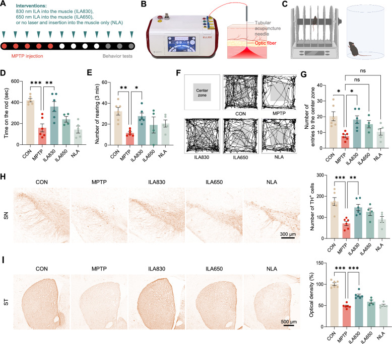
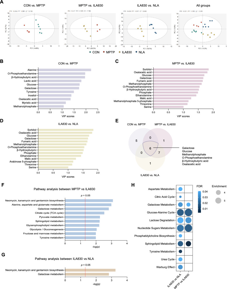
Parkinson's disease (PD) affects 1-2% of the global population and presents significant therapeutic challenges. Due to the limitations of existing treatments, there is a pressing need for alternative approaches. This study investigated the effects of invasive laser acupuncture (ILA), which combines acupuncture and photobiomodulation. In this method, optical fibers are inserted into the muscle layers of the acupoint to enhance therapeutic outcomes. Mice with MPTP-induced PD were treated with ILA at 830 nm or 650 nm. Protective effects of nigrostriatal dopaminergic neurons and fibers were assessed by examining TH immunoreactivity in the brain. Neuroinflammation markers in the brain and muscle metabolomic profiles were also analyzed. Comparisons between invasive and non-invasive laser application, as well as the impact of nerve blocking with lidocaine, were also evaluated. ILA at 830 nm (ILA830) significantly improved motor performance and increased the nigrostriatal TH-positive immunoreactivities. It reduced the levels of α-synuclein, apoptotic proteins, and inflammatory cytokines, while increasing anti-inflammatory in the brain. ILA830 also decreased nigrostriatal astrocyte and microglia activation. Muscle metabolomic analysis showed distinct group clustering and significant changes in metabolites like glucose and galactose, correlating with improved motor functions. Invasive laser treatment was more effective than non-invasive, and lidocaine pre-treatment did not block its effects. ILA at 830 nm effectively ameliorates PD symptoms by protecting dopaminergic neurons, and reducing neuroinflammation in the brain. Muscle metabolomic changes by ILA830, such as increased glucose and galactose, correlate with motor improvement. This approach offers a promising strategy for PD treatment, warranting further research to optimize its use in clinical settings.


Conflict of interest statement

Declarations. Competing interests: The authors declare no competing interests. Ethics approval and consent to participate: All experimental procedures involving animals were conducted in accordance with the guidelines of the National Institutes of Health (NIH) for the care and use of laboratory animals and were approved by the Institutional Animal Ethical Committee at Kyung Hee University. The ethics approval number is KHSASP-23-280.

Figures

Fig. 1
Fig. 1
Effects of ILA on motor functions and dopaminergic neuronal death in MPTP-induced PD mice. A Experimental design. B Image of the novel acupuncture needle integrating with laser technology. C Illustrative demonstration of rotarod and cylinder tests. D Latency to fall off the rotating rod. E Cylinder test results for the number of rearing behaviors. F Representative images of OF test. G Number of entries into the center zone in the OF test. H Representative images and the number of TH-positive cells in the SN. I Representative images and optical density of ST. The results were presented as mean ± SEM, * p < 0.05, * p < 0.01, *** p < 0.001
Fig. 2
Fig. 2
Changes of neuroprotective molecules by ILA830 in the brain. A Representative western blot images of α-synuclein, Akt, Bax, Bcl-2, and β-actin in the SN. B Western blot analysis of phosphorylated α-synuclein normalized to β-actin in the SN. C Western blot analysis of phosphorylated Akt and Akt in the SN. D Statistical analysis of the protein expression levels of Bax, normalized to β-actin in the SN. E Statistical analysis of the protein expression levels of Bcl-2, normalized to β-actin in the SN. F Statistical analysis of the ratio of Bax/Bcl-2 in the SN. Data are shown as the mean ± SEM. * p < 0.05, ** p < 0.01, *** p < 0.001
Fig. 3
Fig. 3
Effects of ILA830 on inflammatory responses and glial activation in the brain. A Representative western blot images of JNK, p38, NF-κB, and β-actin in the SN. B Statistical analysis of the protein expression levels of p-JNK/JNK in the SN. C Statistical analysis of the protein expression levels of p-38/p38 in the SN. D Statistical analysis of the ratio of NF-κB, normalized to β-actin in the SN. E ELISA analysis of proinflammatory cytokine IL-6 in the ST. F ELISA analysis of proinflammatory cytokine IL-1β in the ST. G ELISA analysis of proinflammatory cytokine TNF-α in the ST. H ELISA analysis of anti-inflammatory cytokine IL-10 in the ST. Representative images of immunofluorescence staining of GFAP- and IBA1-positive cells in the SN and ST. Scale bar = 300 (I), 10 (J) μm. K,M Quantitative analysis of GFAP-positive cells in the SN and ST. L,N Quantitative analysis of IBA1-positive cells in the SN and ST. O,R Representative images of astrocytic processes via Sholl analysis in the SN and ST. Scale bar = 100 (I), 20 (J) μm. P,S The number of intersections of astrocytic processes at different distances from the soma in the SN and ST. Q,T The number of total intersections within 50 µm radius from the soma in the SN and ST. Data are shown as the mean ± SEM. * p < 0.05, ** p < 0.01, *** p < 0.001
Fig. 4
Fig. 4
Alterations of metabolites and pathway analysis following ILA treatment in MPTP-induced PD mice. A Score plot of PLS-DA. BD List of metabolites with VIP scores greater than 1. E Venn diagram showing the common metabolites that showed changes in comparisons between groups; galactose, glucose, methanolphosphate, O-phosphoethanolamine, β-hydroxybutyric acid, oxalacetic acid. F,G Bar chart displaying significant pathways. H Bubble chart showing significant pathways of F and G; the size of bubbles represents the enrichment factor of the pathway and the color shows the p value of each pathway
Fig. 5
Fig. 5
Effects of ILA on motor function, dopaminergic neuronal survival, and their correlation with muscle metabolites in MPTP-induced PD mice. A Experimental design. B Latency to fall off the rotating rod. C Number of rearing in the cylinder test. D Number of entries into the center zone recorded in the OF test. E Fluorescence raw traces of HT-22 cells expressing GCaMP6s during laser exposure. F dF/F0 graph of GCaMP6s-expressing HT-22 cells under laser irradiation at 60 Hz, with wavelengths of 830 nm and 650 nm (830 nm: −0.66 ± 0.29%, 650 nm: −0.81 ± 0.46% n = 13 cells). G Heatmap demonstrating the correlations of muscle metabolites and behavioral test results. HJ Bar graphs demonstrating the relative abundance of glucose, galactose, and O-phosphoethanolamine, along with their correlation with behavioral test results. Data are shown as the mean ± SEM. * p < 0.05, ** p < 0.01, *** p < 0.001
Fig. 6
Fig. 6
Schematic diagram of ILA830 treatment effects on PD symptom improvement via muscle metabolic alterations and brain recovery in a PD mouse model. ILA at the GB34 acupoint using an 830 nm wavelength stimulates the muscle layer, inducing therapeutic effects in the MPTP-induced PD mouse model. This targeted stimulation leads to significant alterations in muscle metabolites, such as glucose and galactose, enhancing systemic energy availability. These peripheral metabolic changes are proposed to influence the brain, leading to anti-inflammatory and anti-apoptotic effects. Consequently, ILA treatment contributes to the improvement of both motor and non-motor symptoms observed in PD

References

    1. Mhyre TR, et al. Parkinson’s disease. Subcell Biochem. 2012;65:389–455. - PMC - PubMed
    1. Zaman V, et al. Cellular and molecular pathophysiology in the progression of Parkinson’s disease. Metab Brain Dis. 2021;36(5):815–27. - PMC - PubMed
    1. GBD 2016 Parkinson’s Disease Collaborators. Global, regional, and national burden of Parkinson’s disease, 1990–2016: a systematic analysis for the Global Burden of Disease Study 2016. Lancet Neurol. 2018;17(11):939–53. - PMC - PubMed
    1. Aarsland D, et al. Parkinson disease-associated cognitive impairment. Nat Rev Dis Primers. 2021;7(1):47. - PubMed
    1. Jankovic J. Parkinson’s disease: clinical features and diagnosis. J Neurol Neurosurg Psychiatry. 2008;79(4):368–76. - PubMed

LinkOut - more resources