Review
. 2025 May;58(3):337-351.
doi: 10.5946/ce.2024.263. Epub 2025 May 8.

Classification of image-enhanced endoscopy in colon tumors

Affiliations
Review

Classification of image-enhanced endoscopy in colon tumors

One-Zoong Kim. Clin Endosc. 2025 May.

Abstract

Colorectal cancer accounts for 10% of global cancer cases in each year, making accurate evaluation and resection crucial. Imaging-enhanced endoscopy helps differentiate between hyperplastic polyps and adenomas, guiding treatment decisions. Colon tumors are classified into benign (e.g., serrated and adenomatous polyps) and malignant (e.g., adenocarcinomas). The Paris classification categorizes superficial neoplastic lesions by morphology, while laterally spreading tumors are classified by size and growth pattern. Effective classification aids in determining resectability and appropriate interventions for colon tumors, ultimately improving patient outcomes. Image-enhanced endoscopy improves colon tumor diagnosis using various techniques like dye, optical, and electronic methods. Kudo's pit pattern categorizes lesions based on surface morphology using dye, while Sano, Jikei, and Hiroshima classifications focus on vascular patterns using narrow-band imaging (NBI). The NBI International Colorectal Endoscopic (NICE) classification integrates these methods to identify lesions, especially deep submucosal invasive cancers. The Workgroup Serrated Polyps and Polyposis (WASP) classification targets sessile serrated lesions, and the Japan NBI Expert Team (JNET) classification further refines adenoma categorization with low- and high-grade adenoma. The Colorectal Neoplasia Endoscopic Classification to Choose the Treatment (CONECCT) classification consolidates multiple systems for comprehensive assessment, aiding in treatment decisions and potentially applicable to artificial intelligence for diagnostic validation across imaging modalities like linked color imaging, blue light imaging, or i-scan.

Keywords: Adenomatous polyps; Colonic polyps; Colorectal neoplasms; Endoscopy; Narrow band imaging.


Conflict of interest statement

Conflicts of Interest

The author has no potential conflicts of interest.

Figures

Fig. 1.
Fig. 1.
Classification of the degree of submucosal invasion. We divided submucosal cancers into sm1, 2, and 3, according to the depth of invasion, and further divided sm1 lesions into 1a, 1b, and 1c, according to the width of invasion. Reused from the article of Wada et al. Gastrointest Endosc 2009;70:522–531, with original copyright holder’s permission.
Fig. 2.
Fig. 2.
Schematic representation of the Paris classification of polyp morphology. M, mucosal layer; SM, submucosal layer. Adapted from Johnson et al. Can J Surg 2023;66:E491–E498, according to the Creative Commons license.
Fig. 3.
Fig. 3.
Laterally spreading tumor (LST) classification. SMI, submucosal invasion; LST-G, LST granular; LST-NG, LST non-granular; CI, confidence interval. Adapted from Castillo-Regalado and Uchima. World J Gastrointest Endosc 2022;14:113–128, according to the Creative Commons license.
Fig. 4.
Fig. 4.
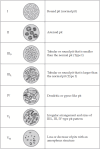
Kudo and Tsuruta pit pattern classification for colorectal neoplasia. Adapted from Tanaka et al. Dig Endosc 2004;16:S161–S164, with original copyright holder’s permission.
Fig. 5.
Fig. 5.
(A) A highly magnified image showing irregular pits that vary in size. (B) In a low magnification colonoscopic image, the pits do not occupy the entire surface; this pattern defined the non-V lesion in the present study. (C, D) Magnifying pictures. Adapted from Ohta et al. Dig Endosc 2004;16:308–314, with original copyright holder’s permission.
Fig. 6.
Fig. 6.
Capillary pattern classification. Adapted from Ikematsu et al. BMC Gastroenterol 2010;10:33, according to the Creative Commons license.
Fig. 7.
Fig. 7.
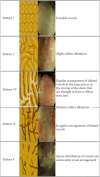
Jikei classification. Adapted from Sano et al. Dig Endosc 2016;28:526–533, with original copyright holder’s permission.
Fig. 8.
Fig. 8.
NBI magnification findings of colorectal lesion. Microvessels are not observed or are extremely opaque (type A); fine microvessels are observed around the pits, and clear pits can be observed via the nest of microvessels (type B); microvessels are irregular, and the vessel diameter or distribution is heterogeneous (type C). Reused from the article of Kanao et al. Gastrointest Endosc 2009;69(3 Pt 2):631–636, with original copyright holder’s permission.
Fig. 9.
Fig. 9.
NBI magnification subclassification of type C. Microvessels comprise an irregular network, pits observed via the microvessels are slightly nondistinct, and vessel diameter or distribution is homogeneous (C1); microvessels comprise an irregular network, pits observed via the microvessels are irregular, and vessel diameter or distribution is heterogeneous (between types C1 and C3) (C2); pits via the microvessels are invisible, irregular vessel diameter is thick, or the vessel distribution is heterogeneous, and avascular areas are observed (C3). Reused from the article of Kanao et al. 2009;69(3 Pt 2):631–636, with original copyright holder’s permission.
Fig. 10.
Fig. 10.
Showa classification. Adapted from Sano et al. Dig Endosc 2016;28:526–533, with original copyright holder’s permission.
Fig. 11.
Fig. 11.
The NBI international colorectal endoscopic (NICE) classification. a)Can be applied using colonoscopes with or without optical (zoom) magnification. b)These structures (regular or irregular) may represent the pits and the epithelium of the crypt opening. c)Type 2 consists of Vienna classification types 3, 4 and superficial 5 (all adenomas with either low or high grade dysplasia, or with superficial submucosal carcinoma). The presence of high grade dysplasia or superficial submucosal carcinoma may be suggested by an irregular vessel or surface pattern, and is often associated with atypical morphology (e.g., depressed area). Adapted from Hayashi et al. Gastrointest Endosc 2013;78:625–632, with original copyright holder’s permission.
Fig. 12.
Fig. 12.
The WASP classification: method for optical diagnosis of hyperplastic polyps, sessile serrated adenomas/polyps and adenomas based on the NICE criteria and the Hazewinkel criteria in a stepwise approach. Reused from the article of IJspeert et al. Gut 2016;65:963–970, with original copyright holder’s permission.
Fig. 13.
Fig. 13.
The Japan NBI Expert Team (JNET) classification. a)If visible, the caliber in the lesion is similar to surrounding normal mucosa. b)Micro-vessels are often distributed in a punctate pattern and well-ordered reticular or spiral vessels may not be observed in depressed lesions. c)Deep submucosal invasive cancer may be included. Adapted from Sano et al. Dig Endosc 2016;28:526–533, with original copyright holder’s permission.
Fig. 14.
Fig. 14.
The Colorectal Neoplasia Endoscopic Classification to Choose the Treatment (CONECCT) table. The words in bold are the ones that require particular attention when analyzing lesions. LST, laterally spreading tumor; NBI, narrow-band imaging; ESD, endoscopic submucosal dissection. Adapted Fabritius et al. Endosc Int Open 2019;7:e1197–e1206, according to the Creative Commons license.

References

    1. Kashida H, Kudo SE. Early colorectal cancer: concept, diagnosis, and management. Int J Clin Oncol. 2006;11:1–8. doi: 10.1007/s10147-005-0550-5. - DOI - PubMed
    1. Dudás B. Human histology: a text and atlas for physicians and scientists. Elsevier; 2023.
    1. Kumar V, Abbas MBBS AK, Aster JC. Robbins and cotran pathologic basis of disease. 10th ed. Elsevier; 2021.
    1. Lamps LW, Bellizzi AM, Frankel WL, et al. Neoplastic gastrointestinal pathology: an illustrated guide. Demos Medical Publishing; 2016.
    1. Kudo S. Endoscopic mucosal resection of flat and depressed types of early colorectal cancer. Endoscopy. 1993;25:455–461. doi: 10.1055/s-2007-1010367. - DOI - PubMed

LinkOut - more resources