

Dual-Energy CT in Breast Cancer: Current Applications and Future Outlooks
- PMID: 40336872
- PMCID: PMC11935073
- DOI: 10.1002/pro6.1213
Dual-Energy CT in Breast Cancer: Current Applications and Future Outlooks
Abstract
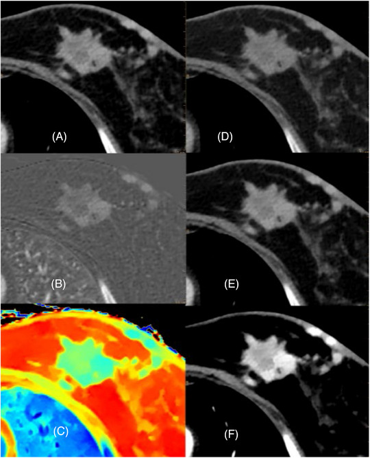
Breast cancer is the most prevalent cancerous tumor in women, characterized by different subtypes and varying responses to treatment. The continued evolution of breast cancer diagnosis and management has resulted in a transition from a one-size-fits-all approach to a new era of personalized treatment plans. Therefore, it is essential to accurately identify the biological characteristics of breast tissue in order to minimize unnecessary biopsies of benign lesions and improve the overall clinical process, leading to reduced expenses and complications associated with invasive biopsy procedures. Challenges for future research include finding ways to predict the response of breast cancer patients to adjuvant systemic treatment. Dual-energy CT (DECT) is a new imaging technology integrating functional imaging and molecular imaging. Over the past decade, DECT has gained relevancy, especially in oncological radiology. This article proposed a literature review of the application and research status of DECT in breast cancer treatment strategy determination and prognosis prediction.
Keywords: Breast cancer; Clinical diagnosis; Dual‐energy CT; Prognosis prediction; Quantitative parameters.
© 2023 The Authors. Precision Radiation Oncology published by John Wiley & Sons Australia, Ltd on behalf of Shandong Cancer Hospital & Institute.
Conflict of interest statement
The authors declare no conflicts of interest.
Figures
References
-
- Giaquinto AN, Sung H, Miller KD, et al. Breast Cancer Statistics, 2022. CA Cancer J Clin. 2022;72(6):524‐541. - PubMed
-
- Chae EJ, Song JW, Seo JB, Krauss B, Jang YM, Song KS. Clinical utility of dual‐energy CT in the evaluation of solitary pulmonary nodules: initial experience. Radiology. 2008;249(2):671‐681. - PubMed
-
- De Cecco CN, Darnell A, Rengo M, et al. Dual‐energy CT: oncologic applications. AJR Am J Roentgenol. 2012;199(5 Suppl):S98‐S105. - PubMed
-
- Lennartz S, Le Blanc M, Zopfs D, et al. Dual‐Energy CT‐derived Iodine Maps: Use in Assessing Pleural Carcinomatosis. Radiology. 2019;290(3):796‐804. - PubMed
-
- Agostini A, Borgheresi A, Mari A, et al. Dual‐energy CT: theoretical principles and clinical applications. Radiol Med. 2019;124(12):1281‐1295. - PubMed
Publication types
LinkOut - more resources
Full Text Sources
Miscellaneous