. 2025 Apr 30;16(22):10010-10018.
doi: 10.1039/d5sc01150g. eCollection 2025 Jun 4.

Azetidinyl Malachite Green: a superior fluorogen-activating protein probe for live-cell and dynamic SIM imaging

Affiliations

Azetidinyl Malachite Green: a superior fluorogen-activating protein probe for live-cell and dynamic SIM imaging

Fei Deng et al. Chem Sci. .

Abstract

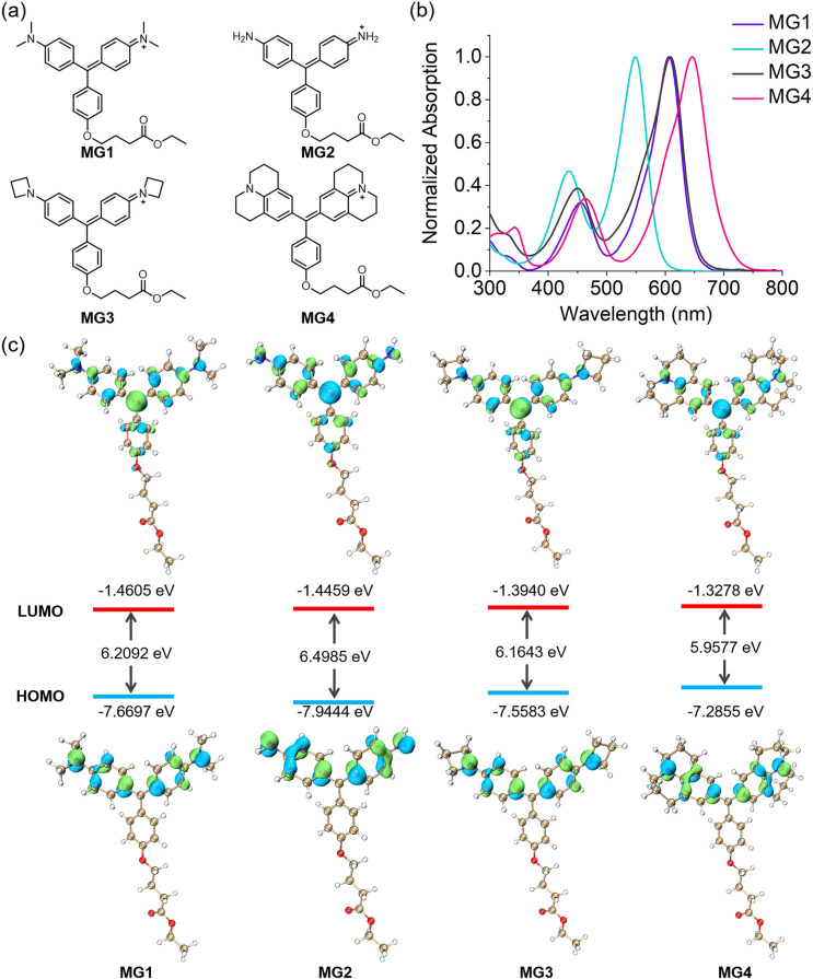
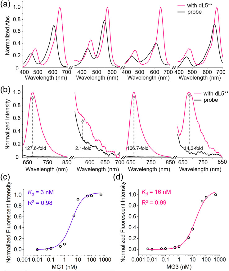
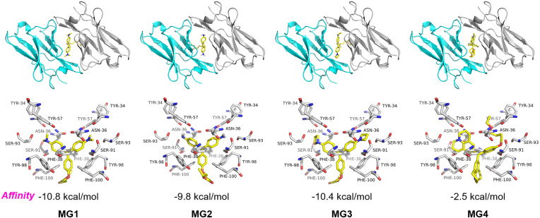
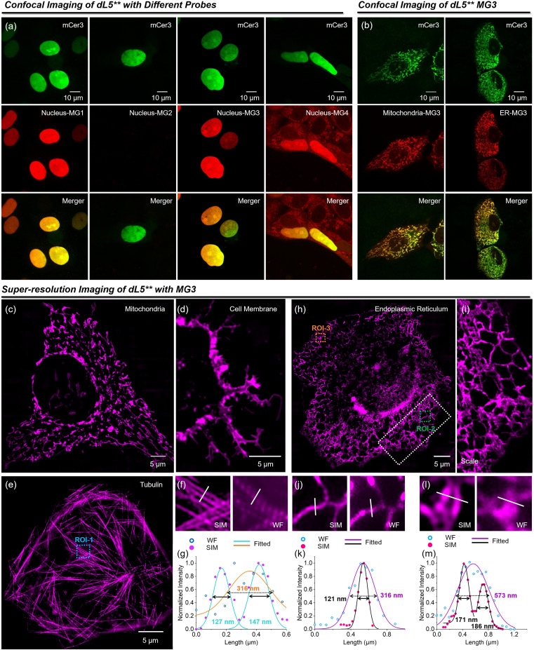
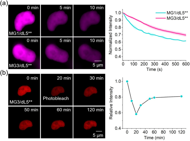
Malachite Green (MG) and its fluorogen-activating protein (FAP) pair are valuable tools for live-cell and super-resolution fluorescence imaging due to their unique near-infrared absorption and signal enhancement. However, the low brightness and photostability of MG have limited its use in dynamic imaging. In this study, we introduce a novel derivative, azetidinly Malachite Green (Aze-MG), which enhances the brightness of the MG-FAP complex by 2.6-fold. This enhancement is achieved by replacing the N,N-dimethylamino group in MG with an azetidine group, which suppresses the twisted intramolecular charge transfer (TICT) effect, leading to improved quantum yield and photostability. Additionally, the reduced binding affinity of Aze-MG for FAP enables a buffering strategy, allowing the reversible exchange of photobleached fluorogens with free fluorogens, thereby ensuring stable fluorescence over time. This combination of improved brightness and buffering capability makes Aze-MG an ideal probe for live-cell and dynamic SIM imaging.


Conflict of interest statement

There are no conflicts to declare.

Figures

Fig. 1
Fig. 1. (a) Molecular structures of MG1–4. (b) Absorption spectra of MG1–4 in PBS (pH 7.40). (c) Computed frontier orbitals and energy levels of MG1–4.
Fig. 2
Fig. 2. (a) Absorption and (b) fluorescence spectra of 0.5 μM MGs in the absence and presence of 2 μM dL5** (MG1–4: from left to right). Binding equilibrium analysis of MG1 (c) and MG3 (d) to dL5**.
Fig. 3
Fig. 3. Molecular docking analysis of MGs. (Top) Frontal view of the MGs/dL5** complex. (Bottom) Spatial distribution of amino acid side chains in dL5** that come in contact with MGs.
Fig. 4
Fig. 4. (a) Confocal images of HeLa cells expressing dL5**-mCer3 in the nucleus with MG1–4. (b) Confocal images of HeLa cells expressing dL5**-mCer3 in mitochondria and ER with the addition of 200 nM MG3. Green fluorescence signals from mCer3; red fluorescence signals from MG/dL5**. SIM imaging of mitochondria (c), cell membrane (d), tubulin (e) and endoplasmic reticulum (h). (i) Enlarged view of ROI-2 in (h). (f) SIM and widefield of ROI-1 in (e). (g) Intensity distributions across tubulin (white line) in (f). SIM and widefield of ROI-2 (j) and ROI-3 (l) in (h). (k) and (m) Intensity distributions across ER (white line) in (j) and (l).
Fig. 5
Fig. 5. (a) Quantification of the relative fluorescent intensity of MG1/dL5** and MG3/dL5** in HeLa cells with continuous irradiation using super-resolution SIM microscopy. (b) Fluorescence recovery of MG3/dL5** in HeLa cells after 20 min of continuous irradiation using a 640 nm laser.
Fig. 6
Fig. 6. Time-lapse SIM images of (a) mitochondrial fission, (b) mitophagy, ER tubule (c) and RER dynamics (d) in living HeLa cells through MG3/dL5** labeling. (e) Time colored outline of the cavity in ROI-4. And the time dependence of area in ROI-4-6. (f) Dual-color time-lapse SIM images of mitochondria and ER with MG3/dL5** and GFP in living HeLa cells.

Similar articles

References

    1. Daugird T. A. Shi Y. Holland K. L. Rostamian H. Liu Z. Lavis L. D. Rodriguez J. Strahl B. D. Legant W. R. Correlative single molecule lattice light sheet imaging reveals the dynamic relationship between nucleosomes and the local chromatin environment. Nat. Commun. 2024;15:4178. doi: 10.1038/s41467-024-48562-0. - DOI - PMC - PubMed
    1. Qiao Q. Liu W. Zhang Y. Chen J. Wang G. Tao Y. Miao L. Jiang W. An K. Xu Z. In Situ Real-Time Nanoscale Resolution of Structural Evolution and Dynamics of Fluorescent Self-Assemblies by Super-Resolution Imaging. Angew. Chem., Int. Ed. 2022;134:e202208678. doi: 10.1002/ange.202208678. - DOI - PubMed
    1. Miao L. Yan C. Chen Y. Zhou W. Zhou X. Qiao Q. Xu Z. SIM imaging resolves endocytosis of SARS-CoV-2 spike RBD in living cells. Cell Chem. Biol. 2023;30:248–260. doi: 10.1016/j.chembiol.2023.02.001. - DOI - PMC - PubMed
    1. Samanta S. Lai K. Wu F. Liu Y. Cai S. Yang X. Qu J. Yang Z. Xanthene, cyanine, oxazine and BODIPY: the four pillars of the fluorophore empire for super-resolution bioimaging. Chem. Soc. Rev. 2023;52:7197–7261. doi: 10.1039/D2CS00905F. - DOI - PubMed
    1. Kozma E. Kele P. Fluorogenic probes for super-resolution microscopy. Org. Biomol. Chem. 2019;17:215–233. - PubMed

LinkOut - more resources