

Niclosamide: CRL4AMBRA1 mediated degradation of cyclin D1 following mitochondrial membrane depolarization
- PMID: 40337304
- PMCID: PMC12054360
- DOI: 10.1039/d5md00054h
Niclosamide: CRL4AMBRA1 mediated degradation of cyclin D1 following mitochondrial membrane depolarization
Abstract
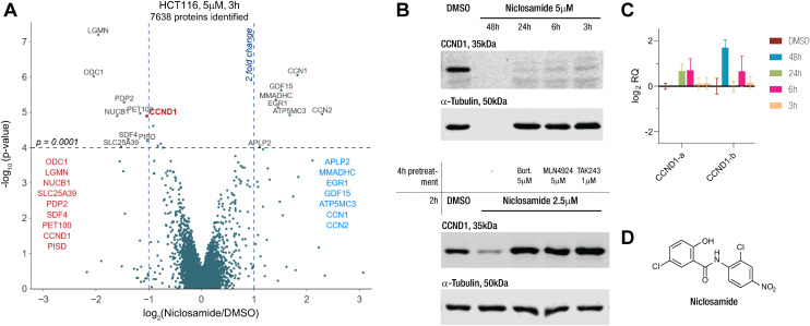
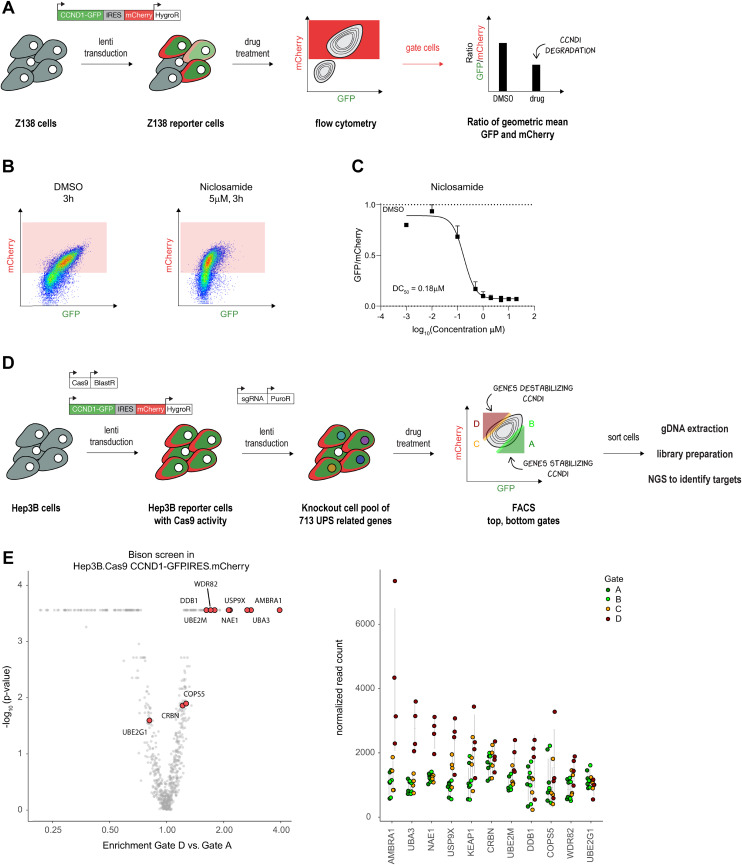
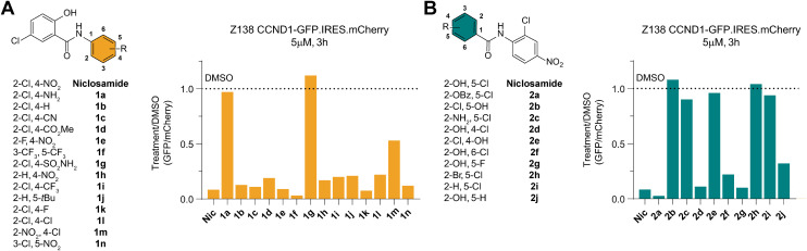
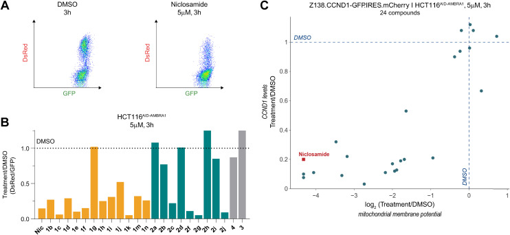
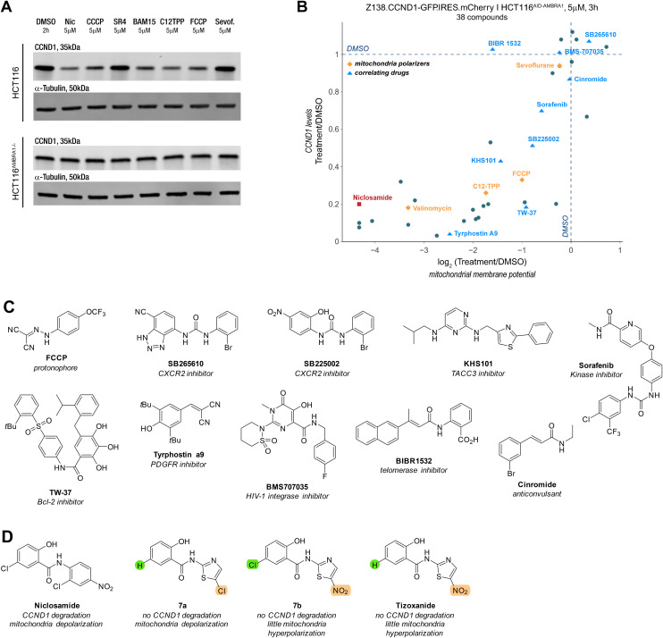
Targeted protein degradation has emerged as a promising approach in drug discovery, utilizing small molecules like molecular glue degraders to harness the ubiquitin-proteasome pathway for selective degradation of disease-driving proteins. Based on results from proteomics screens we investigated the potential of niclosamide, an FDA-approved anthelmintic drug with a 50 year history in treating tapeworm infections, as a molecular glue degrader targeting the proto-oncogene cyclin D1. Proteomics screens in HCT116 colon carcinoma and KELLY neuroblastoma cells, found that niclosamide induces rapid cyclin D1 degradation through a mechanism involving the ubiquitin-proteasome pathway. A genetic CRISPR screen identified the E3 ligase CRL4AMBRA1 as a key player in this process. Structure-activity relationship studies highlighted critical features of niclosamide necessary for cyclin D1 degradation, demonstrating a correlation between mitochondrial membrane potential (MMP) disruption and cyclin D1 downregulation. Notably, various mitochondrial uncouplers and other compounds with similar drug sensitivity profiles share this correlation suggesting that MMP disruption can trigger cyclin D1 degradation, and that the cellular signal driving the degradation differs from previously described mechanism involving CRL4AMBRA1. Our findings underscore the complexities of proteostatic mechanisms and the multitude of mechanisms that contribute to degrader drug action.
This journal is © The Royal Society of Chemistry.
Conflict of interest statement
N. H. T. is a founder and shareholder of Zenith Therapeutics as well a consultant to Ridgeline Discovery and Red Ridge Bio. B. L. E. has received research funding from Novartis and Calico. He has received consulting fees from Abbvie. He is a member of the scientific advisory board and shareholder for Neomorph Inc., TenSixteen Bio, Skyhawk Therapeutics, and Exo Therapeutics. E. S. F. is a founder, scientific advisory board (SAB) member, and equity holder of Civetta Therapeutics, Proximity Therapeutics, Stelexis Biosciences, and Neomorph, Inc. (also board of directors). He is an equity holder and SAB member for Avilar Therapeutics, Photys Therapeutics, and Ajax Therapeutics and an equity holder in Lighthorse Therapeutics, CPD4, and Anvia Therapeutics. E. S. F. is a consultant to Novartis, EcoR1 capital, Odyssey and Deerfield. The Fischer lab receives or has received research funding from Deerfield, Novartis, Ajax, Interline, Bayer and Astellas. K. A. D. receives or has received consulting fees from Neomorph Inc and Kronos Bio. M. S. has received research funding from Calico Life Sciences LLC.
Figures

Similar articles
-
PROteolysis TArgeting Chimera (PROTAC) Estrogen Receptor Degraders for Treatment of Estrogen Receptor-Positive Advanced Breast Cancer.Target Oncol. 2025 May;20(3):431-444. doi: 10.1007/s11523-025-01137-5. Epub 2025 May 6. Target Oncol. 2025. PMID: 40327300 Free PMC article. Review.
-
Short-Term Memory Impairment.2024 Jun 8. In: StatPearls [Internet]. Treasure Island (FL): StatPearls Publishing; 2025 Jan–. 2024 Jun 8. In: StatPearls [Internet]. Treasure Island (FL): StatPearls Publishing; 2025 Jan–. PMID: 31424720 Free Books & Documents.
-
Role of AMBRA1 in mitophagy regulation: emerging evidence in aging-related diseases.Autophagy. 2024 Dec;20(12):2602-2615. doi: 10.1080/15548627.2024.2389474. Epub 2024 Sep 2. Autophagy. 2024. PMID: 39113560 Free PMC article. Review.
-
Equine lentivirus Gag protein degrades mitochondrial antiviral signaling protein via the E3 ubiquitin ligase Smurf1.J Virol. 2025 Jan 31;99(1):e0169124. doi: 10.1128/jvi.01691-24. Epub 2024 Dec 12. J Virol. 2025. PMID: 39665545 Free PMC article.
-
Rab40 GTPases regulate AMBRA1-mediated transcription and cell migration.J Cell Sci. 2025 Apr 1;138(7):jcs263707. doi: 10.1242/jcs.263707. Epub 2025 Apr 11. J Cell Sci. 2025. PMID: 40110710 Free PMC article.
References
Grants and funding
LinkOut - more resources
Full Text Sources
Research Materials