

Diffusion Tensor Imaging Analysis Along the Perivascular Space (DTI-ALPS) in Normal Pressure Hydrocephalus: A Review of Recent Advances
- PMID: 40337579
- PMCID: PMC12057585
- DOI: 10.7759/cureus.81830
Diffusion Tensor Imaging Analysis Along the Perivascular Space (DTI-ALPS) in Normal Pressure Hydrocephalus: A Review of Recent Advances
Abstract
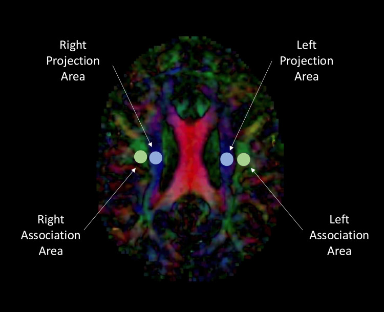
Glymphatic dysfunction is linked to neurodegenerative diseases, and imaging markers of this dysfunction may aid in diagnosis and prognosis. Glymphatic dysfunction has been proposed as a key mechanism in the pathogenesis of normal pressure hydrocephalus (NPH). Advanced magnetic resonance techniques, especially diffusion tensor imaging, have been used to evaluate glymphatic function. Diffusion tensor imaging analysis along the perivascular space (DTI-ALPS) is a noninvasive metric that correlates with glymphatic function and has been recently studied in a variety of neurodegenerative diseases. We aim to summarize studies evaluating the association between DTI-ALPS index values and NPH diagnosis and outcomes. Current studies suggest lower DTI-ALPS index values in NPH patients compared to healthy controls. The DTI-ALPS index correlated with other imaging-based markers of NPH and clinical endpoints. However, limitations of the current literature include small cohort sizes; future studies are needed in larger, heterogeneous cohorts to validate these trends. Thus, the DTI-ALPS index shows promise as a valuable tool for diagnosing NPH, predicting treatment response, and assessing disease progression.
Keywords: biomarkers; brain; diffusion tensor imaging; neuroimaging; normal pressure hydrocephalus.
Copyright © 2025, Vij et al.
Conflict of interest statement
Conflicts of interest: In compliance with the ICMJE uniform disclosure form, all authors declare the following: Payment/services info: All authors have declared that no financial support was received from any organization for the submitted work. Financial relationships: All authors have declared that they have no financial relationships at present or within the previous three years with any organizations that might have an interest in the submitted work. Other relationships: All authors have declared that there are no other relationships or activities that could appear to have influenced the submitted work.
Figures
Similar articles
-
Noninvasive assessment of glymphatic dysfunction in idiopathic normal pressure hydrocephalus with diffusion tensor imaging.J Neurosurg. 2023 Sep 8;140(3):612-620. doi: 10.3171/2023.6.JNS23260. Print 2024 Mar 1. J Neurosurg. 2023. PMID: 37724800
-
Associations of ventriculomegaly and white matter hyperintensities with glymphatic dysfunction in idiopathic normal pressure hydrocephalus.Eur Radiol. 2025 Jul;35(7):4315-4324. doi: 10.1007/s00330-024-11320-3. Epub 2025 Jan 21. Eur Radiol. 2025. PMID: 39836203 Free PMC article.
-
Altered glymphatic system in idiopathic normal pressure hydrocephalus.Parkinsonism Relat Disord. 2021 Jan;82:56-60. doi: 10.1016/j.parkreldis.2020.11.009. Epub 2020 Nov 20. Parkinsonism Relat Disord. 2021. PMID: 33248394
-
Diffusion Tensor Image Analysis ALong the Perivascular Space (DTI-ALPS): Revisiting the Meaning and Significance of the Method.Magn Reson Med Sci. 2024 Jul 1;23(3):268-290. doi: 10.2463/mrms.rev.2023-0175. Epub 2024 Apr 2. Magn Reson Med Sci. 2024. PMID: 38569866 Free PMC article. Review.
-
Diffusion Tensor Imaging Along the Perivascular Space Is a Promising Imaging Method in Parkinson's Disease: A Systematic Review and Meta-Analysis Study.CNS Neurosci Ther. 2025 May;31(5):e70434. doi: 10.1111/cns.70434. CNS Neurosci Ther. 2025. PMID: 40376934 Free PMC article. Review.
References
-
- Glymphatic system evaluation using diffusion tensor imaging in patients with traumatic brain injury. Park JH, Bae YJ, Kim JS, et al. Neuroradiology. 2023;65:551–557. - PubMed
Publication types
LinkOut - more resources
Full Text Sources