

A Rare Case of Marchiafava-Bignami Disease With Reversible Splenial Lesion
- PMID: 40337580
- PMCID: PMC12058231
- DOI: 10.7759/cureus.81845
A Rare Case of Marchiafava-Bignami Disease With Reversible Splenial Lesion
Abstract
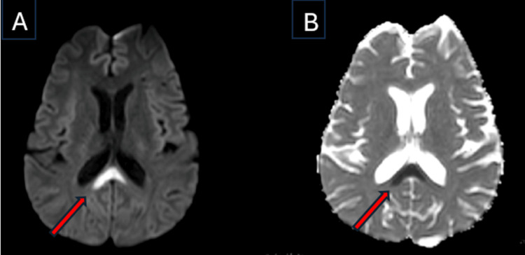
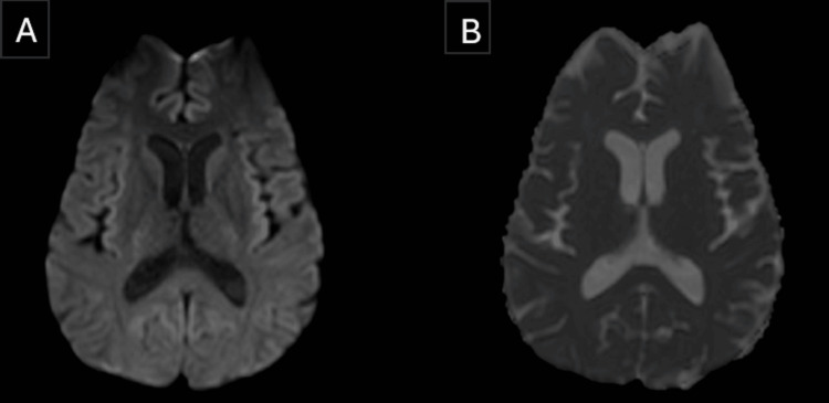
Individuals with a history of chronic alcohol consumption can develop Marchiafava-Bignami disease (MBD), a rare neurological disorder that is thought to occur secondary to thiamine deficiency and alcohol-induced brain damage. It is characterized by the toxic demyelination and necrosis of the corpus callosum. We report the case of a 42-year-old man who developed acute-onset altered sensorium and irrelevant speech output associated with ataxia. The clinical presentation raised a suspicion of MBD, although it was not a top differential diagnosis initially. Magnetic resonance imaging revealed significant demyelination of the splenium of the corpus callosum, confirming the suspicion and prompting immediate intervention aimed at addressing both the neurological manifestations and the possible underlying thiamine deficiency. Relevant history of significant alcohol consumption further supported this diagnosis. Following the initiation of thiamine supplementation and supportive care, the patient exhibited gradual improvement in neurological function, eventually leading to a complete clinical recovery and resolution of radiological findings, suggestive of the type B spectrum of the disease. This report highlights the importance of clinical evaluation and neuroimaging in the diagnosis, treatment, and prognostic stratification of patients with MBD.
Keywords: alcohol related complications; corpus callosum lesion; marchiafava-bignami disease (mbd); splenium of the corpus callosum; thiamine or vitamin b1 deficiency.
Copyright © 2025, Renuji et al.
Conflict of interest statement
Human subjects: Consent for treatment and open access publication was obtained or waived by all participants in this study. Conflicts of interest: In compliance with the ICMJE uniform disclosure form, all authors declare the following: Payment/services info: All authors have declared that no financial support was received from any organization for the submitted work. Financial relationships: All authors have declared that they have no financial relationships at present or within the previous three years with any organizations that might have an interest in the submitted work. Other relationships: All authors have declared that there are no other relationships or activities that could appear to have influenced the submitted work.
Figures


Similar articles
-
Marchiafava-Bignami Disease: A Case Report of a Reversible Cause of Dementia.Cureus. 2024 Nov 6;16(11):e73146. doi: 10.7759/cureus.73146. eCollection 2024 Nov. Cureus. 2024. PMID: 39650996 Free PMC article.
-
Diagnosis and Management of Marchiafava-Bignami Disease, a Rare Neurological Complication of Long-term Alcohol Abuse.Discoveries (Craiova). 2023 Jun 30;11(2):e168. doi: 10.15190/d.2023.7. eCollection 2023 Apr-Jun. Discoveries (Craiova). 2023. PMID: 37559750 Free PMC article. Review.
-
Marchiafava-Bignami Type A Disease: A Rare Neurological Manifestation in Oropharyngeal Cancer.Cureus. 2024 Dec 8;16(12):e75308. doi: 10.7759/cureus.75308. eCollection 2024 Dec. Cureus. 2024. PMID: 39776733 Free PMC article.
-
Marchiafava Bignami disease presenting as a cerebrovascular accident.Am J Emerg Med. 2024 Dec;86:190.e1-190.e3. doi: 10.1016/j.ajem.2024.09.058. Epub 2024 Sep 28. Am J Emerg Med. 2024. PMID: 39366786
-
Diagnosis and management of Marchiafava-Bignami disease: a review of CT/MRI confirmed cases.J Neurol Neurosurg Psychiatry. 2014 Feb;85(2):168-73. doi: 10.1136/jnnp-2013-305979. Epub 2013 Aug 26. J Neurol Neurosurg Psychiatry. 2014. PMID: 23978380 Free PMC article. Review.
References
-
- Clinicoradiologic subtypes of Marchiafava-Bignami disease. Heinrich A, Runge U, Khaw AV. J Neurol. 2004;251:1050–1059. - PubMed
-
- Thiamine deficiency, oxidative metabolic pathways and ethanol-induced neurotoxicity: how poor nutrition contributes to the alcoholic syndrome, as Marchiafava-Bignami disease. Fernandes LM, Bezerra FR, Monteiro MC, et al. Eur J Clin Nutr. 2017;71:580–586. - PubMed
-
- [Marchiafava-Bignami disease] Navarro JF, Noriega S. https://pubmed.ncbi.nlm.nih.gov/10229969/ Rev Neurol. 1999;28:519–523. - PubMed
-
- Marchiafava-Bignami disease: longitudinal MR imaging and MR spectroscopy study. Gambini A, Falini A, Moiola L, Comi G, Scotti G. https://pubmed.ncbi.nlm.nih.gov/12591642/ AJNR Am J Neuroradiol. 2003;24:249–253. - PMC - PubMed
Publication types
LinkOut - more resources
Full Text Sources