Review
. 2025 Mar 20:12:rbaf007.
doi: 10.1093/rb/rbaf007. eCollection 2025.

Analytical methods in studying cell force sensing: principles, current technologies and perspectives

Affiliations
Review

Analytical methods in studying cell force sensing: principles, current technologies and perspectives

Xiaojun Liu et al. Regen Biomater. .

Abstract

Mechanical stimulation plays a crucial role in numerous biological activities, including tissue development, regeneration and remodeling. Understanding how cells respond to their mechanical microenvironment is vital for investigating mechanotransduction with adequate spatial and temporal resolution. Cell force sensing-also known as mechanosensation or mechanotransduction-involves force transmission through the cytoskeleton and mechanochemical signaling. Insights into cell-extracellular matrix interactions and mechanotransduction are particularly relevant for guiding biomaterial design in tissue engineering. To establish a foundation for mechanical biomedicine, this review will provide a comprehensive overview of cell mechanotransduction mechanisms, including the structural components essential for effective mechanical responses, such as cytoskeletal elements, force-sensitive ion channels, membrane receptors and key signaling pathways. It will also discuss the clutch model in force transmission, the role of mechanotransduction in both physiology and pathological contexts, and biomechanics and biomaterial design. Additionally, we outline analytical approaches for characterizing forces at cellular and subcellular levels, discussing the advantages and limitations of each method to aid researchers in selecting appropriate techniques. Finally, we summarize recent advancements in cell force sensing and identify key challenges for future research. Overall, this review should contribute to biomedical engineering by supporting the design of biomaterials that integrate precise mechanical information.

Keywords: biomaterial design; biomaterial–cell interaction; biosensor; cell biomechanics.


Figures

None
Graphical abstract
Figure 1.
Figure 1.
The cell cytoskeleton and force-sensitive machinery. (A) Two cells adhered to the extracellular matrix (ECM) through focal adhesions and adherent junctions via cadherins, with mechanosensitive ion channels on the membrane. Microfilaments, microtubules and intermediate filaments work together to support cell shape alternations under various conditions. (B) Portrayal of the focal adhesion and acto-myosin system. Herein, adaptor proteins talin and vinculin act as force sensors in cell force transmission [9, 18]. Myosin mediates the contraction between actin filaments and the forces propagate toward integrin by talin and vinculin. Only relative higher tension pulling on integrin induces the cluster formation of integrin. (C) Cadherins link to MFs via catenins (the adaptor proteins), and then connect to MFs [19, 20]. Similar to focal adhesions, the cell junctions sense forces from the intracellular cytoskeleton. Myosin pulls actin filaments and MFs thereby stretching talin and α-, β-catenin. (D) Piezo 1 is a tension-sensitive channel, which is opened by membrane stretching and actomyosin traction force [21–23]. (E) Nesprins and SUN1/2 link the nuclear interior to cytoskeletal filaments. Molecular motors interact with these filaments to generate forces, which are transmitted to the nucleus via the linker of nucleoskeleton and cytoskeleton complex (LINC). Genomic regions associated with the lamina, known as lamina-associated chromatin domains (LADs), exhibit low transcriptional activity.
Figure 2.
Figure 2.
Cell mechanical signaling pathways. Here, three examples are shown including integrin-actomyosin-LINC, integrin-YAP, ion channel-Calcium-kinase and contraction [38, 39, 41–43]. Force transmitted along the integrin-actomyosin-LINC directly induces target gene expression. The force drives YAP translocation into the nucleus and thus modulates gene transcription. Mechanosensitive ion channels such as PIEZO-1 will open upon sensing membrane tension, and increased cytosolic calcium mediates downstream signaling. LINC, linker of nucleoskeleton and cytoskeleton complex; NPC, nuclear pore complex; YAP, Yes-associated protein.
Figure 3.
Figure 3.
The clutch model. (A) The relationship between force and average bond lifetime of slip bond and catch bond [44, 45]. (B) The relationship between the force loading rate and force in the corresponded bonding [44]. (C) The changes in the lifetime of integrin–ECM and talin–(F-actin) as the force increases [48–52]. (a) The force is small, which is conducive to the formation of talin–(F-actin) bond, and the lifetime is the maximum value. However, the integrin–ECM bond is easy to dissociate, and the integrin activation ratio is very low, making it difficult to pass down to talin and F-actin. (b) Right of the focus of the two curves. Here is the optimal force transduction threshold. When the lifetimes of integrin–ECM bond and talin–(F-actin) bond match, the force transduction process of ECM–integrin–talin–(F-actin) is the smoothest and stable. (c) This is the optimal force of the integrin–ECM bond, but the stability of talin–(F-actin) here has dropped a lot, and the probability that the cell–ECM can form a stable structure is lower than that at b. (d) If the force is too large, the stability of both bonds will be reduced, which is not conducive to cell–ECM connection and force conduction. (D) Briefly describes the three binding events that systemically determines the force chain transduction and may work as one clutch or two. The clutch cycle largely depends on the binding lifetime, force level, affected by force loading rate and electrostatic interaction at binding domains [48–52]. (Binding 1: integrin to ECM; binding 2: talin/vinculin to F-actin; binding 3: myosin to F-actin.)
Figure 4.
Figure 4.
The principle of TFM. The cell displacement and bead dislocation were imaged in Z-stack mode [154]. The raw images were used to interpret the force at certain region, where the stress-strain relationship could be calculated according Hooke’s law. We acknowledge permission from the copyright clearance center of the RSC to reuse the article [154].
Figure 5.
Figure 5.
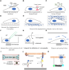
Schematic of different analytical methods for cell force studies. (A) Atomic force microscope probing the cell stiffness [106]. (B) Optical tweezers measuring the dynamics of the cell–ligand interactions via trapped beads [143]. (C) Magnetic tweezer records the torque and normal tension between beads and ligands [110]. (D) µFSA monitors the displacement of micropillars as they are pulled by the cell [143]. (E) TFM detects the cell force in 2D and 3D, depending on the displacement of labeled beads [103, 104]. (F) Micropipette aspiration can quantify the cell–cell interaction, the experienced force will be computed according the suction pressure [114]. (G) Micropipette force sensor enables the measurement of microalgae adhesion force onto substrate [118]. (H) Fluidic systems favor the observation of cell status in the flow chamber of various dimensions [122]. (I) Real-time deformability cytometer [126]. (J) Force tension sensors based on DNA fragments [161]. (K) Membrane tension sensor based on elastic peptide [162].
Figure 6.
Figure 6.
Optical tweezers in cell force sensing. (A) A single-molecule optical trap assay is used to investigate the bonding property between vinculin and actin filament [49]. Beads are immobilized with T12 vinculin and fixed on a stage. The stage is translated which leads to displacement of trapped beads. The authors suggest that the directional and force-stabilized binding of vinculin to F-actin may help understand how adhesion complexes keep front-rear asymmetry in migrating cells. (B) Studying microtubules dynamics and interaction with dynein. Microtubules nucleate from an axoneme, linked to a bead and held by a ‘keyhole’ optical trap, grow against a microfabricated barrier. Microtubule-associated proteins (MAPs) and motor proteins may be added in solution and a mechanical signal is generated [167]. (C) Studying cell membrane tension/cell microrheology. Huh7 cell membrane tension is measured by tether-pulling. Initially, the bead is trapped by; after contacting with cell membrane for 30 seconds to form a lipid tether, the bead is pulled away from the cell [168].
Figure 7.
Figure 7.
The principle of MT. (A) The magnetic beads movement in a magnetic field. The displacement can be divided in to δx, δy, δz. (B) Measuring the DNA plectonems formation upon positive supercoiling using magnetic tweezer. RNA polymerase (RNAP)–promoter open complex formation on a positively supercoiled DNA. Promoter opens DNA and n positive supercoils addition occur, and the bead downward movement was recorded by n·Δz [195].
Figure 8.
Figure 8.
AFM used in cell force sensing. (A) Bacteria–host binding process investigation where WT LGG bacterium is attached to the cantilever probe and brought into contact with cell surface. The formation of membrane nanotethers and bonding property are investigated [207]. For virus–cell binding process [208], this beginning step of binding between the virus to the cell involves EnvA glycoproteins to complementary tumor virus A (TVA) receptors. The AFM cantilever conjugated with virus approaches and retracts from the cell. (B) Cell–ligand interaction where a single fibroblast was immobilized on ConA- or VN-coated cantilever, approached to the substrate-coated support, and retracted vertically after a few seconds contact time. The adhesion force between fibroblast and support was measured during this process [52]. (C) Cell elasticity measurement where the cell is indented by a bead probe, and the mechanical response of contacted areas are recorded. Cortical stiffness of a cell is analyzed according to force–distance curves generated in this assay [151]. (D) Ligand–receptor interaction in vitro. Biotin is attached to the cantilever and streptavidin to the support. The force curve monitors the separating process along the chain, which includes the initial elongation of PEG linker as well as eventual rupture force of the bond [209].
Figure 9.
Figure 9.
The micropipette aspiration setup and cell membrane tension measurement by MPA. (A) Schematic diagram. The micropipette is connected to a liquid reservoir, mounted on a stage and the pressure can be modulated precisely. (B) The equation used in cell membrane tension analysis. The parameters and equations are explained in the text. (We acknowledge permission from the Copyright Clearance Center of the Elsevier to reuse the article [228].) The micropipette approach in MPA demonstrates its application in cell mechanics studies, but the force-sensing method of micropipette force sensing (MFS) is conceptually different. Instead of probing suction pressure, MFS measures the deflection of a micropipette force sensor to assess force using Hooke’s law [119, 243] (Figure 5G). This technique evolved from earlier flexible needles used for force sensing in biological cells and molecules [118, 149, 244, 245].
Figure 10.
Figure 10.
Real-time deformability cytometry (RT-DC). (A) Setup and measurement principle. (B) Scatter plot of deformation versus cell size. Color indicates a linear density scale; black line, 50%-density contour.
Figure 11.
Figure 11.
Digital reversible tension sensor (DTS). (A) PEG-based TS, adjusting the number of PEG units in the elastic polymer [258, 259]. Different forces yielded FRET index is calibrated, and then cell membrane receptor tensions are investigated. (B) (GPGGA)8-based TS, elastic repeat GPGGA from silk protein is recruited and expressed in E. coli with RGD motif for binding integrin. Further modifications are added for quencher-fluorophore according to click chemistry reaction [260]. (C) Talin-based TS (Talin- FL-TSM (tension sensor modules)), which can be used in the study of force transduction. Paired FRET fluorescent proteins were inserted inside of talin molecule after expression in mammalian cells [261]. Here FL mainly unfolds at 3–5 pN at a pulling speed of 500 nm/s. (D) Hairpin DNA TS linked with RGD for integrin binding and modified with FRET pairs, is very useful to measure real-time signal for integrin tension. The lower limit reaches up to 4.7 pN [161]. Redrafted from the following articles [128, 258, 269–279]. We acknowledge permission from Springer Nature and ACS.
Figure 12.
Figure 12.
Binary tension sensors. (A) ITS, conjugating quencher-fluorophore pairs into TGT module, and thus providing a better signal-to-noise ratio [262]. Integrin tensions of activated platelets are examined with high throughput. (B) TGT, a dsDNA fragment conjugated with RGD ligand where the force signal is detected as long as the fluorescence is lost [263]. (C) YFP-based TS, engineered with RGD coding sequence and transfected into bacteria, helps map integrin tensions without additional chemical modification [264]. (D) Nano YoYo, an elegant design inspired from SSB–ssDNA interaction, acquires sensitivity up to 4 pN [265]. (E) I27-based TS, linked with RGD and green fluorescence protein (GFP), can be unfolded at high force and reports cell traction forces by integrin [266]. Redrafted from the following articles [229, 232, 235–237]. We acknowledge permission from Springer Nature, RSC, ACS, Wiley and AA.
Figure 13.
Figure 13.
Combo tension sensors. (A) The design of RSDTP (reversible shearing DNA-based tension probe), which adapts from ITS and hairpin TS. The hairpin structure makes it reversible between folding and unfolding states, while ITS moiety imbues the upper threshold of rupturing RSDTP. Altogether RSDTP has a broad measuring range of between 4 and 60 pN [290]. (B) Representative images of reflection interference contrast microscopy (RICM) and integrin-mediated tension of a cell before and after treated with cytochalasin D. (C) Schematic of real-time force images with multiplexed RSDTPs. (D) Representative total internal reflection fluorescence (TIRF) images of GFP–paxillin, tension signals of 17 pN (Cy3B) and 56 pN (Atto647N) and overlay of all channels. (E) Schematic of photocleavable RSDTP. A photocleavable linker and a non-nucleotide moiety were incorporated into the sugar-phosphate backbone within the loop region, linking two nucleotide sequences through a short, UV-photocleavable C3 spacer arm. (F) Upper: schematic of the periodic pulse of UV illumination. Lower: time-lapse TIRF images of GFP–paxillin and single-molecule tension signals of a GFP–paxillin-expressing cell adhered to a mixed-sensor surface (2% 56-pN photocleavable RSDTP and 98% 56-pN TGT) show dynamic changes of FAs before and after photocleavage. Scale bar: 10 μm. We acknowledge permission from the Copyright Clearance Center of the Springer Nature to reuse the article [290].
Figure 14.
Figure 14.
Membrane tension sensors. (A) Cell membrane tension sensor FliptR (fluorescent lipid tension reporter). The FliptR can be used to monitor cell membrane tension since the FliptR molecule changes its optical property under lateral pressure from cell membrane [291]. (B) The engineered protein membrane TS based on (GPGGA)8. Membrane shear stress sensor (GPGGA)8-MSS, in which two membrane-binding proteins lyn and K-Ras were engineered at the two ends. MSS and K-MSS expressed cells are treated with sucrose and then imaged [162]. ECFP, cyan fluorescence protein; KMSS is a head-less mutant, the control probe; MSS, membrane-bound FRET-based tension sensor; YPet, yellow fluorescent protein for energy transfer. Redrafted from the following articles [162, 291]. We acknowledge permission from Springer Nature, and Cell Press.

Similar articles

References

    1. Cao D, Ding J. Recent advances in regenerative biomaterials. Regen Biomater 2022;9:rbac098. - PMC - PubMed
    1. He J, Liu Q, Zheng S, Shen R, Wang X, Gao J, Wang Q, Huang J, Ding J. Enlargement, reduction, and even reversal of relative migration speeds of endothelial and smooth muscle cells on biomaterials simply by adjusting RGD nanospacing. ACS Appl Mater Interfaces 2021;13:42344–56. - PubMed
    1. Liu Q, Zheng S, Ye K, He J, Shen Y, Cui S, Huang J, Gu Y, Ding J. Cell migration regulated by RGD nanospacing and enhanced under moderate cell adhesion on biomaterials. Biomaterials 2020;263:120327. - PubMed
    1. Ehrlicher AJ, Nakamura F, Hartwig JH, Weitz DA, Stossel TP. Mechanical strain in actin networks regulates FilGAP and integrin binding to filamin A. Nature 2011;478:260–3. - PMC - PubMed
    1. Haynie DT. Molecular physiology of the tensin brotherhood of integrin adaptor proteins. Proteins 2014;82:1113–27. - PubMed

LinkOut - more resources