. 2025 Apr 10;15(4):559.
doi: 10.3390/biom15040559.

Peptide-Based Regulation of TNF-α-Mediated Cytotoxicity

Affiliations

Peptide-Based Regulation of TNF-α-Mediated Cytotoxicity

Betul Zehra Temur et al. Biomolecules. .

Abstract

Tumor necrosis factor alpha (TNF-α) is a pro-inflammatory cytokine associated with TNF receptor 1 (TNFR1) and TNF receptor 2 (TNFR2), which play important roles in several inflammatory diseases. There is a growing interest in developing alternative molecules that can be used as TNF blockers. In this study, we focused on TNF-α-, TNFR1-, and TNFR2-mimicking peptides to inhibit TNF-α receptor binding in various ways. Six peptides (OB1, OB2, OB5, OB6, OB7, and OB8) were developed to bind TNFR1, TNFR2, and TNF-α. OB1 and OB2 bound to TNF-α with lower Kd values of 300 and 46.7 nM, respectively, compared to previously published sequences. These synthetic peptides directly and indirectly inhibited TNF-α in vitro without cytotoxicity to L929 cells, and OB1 significantly inhibited apoptosis in the presence of hTNF-α. Peptides developed in this study may prove to be useful for therapeutic inhibition of TNF-α.

Keywords: TNF-α; TNF-α blocker; TNF-α inhibition; TNF-α receptors; TNF-α-binding peptide; TNFR1-binding peptide; TNFR2-binding peptide.


Conflict of interest statement

Ahmet Emin Atik is employed by the company Turgut Ilaclari A.S. The remaining authors declare that the research was conducted in the absence of any commercial or financial relationships that could be construed as a potential conflict of interest.

Figures
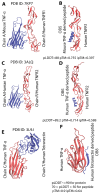
Figure 1
Figure 1
Structures of protein–protein interactions that gave rise to OB5, OB6, and OB8 peptides and target-protein structures retrieved through AlphaFold. (A,C,E) Structures from which the peptides were predicted. (B,D,F) Structure of the AlphaFold 2 (B,D)- and AlphaFold 3 (F)-derived complexes with cartoon representation of the backbone. For all AlphaFold models, the model with the highest pLDDT, pTM, and iPTM scores was selected. As ipTM values were relatively low for the model, MD simulations were then conducted, followed by comparing MM-PBSA-derived relative binding free energy values between peptide and target with MM-PBSA-derived relative binding energy values of the positive control.
Figure 2
Figure 2
Conformational stability, total relative binding free energy (RBFE), and enthalpic contribution to RBFE retrieved from MD simulations of the structures. (A) RMSD-based conformational stability analyses of 100 ns MD simulations indicated that the last 10 ns of each simulation could be considered stable. Thus, a trajectory encompassing the last 10 ns was used for estimating RBFE for each simulation using the MM-PBSA method. (B) Based on RBFE analyses, the affinities of peptides capable of binding TNFR1 (peptide in the 7KP7_PEP complex) and TNFR2 (peptide in the 3ALQ_PEP complex) were higher than those of their corresponding positive control complexes (7KP7_PC and 3ALQ_PC). TNF-α-binding peptide (peptide in the 3L9J_PEP complex) also returned comparable affinity with its positive control complex (3L9J_PC). (C) Given that the entropic term was estimated via error-prone quasi-harmonic approximation, the enthalpic contribution to RBFE was also computed and shown to have a similar pattern.
Figure 3
Figure 3
Characterization of OB5 peptide: (A) HPLC chromatogram and (B) MS/MS spectrum.
Figure 4
Figure 4
Binding affinities of the peptides for rhTNF-α via microscale thermophoresis. Binding curves of (A) adalimumab, (B) OB1, (C) OB2, and (D) OB8. MST experiments were performed using labeled rhTNF-α at 20 nM and variable concentrations of adalimumab and synthesized peptides. TNFR1 and rhTNF-α MST experiments were performed using 1 nM of labeled TNFR1. Binding data are plotted as the mean ± SD of three replicates. (E) Dissociation constant (Kd) of indicated interactions given as mean ± SD of three replicates.
Figure 5
Figure 5
Binding affinities of the peptides for TNFR1 and TNFR2 measured by MST. Binding curves of (A) rhTNF-α, (B) OB5, (C) OB6, and (D) OB7. MST experiments were performed using labeled TNFR1 and TNFR2 at 20 nM and variable concentrations of synthesized peptides. TNFR1 and rhTNF-α MST experiments were performed using 1 nM of labeled TNFR1. Binding data are plotted as the mean ± SD of three replicates. (E) Kd values of indicated interactions are presented as mean ± SD of three replicates.
Figure 6
Figure 6
Inhibition of TNF-α activity on L929 cells by TNF-α-binding peptides compared to adalimumab. (A) Cytotoxicity of synthesized peptides and adalimumab and (B) neutralization of TNF-α-induced cytotoxicity. All data are presented as means ± SEM of triplicate experiments. (C) The effect of TNF-α-binding peptides on caspase 3/7 activation. Results are presented as means ± SEM. ns: not significant; * p < 0.05; ** p < 0.01; *** p < 0.001; **** p ≤ 0.0001 vs. the control group.
Figure 7
Figure 7
The effect of TNFR1 applied with TNF-α on caspase 3/7 activity. Results are presented as means ± SEM. ns, not significant; ** p < 0.01; **** p ≤ 0.0001 vs. the control group.
Figure 8
Figure 8
Morphological analysis of L929 cells after treatment with different molecules.
Figure 9
Figure 9
Inhibition of TNF-α activity on L929 cells by receptor-binding peptides. (A) Cytotoxicity of synthesized peptides and (B) neutralization of TNF-α-induced cytotoxicity measured by MTT assay. All data are presented as means ± SEM of triplicate experiments. (C) The effect of receptor-binding peptides on caspase 3/7 activation. Results are presented as means ± SEM. ns: not significant; * p < 0.05; ** p < 0.01; *** p < 0.001; **** p ≤ 0.0001 vs. the control group.

References

    1. Hehlgans T., Pfeffer K. The intriguing biology of the tumour necrosis factor/tumour necrosis factor receptor superfamily: Players, rules and the games. Immunology. 2005;115:1–20. doi: 10.1111/j.1365-2567.2005.02143.x. - DOI - PMC - PubMed
    1. Kriegler M., Perez C., DeFay K., Albert I., Lu S.D. A novel form of TNF/cachectin is a cell surface cytotoxic transmembrane protein: Ramifications for the complex physiology of TNF. Cell. 1988;53:45–53. doi: 10.1016/0092-8674(88)90486-2. - DOI - PubMed
    1. Li P., Zheng Y., Chen X. Drugs for Autoimmune Inflammatory Diseases: From Small Molecule Compounds to Anti-TNF Biologics. Front. Pharmacol. 2017;8:460. doi: 10.3389/fphar.2017.00460. - DOI - PMC - PubMed
    1. Black R.A., Rauch C.T., Kozlosky C.J., Peschon J.J., Slack J.L., Wolfson M.F., Castner B.J., Stocking K.L., Reddy P., Srinivasan S., et al. A metalloproteinase disintegrin that releases tumour-necrosis factor-alpha from cells. Nature. 1997;385:729–733. doi: 10.1038/385729a0. - DOI - PubMed
    1. Locksley R.M., Killeen N., Lenardo M.J. The TNF and TNF receptor superfamilies: Integrating mammalian biology. Cell. 2001;104:487–501. doi: 10.1016/S0092-8674(01)00237-9. - DOI - PubMed

MeSH terms

Substances

LinkOut - more resources