. 2025 May 8;5(1):33.
doi: 10.1007/s44154-025-00223-y.

Roles of NADPH oxidases in regulating redox homeostasis and pathogenesis of the poplar canker fungus Cytospora chrysosperma

Affiliations

Roles of NADPH oxidases in regulating redox homeostasis and pathogenesis of the poplar canker fungus Cytospora chrysosperma

Quansheng Li et al. Stress Biol. .

Abstract

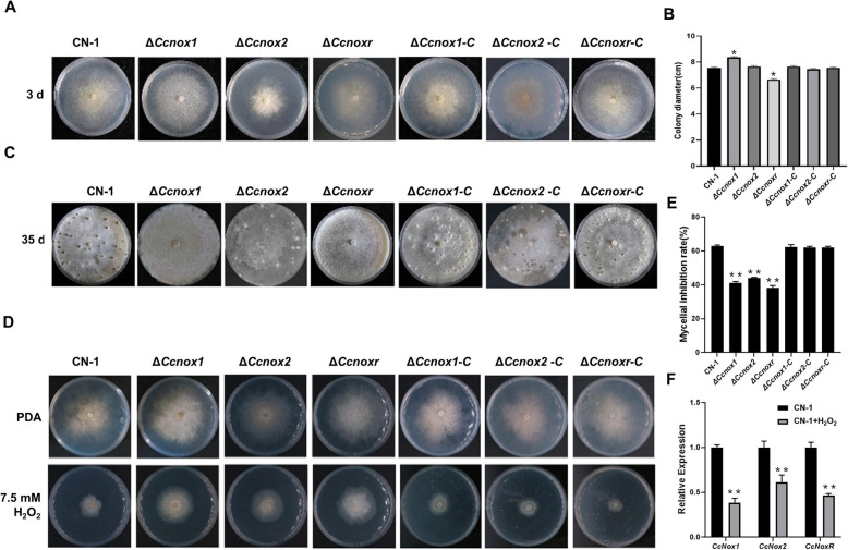
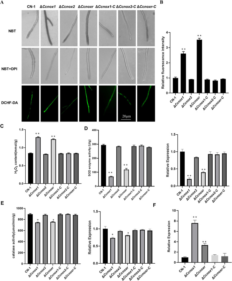
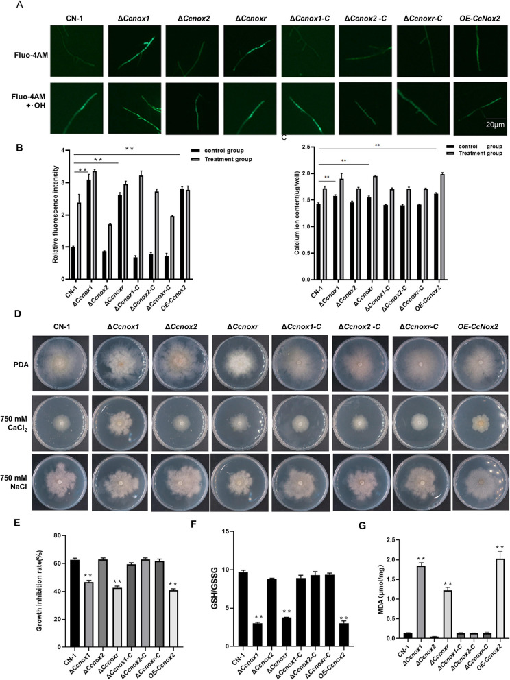
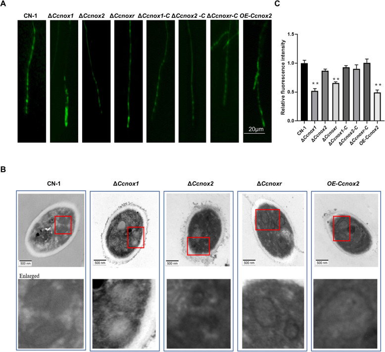
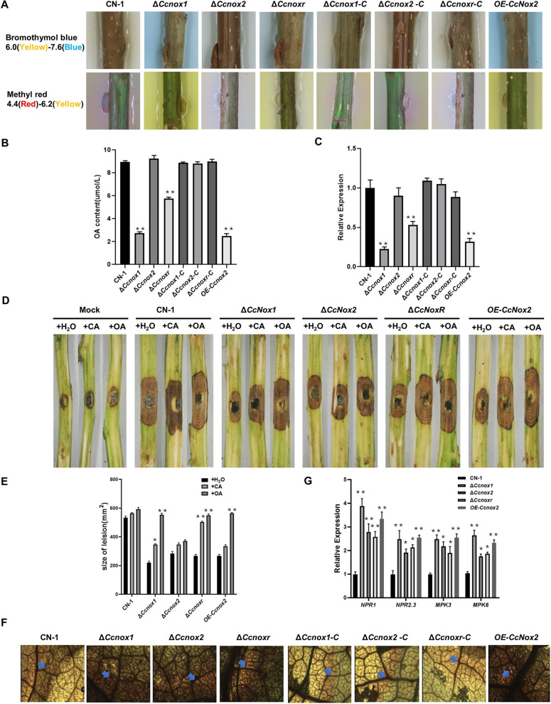
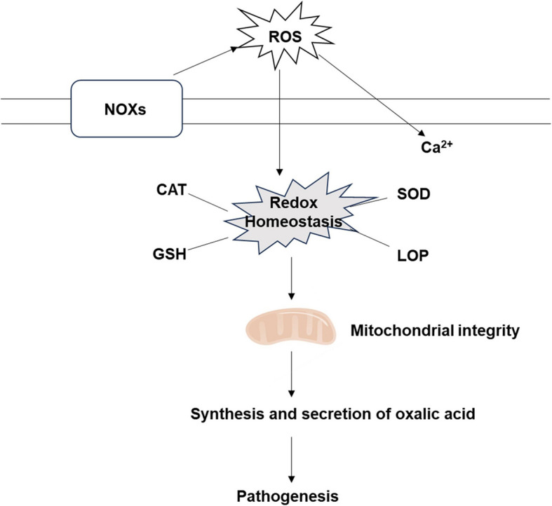
Poplar canker, caused by the fungus Cytospora chrysosperma, results in tremendous losses in poplar plantations in China. Although NADPH oxidases (NOXs) play important roles in the development and pathogenicity of several pathogenic fungi, their roles in C. chrysosperma remain unclear. In this study, we characterized three NOX genes (CcNox1, CcNox2, and CcNoxR) in C. chrysosperma. All three genes were highly upregulated during poplar branch infection, and deletion of any of them severely reduced virulence on poplar branches. Furthermore, deletion of either CcNox1 or CcNoxR resulted in a significant increase in endogenous reactive oxygen species production in hyphae, enhanced influx of Ca2+, the disruption of redox homeostasis and compromised mitochondrial integrity. Moreover, biosynthesis and secretion of a known virulence factor oxalic acid was obviously defective and exogenous oxalic acid supplementation rescued the virulence of the mutants. Taken together, our findings reveal that NOXs play important roles in redox homeostasis, mitochondrial integrity and pathogenicity in C. chrysosperma.

Keywords: Cytospora chrysosperma; Mitochondrial integrity; NADPH oxidases; Pathogenicity; Poplar canker; Redox homeostasis.


Conflict of interest statement

Declarations. Ethics approval and consent to participate: Not applicable. Consent for publication: Not applicable. Competing interests: The authors have no relevant financial or non-financial interests to disclose.

Figures

Fig. 1
Fig. 1
Indispensable role of NADPH oxidases in virulence of Cytospora chrysosperma. A The relative expression levels of CcNox1, CcNox2, and CcNoxR during C. chrysosperma infection was quantified at 0, 12, 24, and 48 h post inoculation (hpi), and the expression was normalized to that of the CcAct1 gene. B Virulence of NOX mutants on poplar twigs. Agar plugs without fungal mycelia were used as a mock treatment. Disease symptoms were photographed 4 days post inoculation (dpi). C The average lesion sizes on twigs at 4 dpi. The bars indicate the standard deviations of the means of 25 individual poplar twigs. The experiment consisted of three biological replicates. The data are presented as the means ± SDs and asterisks represent significant differences (**p < 0.01)
Fig. 2
Fig. 2
Vegetative growth, conidiation, and resistance to oxidative stress of the ∆Ccnox1, ∆Ccnox2, and ∆Ccnoxr strains. A Colony morphology of CN-1, NOXs mutant, and complemented strains after they had grown on PDA media for 3 days at 25 °C. B Diameters of the colonies of the strains on PDA media. C Observations of the pycnidia at 35 dpi on PDA. D Growth phenotype on PDA media supplemented with or without H2O2 for 3 days at 25 °C. E Statistical analysis on the inhibition rate of all strains under H2O2 stress. F Expression levels of CcNox1, CcNox2, and CcNoxR in CN-1 in the presence or absence of hydrogen peroxide. The experiment consisted of three biological replicates. The data are presented as the means ± SDs and asterisks represent significant differences (**p < 0.01, *p < 0.05)
Fig. 3
Fig. 3
Deletion of either CcNox1 or CcNoxR increased ROS accumulation. A The hyphae of all strains were immersed in NBT staining solution for 2 h in the presence or absence of 20 μM DPI before observation under a light microscope. The fluorescent probe DCFH-DA was used to stain the mycelia, and the accumulation of reactive oxygen species was observed under a universal fluorescence microscope. Bar: 20 μm. B Fluorescence intensity analysis of DCFH-DA-stained mycelium using ImageJ software. C Quantification of intracellular H2O2 production. Mycelia were cultured in PDB for 2 days and then ground with liquid nitrogen to extract H2O2 for determination. D Determination of SOD enzyme activity and the expression of CcSod1 in the different strains. E Determination of catalase activity and the expression of CcCat1 in the different strains. F The relative expression level of CcNox2 in the CcNox1 or CcNoxR strain. Error bars represent the standard deviations based on three independent replicates. The experiment consisted of three biological replicates. The data are presented as the means ± SDs and asterisks represent significant differences (**p < 0.01, *p < 0.05)
Fig. 4
Fig. 4
Overexpression of CcNox2 phenocopied the ΔCcnox1 and ΔCcnoxr strains. A Spore production by the OE-Ccnox2 and CN-1 strains. Virulence of OE-Ccnox2 and CN-1 strains on poplar twigs. Agar plugs without fungal mycelia were used as a mock treatment. Disease symptoms were photographed at 4 dpi. The hyphae of all strains were immersed in NBT staining solution for 2 h in the presence or absence of 20 μM DPI before observation under a light microscope (DM2500, Leica). The fluorescent probe DCFH-DA was used to stain the mycelia, and the accumulation of reactive oxygen species was observed under a universal fluorescence microscope. Bar: 20 μm. D Fluorescence intensity analysis of DCFH-DA-stained mycelium using ImageJ software. E Quantification of intracellular H2O2 production. Mycelia were cultured in PDB for 2 days and then ground with liquid nitrogen to extract H2O2 for determination. F Determination of SOD activity. G Determination of catalase activity. Error bars represent the standard deviations based on three independent replicates. The data are presented as the means ± SDs and asterisks represent significant differences (**p < 0.01)
Fig. 5
Fig. 5
Deletion of either CcNox1 or CcNoxR led to an increase in Ca2+ influx and the disruption of redox homeostasis. A The content of free calcium ions (Ca2⁺) in different strains was detected using the fluorescent indicator Fluo-4 AM Fluorescence. Under untreated conditions, ∆Ccnox1, ∆Ccnoxr and OE-Ccnox2 strains exhibited higher fluorescence intensity. The fluorescence intensity of all strains was further enhanced by the introduction of hydroxyl radicals (·OH) produced by a mixture of 2 mM H₂O₂, 0.5 mM Cu2⁺ and 0.5 mM ascorbic acid. B Fluorescence intensity analysis of Fluo-4 AM-stained mycelium using ImageJ software. Significance analysis was performed for each strain in the control group. The Ca.2⁺ content was quantified using a calcium ion detection kit in the presence and absence of -OH treatment. Significance analysis was performed for each strain in the control group. D In total, 750 mM CaCl2 was added to PDA for 3 days of culture to observe the morphology of all the strains. E Mycelia inhibition rates. F Measurement of glutathione (GSH) and oxidized glutathione (GSSG) contents. G Determination of MDA content. Error bars represent the standard deviations based on three independent replicates. The experiment consisted of three biological replicates. The data are presented as the means ± SDs and asterisks represent significant differences (**p < 0.01)
Fig. 6
Fig. 6
CcNox1 and CcNoxR are critical for mitochondrial integrity. A Mito-Tracker Green, a mitochondrial probe that emits green fluorescence, was used to stain the mitochondria in mycelial cells, which were then observed using a universal fluorescence microscope. B After culturing the mycelium in PDB for 3 days, it was fixed with 2.5% malondialdehyde for 24 h. The mitochondria were observed using transmission electron microscopy. C The mitochondrial membrane potential was measured using TMRE, a tangerine cationic fluorescence probe that can penetrate the cell membrane. The change in the mitochondrial membrane potential was determined by measuring the intensity of the fluorescence signal with a fluorescence microplate analyzer. The experiment consisted of three biological replicates. The data are presented as the means ± SDs and asterisks represent significant differences (**p < 0.01)
Fig. 7
Fig. 7
CcNox1 and CcNoxR are involved in oxalic acid biosynthesis. A Plate acid–base indicator experiment. A representative color change was observed on different plates supplemented with methyl red and bromophenol blue. B Oxalic acid production in potato dextrose broth (PDB). Each strain was cultured for 3 days, and the resulting liquid culture was analyzed for oxalate accumulation. C Expression level of the oxalate synthase gene CcOah. D The development of diseased spots on poplar branches. OA and CA were used to treat the branches before vaccination. Each strain was inoculated with an agar plug with a diameter of 5 mm; sterile water served as a control. Photos were taken at 3 days after vaccination. E Quantitative analysis of lesion areas. Scalded poplar leaves were inoculated with fungal strains, and after 24 h of inoculation, the detached leaves were stained with 0.01% 3,3′-diaminobenzidine (DAB) for ROS detection and decolorized with 95% alcohol. The blue arrow indicates the ROS activated by the strain. Expression of defense genes (NPR1, NPR2.3, MPK3, and MPK6) in poplar trees. RNA of the poplar branches was extracted at 24 hpi for qRT‒PCR analysis. The experiment consisted of three biological replicates. The data are presented as the means ± SDs and asterisks represent significant differences (**p < 0.01, *p < 0.05)
Fig. 8
Fig. 8
A working model of NOXs for pathogenesis in Cytospora chrysosperma. Under normal circumstances, two NOX enzymes (NOX1 and NOX2) may play different roles. NOX1 and NOXR play a role in the production of reactive oxygen species, while NOX2 plays an iron redox role. When NOX1 or NOXR is disrupted, it will induce overexpression of NOX2, produce excessive reactive oxygen species, and cause cells to be in a state of oxidative stress. When NOX1 or NOXR is disrupted, it will induce overexpression of NOX2, produce excessive reactive oxygen species, and cause cells to be in a state of oxidative stress. Severe oxidative stress, on the one hand, leads to lipid peroxidation, on the other hand, leads to mitochondrial abnormalities, such as mitochondrial swelling, membrane potential decline, and more mitochondrial reactive oxygen species. The abnormality of mitochondria not only disrupts the normal progress of the tricarboxylic acid cycle, leading to a decrease in oxalic acid synthesis, but also causes attenuated virulence

Similar articles

References

    1. Dikalov S (2011) Cross talk between mitochondria and NADPH oxidases. Free Radical Biol Med 2011(51):1289–1301. 10.1016/j.freeradbiomed.2011.06.033 - PMC - PubMed
    1. Ewald CY, Hourihan JM, Bland MS, et al. NADPH oxidase-mediated redox signaling promotes oxidative stress resistance and longevity through memo-1 in C. elegans. eLife 2017:6. 10.7554/eLife.19493 - PMC - PubMed
    1. Goswami RS (2012) Targeted Gene Replacement in Fungi Using a Split-Marker Approach. Methods Mol Biol 835:255–269. 10.1007/978-1-61779-501-516 - PubMed
    1. Han Z, Xiong D, Xu Z|, et al. The Cytospora chrysosperma virulence effector CcCAP1 mainly localizes to the plant nucleus to suppress plant immune responses. mSphere 2021:6. 10.1128/mSphere.00883-20 - PMC - PubMed
    1. Kim HJ, Chen C, Kabbage M et al (2011) Identification and characterization of Sclerotinia sclerotiorum NADPH oxidases. Appl Environ Microbiol. 77:7721–7729. 10.1128/aem.05472-11 - PMC - PubMed

LinkOut - more resources