. 2025 May 8;20(5):e0318178.
doi: 10.1371/journal.pone.0318178. eCollection 2025.

Fatty acid metabolism suppresses neonatal cardiomyocyte proliferation by increasing PDK4 and HMGCS2 expression through PPARδ

Affiliations

Fatty acid metabolism suppresses neonatal cardiomyocyte proliferation by increasing PDK4 and HMGCS2 expression through PPARδ

Shota Tanaka et al. PLoS One. .

Abstract

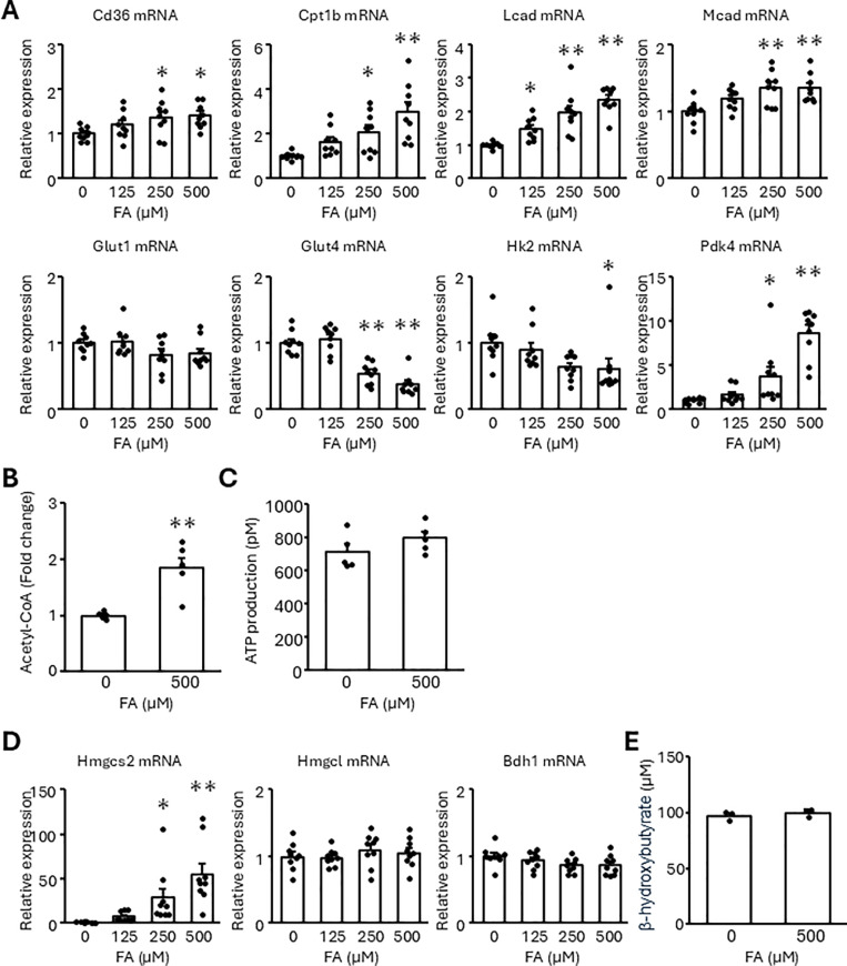
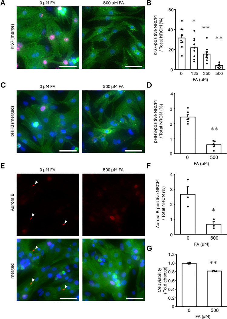
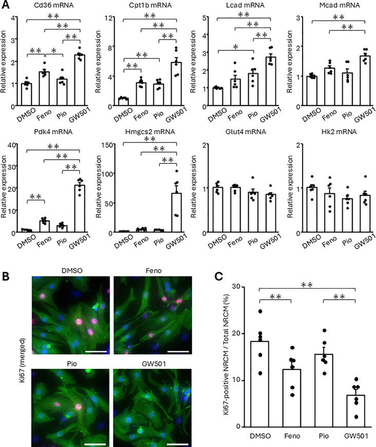
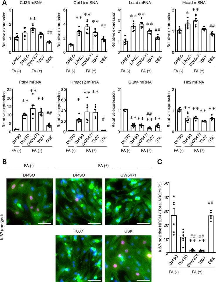
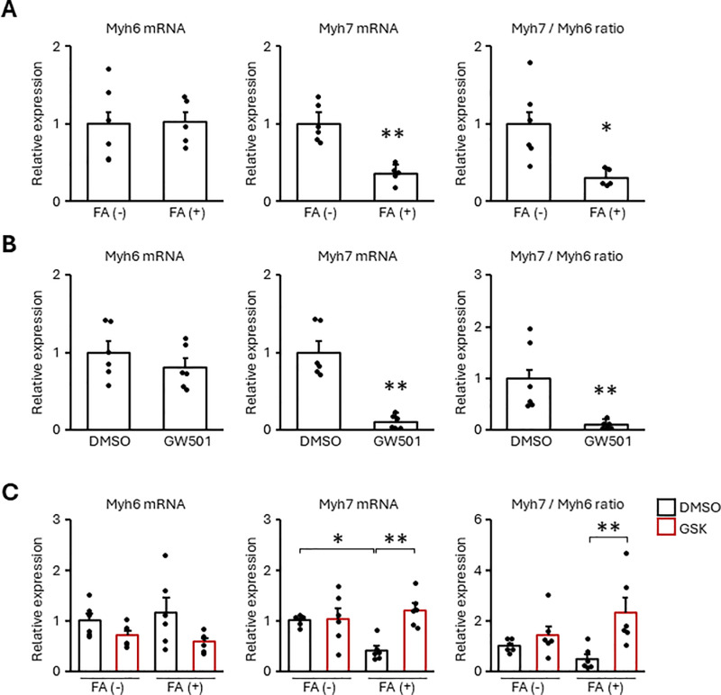
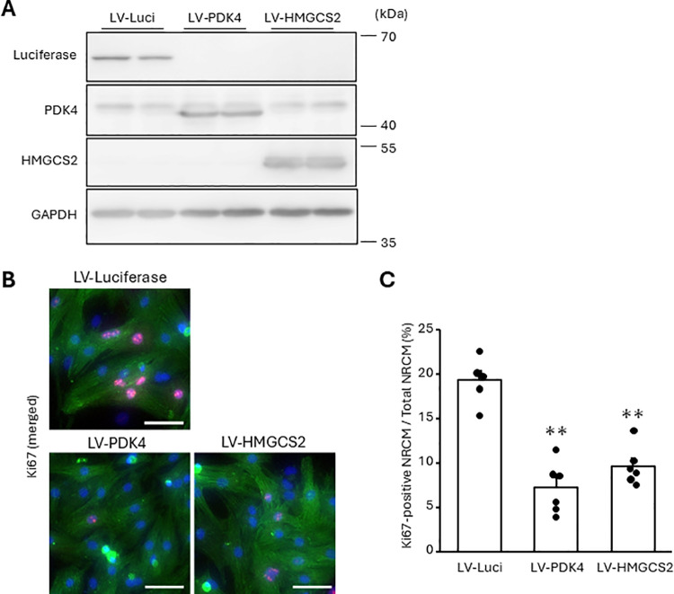
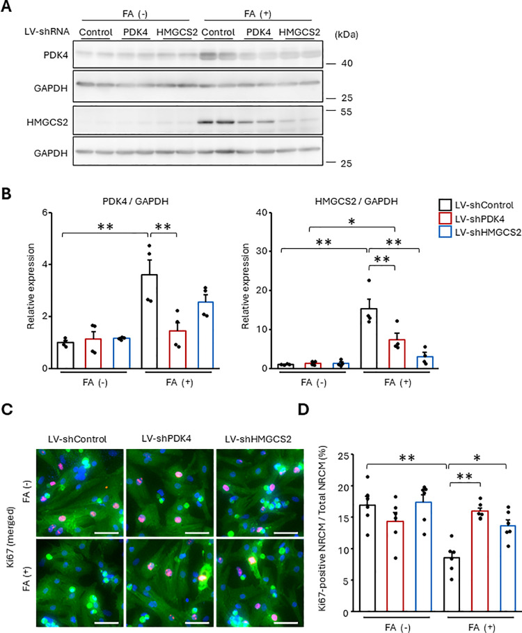
Cardiomyocytes lose their capacity to regenerate immediately after birth. Simultaneously, cardiomyocytes change energy metabolism from glycolysis to oxidative phosphorylation, especially using fatty acids. Accumulating evidence has revealed that fatty acid metabolism weakens the proliferative ability of cardiomyocytes. However, its underlying molecular mechanism remains unclear. In this study, we investigated how fatty acid metabolism contributes to cell cycle regulation in neonatal cardiomyocytes. Cultured neonatal rat cardiomyocytes (NRCMs) were treated with a fatty acid mixture (FA) consisting of palmitic and oleic acids containing L-carnitine. The FA treatment increased not only β-oxidation-related enzymes but also pyruvate dehydrogenase kinase 4 (PDK4), a fatty acid metabolism regulator, and HMG-CoA synthase 2 (HMGCS2), a ketogenic factor. Moreover, Ki67-positive proliferative NRCMs were reduced by the FA, indicating that fatty acids suppress the NRCM cell cycle. GW501516, a peroxisome proliferator-activated receptor δ (PPARδ) activator, also upregulated fatty acid metabolism genes and disturbed NRCM proliferation, whereas GSK3787, a PPARδ inhibitor, recovered FA-induced the cell cycle arrest. Furthermore, overexpression of PDK4 or HMGCS2 using a lentiviral vector suppressed cell cycle activity in NRCMs, and silencing either gene regained cell cycle even in FA-rich condition. In conclusion, fatty acid metabolism increased PDK4 and HMGCS2 via PPARδ activation and suppressed NRCM proliferation.


Conflict of interest statement

The authors have no conflicts of interest associated with this manuscript.

Figures

Fig 1
Fig 1. FA treatment upregulates the expression of fatty acid metabolism-related genes without ATP production in NRCMs.
NRCMs were treated with the indicated concentrations of the FA for 24 h. (A, D) Transcript expression was measured using real-time RT-PCR. (B, C, E) Amounts of acetyl-CoA, ATP, and β-hydroxybutyrate were quantified. Results are shown as the mean ± SEM (A, D: n = 9, B, C: n = 5, E: n = 3). *p < 0.05, **p < 0.01 vs. 0 μM FA by Dunnett test (A, D) and Student’s t-test (B, C, E).
Fig 2
Fig 2. FA suppressed NRCM proliferation.
NRCMs were treated with the indicated concentrations of the FA for 24 h (A-F) or 48 h (G). (A-F) The proportion of Ki67, phospho-Histone H3 (pHH3), or Aurora B-positive NRCMs was analyzed using immunostaining. Cells were stained with anti-Ki67, pHH3, or Aurora B-antibodies (red). Cardiomyocytes and nuclei were labeled with anti-α-actinin antibody (green) and DAPI (blue), respectively. (A, C, E) Representative images. (B, D, F) Quantitative data. The bars indicate 100 μm. Arrowheads indicate Aurora B. (G) The amount of NRCMs was analyzed by cell counting assay using WST-8. Results are shown as the mean ± SEM (B: n = 9, D: n = 6, F: n = 3, G: n = 5). *p < 0.05, **p < 0.01 vs. 0 μM FA by Dunnett test (B) and Student’s t-test (D, F, G).
Fig 3
Fig 3. The activation of PPAR
δ upregulates fatty acid metabolism-related factors and suppresses NRCM proliferation. NRCMs were treated with fenofibrate (Feno), pioglitazone (Pio), or GW501516 (GW501) at 10 μM for 24 h. (A) Transcript expression was measured using real-time RT-PCR. (B, C) The proportion of Ki67-positive NRCMs was analyzed using immunostaining. Cells were stained with an anti-Ki67 antibody (red). Cardiomyocytes and nuclei were labeled with anti-α-actinin antibody (green) and DAPI (blue), respectively. (B) Representative images. (C) Quantitative data. The bars indicate 100 μm. Results are shown as the mean ± SEM (n = 6). *p < 0.05, **p < 0.01 by Tukey-Kramer test.
Fig 4
Fig 4. Inhibition of PPAR
δ represses the increase of fatty acid metabolism-related gene products and recovers the cell cycle arrest in response to FA treatment. NRCMs were pretreated with GW6471, T0070907 (T007), or GSK3787 (GSK) at 10 μM for 24 h, followed by the treatment of 500 μM FA for 24 h. (A) Transcript expression was measured using real-time RT-PCR. (B, C) The proportion of Ki67-positive NRCMs was analyzed using immunostaining. Cells were stained with an anti-Ki67 antibody (red). Cardiomyocytes and nuclei were labeled with anti-α-actinin antibody (green) and DAPI (blue), respectively. (B) Representative images. (C) Quantitative data. The bars indicate 100 μm. Results are shown as mean ± SEM. *p < 0.05, **p < 0.01 vs. FA (-), DMSO and #p < 0.05, ##p < 0.01 vs. FA (+), DMSO by Tukey-Kramer test.
Fig 5
Fig 5. FA promoted NRCM maturation through PPAR
δ. NRCMs were treated with 500 μM FA (A) or 10 μM GW501516 (GW501) (B) for 48 h. Otherwise, NRCMs were pretreated with GSK3787 (GSK) at 10 μM for 24 h, followed by the treatment of 500 μM FA for 48 h (C). The expression of the transcripts was measured by real-time RT-PCR. Results are shown as the mean ± SEM (A: n = 5-6, B, C: n = 6). *p < 0.05, **p < 0.01 vs. 0 μM FA by Student’s t-test (A, B) and Tukey-Kramer test (C).
Fig 6
Fig 6. The overexpression of PDK4 or HMGCS2 suppresses NRCM proliferation.
The NRCMs were infected with the indicated lentiviral vectors for 72 h. (A) Protein expression was measured using western blotting with anti-PDK4 and anti-HMGCS2 antibodies. Representative images are shown. (B, C) The proportion of Ki67-positive NRCMs was analyzed using immunostaining. Cells were stained with an anti-Ki67 antibody (red). Cardiomyocytes and nuclei were labeled with anti-α-actinin antibody (green) and DAPI (blue), respectively. (B) Representative images. (C) Quantitative data. The bars indicate 100 μm. Results are shown as mean ± SEM (n = 6). **p < 0.01 vs. LV-Luciferase by Dunnet test.
Fig 7
Fig 7. The suppression of PDK4 or HMGCS2 abrogates FA-induced cell cycle arrest.
The NRCMs were infected with the indicated lentiviral vectors for 48 h, followed by the stimulation of 500 μM FA. (A, B) Protein expression was measured using western blotting with anti-PDK4 and anti-HMGCS2 antibodies. (C, D) The proportion of Ki67-positive NRCMs was analyzed using immunostaining. Cells were stained with an anti-Ki67 antibody (red). Cardiomyocytes and nuclei were labeled with anti-α-actinin antibody (green) and DAPI (blue), respectively. (A, C) Representative images. (B, D) Quantitative data. The bars indicate 100 μm. Results are shown as mean ± SEM (B: n = 4, D: n = 6). *p < 0.05, **p < 0.01 by Tukey-Kramer test.

Similar articles

References

    1. Poss KD, Wilson LG, Keating MT. Heart regeneration in zebrafish. Science. 2002;298(5601):2188–90. doi: 10.1126/science.1077857 - DOI - PubMed
    1. Liao S, Dong W, Lv L, Guo H, Yang J, Zhao H, et al.. Heart regeneration in adult Xenopus tropicalis after apical resection. Cell Biosci. 2017;7:70. doi: 10.1186/s13578-017-0199-6 PMCID: - DOI - PMC - PubMed
    1. Walsh S, Pontén A, Fleischmann BK, Jovinge S. Cardiomyocyte cell cycle control and growth estimation in vivo--an analysis based on cardiomyocyte nuclei. Cardiovasc Res. 2010;86(3):365–73. doi: 10.1093/cvr/cvq005 - DOI - PubMed
    1. Porrello ER, Mahmoud AI, Simpson E, Hill JA, Richardson JA, Olson EN, et al.. Transient regenerative potential of the neonatal mouse heart. Science. 2011;331(6020):1078–80. doi: 10.1126/science.1200708 PMCID: PMCPMC3099478 - DOI - PMC - PubMed
    1. Mollova M, Bersell K, Walsh S, Savla J, Das LT, Park S-Y, et al.. Cardiomyocyte proliferation contributes to heart growth in young humans. Proc Natl Acad Sci U S A. 2013;110(4):1446–51. doi: 10.1073/pnas.1214608110 PMCID: - DOI - PMC - PubMed

MeSH terms

LinkOut - more resources