. 2025 May 8;20(5):e0319350.
doi: 10.1371/journal.pone.0319350. eCollection 2025.

Gene delivery of AGAT and GAMT boosts creatine levels in creatine transporter deficiency patient fibroblasts

Affiliations

Gene delivery of AGAT and GAMT boosts creatine levels in creatine transporter deficiency patient fibroblasts

Chloe Wells et al. PLoS One. .

Abstract

Creatine is a critical metabolite used to buffer cellular energy demands in highly energetic tissues such as the brain and muscle. Genetic defects in endogenous creatine synthesis or transport across cellular membranes lead to a common set of phenotypes referred to as Cerebral Creatine Deficiency Syndrome (CCDS). The most common form of CCDS is Creatine Transporter 1 (CT1) Deficiency (CTD). It accounts for ~ 70% of cases and results from loss-of-function mutations in the X-linked gene SLC6A8. Affected individuals suffer from intellectual disability, autistic-like behaviors, and epilepsy. There are currently no effective therapies for this disorder, but gene therapy has emerged as a potential approach. The two enzymes which comprise the endogenous creatine synthetic pathway (AGAT and GAMT) are selectively expressed by specific cell types throughout the body. However, after synthesized, creatine uptake relies on the protein product of SLC6A8, CT1, to transport creatine into target cell types. We hypothesized that gene delivery of GATM (encoding AGAT) and GAMT into end-user cell types would bypass the need for CT1, allowing for intracellular synthesis of creatine. We tested this strategy in two human cell types: HEK293T cells and primary fibroblasts. Co-delivery of GATM and GAMT increased internal creatine concentrations by 7.6-fold in HEK293T cells and 12.3-fold in healthy control fibroblasts. We then employed this approach to primary fibroblasts from patients with CTD. This resulted in an up to 11.6-fold increase in intracellular creatine concentrations, far exceeding the intracellular concentration of creatine in healthy control fibroblasts. Importantly, overexpression of AGAT and GAMT resulted in proper targeting of these enzymes to their natural cellular compartment and did not impair the growth of patient fibroblasts. These findings establish gene therapy with GATM and GAMT as a potential strategy for patients with CTD.


Conflict of interest statement

I have read the journal's policy and the authors of this manuscript have the following competing interests: JR is a founder of Vettore Biosciences and a member of its scientific advisory board. All other authors declare no competing interests.

Figures
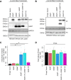
Fig 1
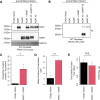
Fig 1. Expression of AGAT and GAMT in HEK293T cells.
(A) HEK293T cells were transfected with the indicated plasmid. Cells were then lysed and analyzed by western blotting with the indicated antibody. N = 3. (B) Same lysates as in (A) were probed for the V5 tagged form of AGAT and GAMT by western blot. N = 3. (C) Cells transfected as in (A) were cultured in creatine-free media supplemented with amino acid precursors for 24 hours. Cells were lysed and the total cellular creatine was quantified. N = 4. Two-sided t-test * indicates P < 0.05. (D) Same experiment as in (C) plotting the total cellular protein measured. N = 4. n.s. indicates not significant for one-way ANOVA. Error bars show SEM.
Fig 2
Fig 2. Total cellular expression of AGAT and GAMT in WT fibroblasts.
(A) WT control fibroblasts were infected with the indicated lentivirus. Cells were then lysed and analyzed by western blotting with the indicated antibody. N = 3. (B) Same lysates as in (A) were probed for the V5 tagged form of AGAT and GAMT by western blot. N = 3. (C) WT fibroblasts stably expressing the indicated transgenes were cultured in creatine-free media supplemented with amino acid precursors for 3 days. Cells were lysed and the total cellular creatine was quantified. N = 3. Two-sided t-test * indicates P < 0.05. (D) Same experiment as in (C) plotting the media creatine concentration measured. N = 3. Two-sided t-test ** indicates P < 0.01. (E) Same experiment as in (C) plotting the total cellular protein measured. N = 3. Two-sided t-test n.s. indicates not significant. Error bars show SEM.
Fig 3
Fig 3. Localization of delivered AGAT and GAMT in WT fibroblasts.
Immunofluorescence (IF) of WT fibroblasts stably expressing AGAT + GAMT and empty vector infected controls imaged at 63x magnification. N = 4. Scale bars indicate 10 µm.
Fig 4
Fig 4. Total cellular expression of AGAT and GAMT in CT1 mutant fibroblasts.
(A) Fibroblasts from a patient with CT1 loss-of-function (SLC6A8Δex10-11/y) were infected with the indicated lentivirus. Cells were then lysed and analyzed by western blotting with the indicated antibody. N = 3. (B) Same lysates as in (A) were probed for the V5 tagged form of AGAT and GAMT by western blot. N = 3. (C) The same CT1 loss-of-function fibroblasts stably expressing the indicated transgenes were cultured in creatine-free media supplemented with amino acid precursors for three days. Cells were lysed and the total cellular creatine was quantified. N = 3. Two-sided t-test P = 0.06. (D) Same experiment as in (C) plotting the media creatine concentration measured. N = 2. Two-sided t-test n.s. indicates not significant. (E) Same experiment as in (C) plotting the total cellular protein measured. N = 3. Two-sided t-test n.s. indicates not significant. Error bars show SEM. (F) A second line of CT1 loss-of-function fibroblasts (SLC6A8W556X/y) stably expressing the indicated transgenes were cultured in creatine-free media supplemented with amino acid precursors for three days. Cells were lysed and the total cellular creatine was quantified. N = 3. Two-sided t-test * indicates P < 0.05. (G) Same experiment as in (F) plotting the media creatine concentration measured. N = 3. Two-sided t-test * indicates P < 0.05. (H) Same experiment as in (F) plotting the total cellular protein measured. N = 3. Two-sided t-test n.s. indicates not significant. Error bars show SEM.
Fig 5
Fig 5. Localization of delivered AGAT and GAMT in CT1 mutant (SLC6A8
Δex10-11/y) fibroblasts. IF of CT1 loss-of-function patient fibroblasts stably expressing AGAT + GAMT and empty vector infected controls imaged at 63x magnification. N = 4. Scale bars indicate 10 µm.
Fig 6
Fig 6. Localization of delivered AGAT and GAMT in CT1 mutant (SLC6A8W556X/y) fibroblasts.
IF of a second line of CT1 loss-of-function patient fibroblasts stably expressing AGAT + GAMT and empty vector infected controls imaged at 63x magnification. N = 4. Scale bars indicate 10 µm.
Fig 7
Fig 7. Growth characteristics of fibroblasts stably expressing AGAT and GAMT.
(A) A model depicting the two sources of intracellular creatine. Creatine is either imported via CT1 or synthesized from its amino acid precursors: L-arginine; glycine; and methionine. The latter is converted into SAM via MAT2. Created with BioRender.com. (B) Growth of WT fibroblasts stably expressing the indicated constructs in standard media (DMEM + 15% FBS + P/S). N = 6. Two-sided K-S test n.s. indicates not significant. (C) Growth of CT1 loss-of-function (SLC6A8Δex10-11/y) patient fibroblasts stably expressing the indicated constructs in standard media. N = 6. Two-sided K-S test **** indicates P < 0.0001. (D) Similar experiment to (C) except media was supplemented with creatine precursor amino acids to 1 mM above standard media. N = 6. Two-sided K-S test *** indicates P < 0.001. (E) Growth of CT1 loss-of-function (SLC6A8W556X/y) patient fibroblasts stably expressing the indicated constructs in standard media. N = 6. Two-sided K-S test * indicates P < 0.05. (F) Similar experiment to (E) except media was supplemented with creatine precursor amino acids. N = 6. Two-sided K-S test **** indicates P < 0.0001. Lines indicate a LOESS curve fit to the mean at each timepoint. Error bars show mean + /- SEM.

Similar articles

References

    1. Joncquel-Chevalier Curt M, Voicu P-M, Fontaine M, Dessein A-F, Porchet N, Mention-Mulliez K, et al.. Creatine biosynthesis and transport in health and disease. Biochimie. 2015;119:146–65. doi: 10.1016/j.biochi.2015.10.022 - DOI - PubMed
    1. Qin W, Khuchua Z, Cheng J, Boero J, Payne RM, Strauss AW. Molecular and Cellular Biochemistry. 1998;184(1/2):153–67. doi: 10.1023/a:1006807515892 - DOI - PubMed
    1. Brosnan JT, Brosnan ME. Creatine: endogenous metabolite, dietary, and therapeutic supplement. Annu Rev Nutr. 2007;27:241–61. doi: 10.1146/annurev.nutr.27.061406.093621 - DOI - PubMed
    1. Wyss M, Kaddurah-Daouk R. Creatine and creatinine metabolism. Physiol Rev. 2000;80(3):1107–213. doi: 10.1152/physrev.2000.80.3.1107 - DOI - PubMed
    1. Mercimek-Andrews S, Salomons GS. Creatine Deficiency Syndromes. University of Washington, Seattle, Seattle (WA); 2022. Available: http://europepmc.org/abstract/MED/20301745 - PubMed

MeSH terms

Supplementary concepts