. 2025 May 8;388(6747):eadk3487.
doi: 10.1126/science.adk3487. Epub 2025 May 8.

Pancreatic cancer-restricted cryptic antigens are targets for T cell recognition

Affiliations

Pancreatic cancer-restricted cryptic antigens are targets for T cell recognition

Zackery A Ely et al. Science. .

Abstract

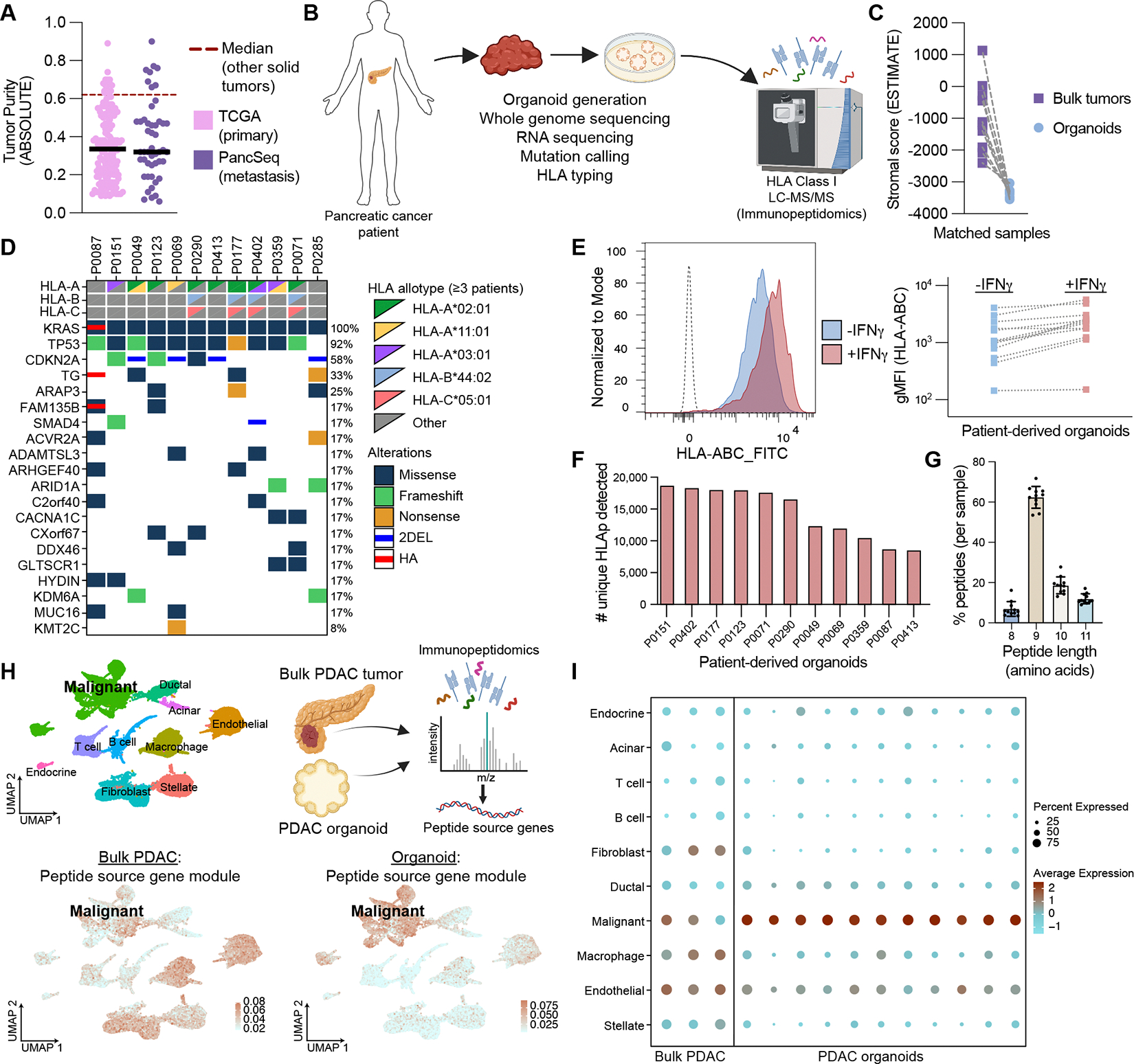
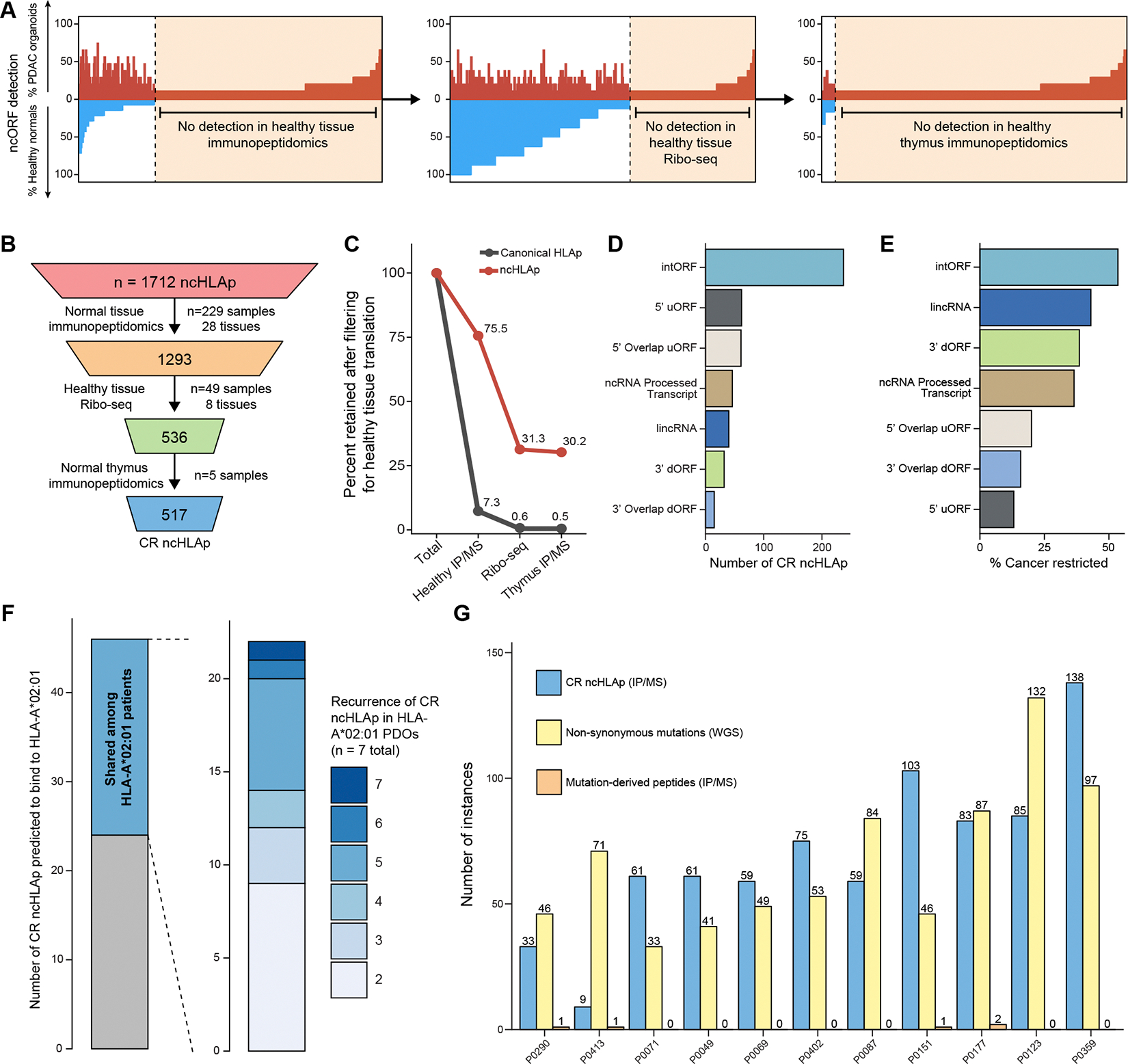
Translation of the noncoding genome in cancer can generate cryptic (noncanonical) peptides capable of presentation by human leukocyte antigen class I (HLA-I); however, the cancer specificity and immunogenicity of noncanonical HLA-I-bound peptides (ncHLAp) are incompletely understood. Using high-resolution immunopeptidomics, we discovered that cryptic peptides are abundant in the pancreatic cancer immunopeptidome. Approximately 30% of ncHLAp exhibited cancer-restricted translation, and a substantial subset were shared among patients. Cancer-restricted ncHLAp displayed robust immunogenic potential in a sensitive ex vivo T cell priming platform. ncHLAp-reactive, T cell receptor-redirected T cells exhibited tumoricidal activity against patient-derived pancreatic cancer organoids. These findings demonstrate that pancreatic cancer harbors cancer-restricted ncHLAp that can be recognized by cytotoxic T cells. Future therapeutic strategies for pancreatic cancer, and potentially other solid tumors, may include targeting cryptic antigens.


Conflict of interest statement

Competing interests: WAF-P has consulted for Longitude Capital and Third Rock Ventures. He holds equity in Regeneron, CRISPR Therapeutics and Editas. WAF-P receives research support (drug-only) from Arcus Biosciences, Pyxis Oncology, Dragonfly Therapeutics, and Philogen. TJ is a member of the Board of Directors of Amgen and Thermo Fisher Scientific, and a co-founder of Dragonfly Therapeutics and T2 Biosystems. TJ serves on the Scientific Advisory Board of Dragonfly Therapeutics, SQZ Biotech, and Skyhawk Therapeutics. TJ is also the President of Break Through Cancer. His laboratory received funding from the Johnson & Johnson Lung Cancer Initiative, but this funding did not support the research described in this manuscript. ZAE is an employee of Longitude Capital. AJA has consulted for Anji Pharmaceuticals, Affini-T Therapeutics, Arrakis Therapeutics, AstraZeneca, Boehringer Ingelheim, Kestrel Therapeutics, Merck & Co., Inc., Mirati Therapeutics, Nimbus Therapeutics, Oncorus, Inc., Plexium, Quanta Therapeutics, Revolution Medicines, Reactive Biosciences, Riva Therapeutics, Servier Pharmaceuticals, Syros Pharmaceuticals, Taiho Pharmaceuticals, T-knife Therapeutics, Third Rock Ventures, and Ventus Therapeutics. AJA holds equity in Riva Therapeutics and Kestrel Therapeutics. AJA has research funding from Amgen, AstraZeneca, Boehringer Ingelheim, Bristol Myers Squibb, Deerfield, Inc., Eli Lilly, Mirati Therapeutics, Nimbus Therapeutics, Novartis, Novo Ventures, Revolution Medicines, and Syros Pharmaceuticals. BMW is a consultant for or sits on the advisory board of Agenus, Bristol Myers Squibb/Mirati, EcoR1 Capital, GRAIL, Harbinger Health, Ipsen, Lustgarten Foundation, Revolution Medicines, Tango Therapeutics, Third Rock Ventures. JMC receives research funding to his institution from Amgen, Merus, Servier, and Bristol Myers Squibb. He receives research support from Merck, AstraZeneca, Esperas Pharma, Bayer, Tesaro, Arcus Biosciences, and Pyxis; he has also received honoraria for being on the advisory boards of Incyte and Blueprint Medicines and for serving on the data safety monitoring committee for Astrazeneca. SRH is inventor of licensed patents related to T cell detection technologies. JGA is a paid consultant to Enara Bio and Moderna. SAC is a member of the scientific advisory boards of Kymera, PTM BioLabs, Seer and PrognomIQ. PDG is a co-founder of Affini-T Therapeutics and is on the Scientific Advisory Boards of Affini-T Therapeutics, Immunoscape, RAPT, Earli, Catalio, Nextech, Metagenomi, and Elpiscience. TMS is a co-founder of Affini-T Therapeutics and is on the Scientific Advisory Boards of Affini-T Therapeutics and Metagenomi. ELS has consulted for ArsenalBio, Blackstone Life Sciences, Chroma Medicine, Clade Therapeutics, Eureka Therapeutics, ImmuneBridge, Legend Biotech, Overland Pharmaceuticals, Predicta Biosciences and Sana Biotech. ELS is an inventor on licensed patents and receives royalties from Bristol Myers Squibb and Sanofi. ELS is on the Scientific Advisory Boards of Bristol Myers Squibb, Chimeric Therapeutics and Sanofi. ELS holds equity in Predicta Biosciences and receives research funding from Sanofi. AMJ is a consultant for and holds equity in RyboDyn. SR receives research support from Microsoft and holds equity in Amgen. WAF-P, TJ, ZAE, ZJK are listed as inventors on provisional patent application number 63/747,859 submitted by Dana-Farber Cancer Institute that covers the use of non-canonical peptide antigens as therapeutic targets in cancer. All other authors declare no competing interests. None of these affiliations represent a conflict of interest with respect to the design or execution of this study or the interpretation of data presented in this manuscript.

Figures

Fig. 1.
Fig. 1.. Organoids enable high-resolution characterization of the pancreatic cancer immunopeptidome.
(A) ABSOLUTE tumor purity in indicated PDAC patient cohorts. The median purity across ten other solid tumor types (40) is depicted with a red dashed line (see Methods). (B) Workflow depicting the proteogenomic characterization of pancreatic cancer patient-derived organoids (PDOs), including whole genome sequencing, RNA-Sequencing, and immunopeptidomics. (C) Quantification of stromal gene program scores calculated by the ESTIMATE algorithm (see Methods) based on RNA-Seq of organoids and matched (autologous) bulk tumor samples. (D) Co-mutation plot depicting the mutational landscape and HLA allotypes of the profiled PDOs. Recurrent HLAs are highlighted to the right of the co-mutation plot. 2DEL refers to deletion of both alleles. HA refers to high-level amplification of the locus. (E) Flow cytometric confirmation of surface HLA-I, representative histogram (left) and individual geometric mean fluorescence intensity (gMFI, right). (F) Number of unique HLAp detected by immunopeptidomics for each PDO. (G) Peptide length distribution of all unique HLAp detected per PDO. Data and error bars represent the mean and standard deviation across all eleven profiled PDOs. (H) Uniform Manifold Approximation and Projection (UMAP) depicting annotated cell types derived from scRNA-Seq of >20 individual patient PDAC samples (top left; see Methods). Expression of peptide source gene modules (PSGM) derived from canonical source genes encoding peptides detected in the immunopeptidome of one bulk PDAC sample, P0625 (bottom left), and one PDO, P0413 (bottom right). PSGM scores represent the average expression of genes in each module relative to a control gene set (see Methods). (I) Average expression of PSGMs derived from the immunopeptidomes of three bulk PDAC tumors (left) and eleven organoid samples (right). Organoid-derived PSGMs show specific enrichment in the malignant population, indicating higher expression in cancer cells relative to non-malignant cell types. “Percent Expressed” refers to the percentage of cells of the annotated type that express the module.
Fig. 2.
Fig. 2.. Empiric detection of cryptic epitopes in the pancreatic cancer immunopeptidome.
(A) Number of non-synonymous variants (missense, frameshift, and in-frame indels) detected by whole-genome sequencing compared to the number of mutation-derived neoepitopes (mutHLAp) detected by immunopeptidomics. (B) Rank-ordered HLA-I affinity for mutation-derived peptide:MHC pairs. Affinity was predicted using a combination of NetMHC-4.0, NetMHCpan-4.0, SMM, and SMMPMBEC (see Methods). Gray dots represent the subset of all pairs with a median predicted peptide:MHC affinity < 500 nM (unique peptide:MHC pairs = 1,242; unique peptides = 1,133). All exonic mutations from the 11 PDOs, regardless of predicted affinity, were included in the immunopeptidomics search space, but only the neoepitopes represented by colored dots were detected. (C) Proportion of unique HLAp specifically mapping to either canonical genes or nuORFs per PDO (ncHLAp median proportion = 5.1%; range = 2.2 – 10.1%). (D) Number of ncHLAp observed in PDAC PDOs derived from indicated nuORF biotypes/categories. Bars represent the sum of each unique PDO-ncHLAp pairing. (E) Recurrence of uniquely mapping ncHLAp across PDAC PDOs with indicated HLAs. (F-J) Canonical and non-canonical HLAp characteristics from PDAC PDOs: (F) Distribution of detected HLAp lengths for canonical (gray) and non-canonical (red) HLAp; (G-H) Peptide sequence profile plots representing the amino acid profile of 9mers predicted to bind A*02:01 (G) or B*44:02 (H) across all relevant organoid samples. HLA allele binding predictions were made with HLAthena; (I) Normalized position on the source ORF from which non-canonical HLAp arise; (J) Overall percentage (of predicted unique binders) of canonical or non-canonical peptides predicted to bind different HLA alleles. Only HLAp predicted to exclusively bind to indicated HLA allele included in analysis.
Fig. 3.
Fig. 3.. Cancer-restriction of ncHLAp.
(A-B) Translation-centric filtering pipeline used to nominate PDAC-restricted ncHLAp. (A) Gating strategy to identify parental ORFs encoding ncHLAp that are not detected in any healthy tissue, including thymus, via HLA-I immunopeptidomics or Ribo-seq. Each vertical bar represents the percentage of samples (healthy tissue samples or PDAC organoids) in which expression of a ncHLAp-encoding ORF was identified. (B) Number of noncanonical HLA-I peptides retained after each filtration step in the translation-centric pipeline. (C) Percentage of noncanonical or canonical HLAp retained after each step of the translation-centric filtering pipeline. IP/MS=immunopeptidomics. (D) Number of CR ncHLAp detected in PDAC PDOs by category/biotype (only ncHLAp categories with ≥10 CR ncHLAp included). (E) Percent of detected ncHLAp that exhibit cancer-restriction within each category/biotype (only ncHLAp categories with ≥10 CR ncHLAp included). (F) Frequency of shared and patient-specific HLA-A*02:01-restricted CR ncHLAp across PDOs. (G) Number of CR ncHLAp and mutation-derived HLAp empirically detected in each PDO line.
Fig. 4.
Fig. 4.. PDAC-restricted ncHLAp exhibit immunogenic potential.
(A) Workflow of ex vivo approach to enable priming and expansion of antigen-specific T cells. (B) Immunogenic potential of empirically detected ncHLAp (CR ncHLAp = top; nonCR ncHLAp = bottom), following priming and two re-stimulations with autologous peptide-loaded APCs. Percent positivity over background in each CTL line indicated in heatmap. Threshold for considering a positive response: >2% over the negative control, assessed by simultaneous staining of autologous pre-stimulated CD8 T cells (tetramer) or no peptide control (ICS). Table indicating #CTL lines evaluated, #CTL lines positive (red), HLA restriction tested, CR/nonCR, and recurrence (recurrent or private among PDOs, number of organoid lines in which a peptide:HLA was empirically detected is indicated) for each ncHLAp tested. Private/recurrent designations are based on the pairing of each ncHLAp and its tested allele across all profiled PDOs. (C) Sankey plot depicting empirically identified CR and nonCR ncHLAp, detection in healthy tissues (including thymus), and immunogenicity. (D) Number of immunogenic or non-immunogenic CR ncHLAp from indicated biotypes. Here, 5’ uORF category is inclusive of 5’ uORFs and 5’ overlap uORFs. ncRNA is inclusive of ncRNA processed transcript and ncRNA retained intron biotypes. 3’ dORF is inclusive of 3’ dORFs and 3’ overlap dORFs. (E) Schematic of BEAM-T to enable single-cell TCR sequencing of pooled ncHLAp-reactive CTLs and deconvolution of their antigen-specificity. (F) Candidate ncHLAp-reactive TCRs identified from BEAM-T with indicated HLA restriction, antigen-specificity score, and CDR3αβ sequences. (G) Functional avidity (as measured by cytotoxicity against peptide-loaded T2_eGFP_ffLuc cells) of HLA-A*02:01-restricted, ncHLAp-directed TCR-T cells. Individual biological replicate values displayed (n=2 biological replicates). Normalized to T2 cell viability alone.
Fig. 5.
Fig. 5.. CR ncHLAp-specific T cells can recognize and kill pancreatic cancer organoids ex vivo and in vivo.
(A) Workflow/approach for organoid:TCR-T cell co-culture. (B) IFN-γ ELISA from co-culture of P0071_eGFP-ffLuc PDOs with the indicated ncHLAp-reactive TCR-T cells (10:1 E:T). Mean −/+ SD (of n=3) (C) CD137 surface expression on indicated ncHLAp-reactive TCR-T cells following co-culture with indicated PDOs. P0151 (HLA-mismatched) PDOs were used as a negative control. Representative experiment of n=3 biological replicates. Mean −/+ SD of n=3 technical replicates displayed. (D) Cytotoxicity after co-culture of P0071_eGFP-ffLuc PDOs with the indicated ncHLAp-reactive TCR-T cells at indicated E:T ratios. Mean −/+ SD (of n=3). (E) Alanine scanning of TCR001 TCR-T cells. Individual biological replicate values displayed (n=2 biological replicates). (F) Workflow for in vivo ncHLAp-directed TCR-T studies. (G) Longitudinal tumor volume assessment of subcutaneously transplanted P0071_eGFP-ffLuc PDOX in NSG mice (n = 8 per arm), block randomized to receive two administrations of indicated TCR-T cells (Day 0 [2 × 10⁷] and Day 10 [1.5 × 10⁷]). Mean −/+ SD. Statistics: (B-D) unpaired t-test; (G) linear mixed effects regression model.

Comment in

References

    1. Leko V, Rosenberg SA, Identifying and Targeting Human Tumor Antigens for T Cell-Based Immunotherapy of Solid Tumors. Cancer Cell 38, 454–472 (2020). - PMC - PubMed
    1. Chong C, Müller M, Pak HS, Harnett D, Huber F, Grun D, Leleu M, Auger A, Arnaud M, Stevenson BJ, Michaux J, Bilic I, Hirsekorn A, Calviello L, Simó-Riudalbas L, Planet E, Lubiński J, Bryśkiewicz M, Wiznerowicz M, Xenarios I, Zhang L, Trono D, Harari A, Ohler U, Coukos G, Bassani-Sternberg M, Integrated proteogenomic deep sequencing and analytics accurately identify non-canonical peptides in tumor immunopeptidomes. Nat Commun 11, 1293 (2020). - PMC - PubMed
    1. Laumont CM, Daouda T, Laverdure JP, Bonneil É, Caron-Lizotte O, Hardy MP, Granados DP, Durette C, Lemieux S, Thibault P, Perreault C, Global proteogenomic analysis of human MHC class I-associated peptides derived from non-canonical reading frames. Nat Commun 7, 10238 (2016). - PMC - PubMed
    1. Laumont CM, Vincent K, Hesnard L, Audemard É, Bonneil É, Laverdure JP, Gendron P, Courcelles M, Hardy MP, Côté C, Durette C, St-Pierre C, Benhammadi M, Lanoix J, Vobecky S, Haddad E, Lemieux S, Thibault P, Perreault C, Noncoding regions are the main source of targetable tumor-specific antigens. Sci Transl Med 10 (2018). - PubMed
    1. Lozano-Rabella M, Garcia-Garijo A, Palomero J, Yuste-Estevanez A, Erhard F, Farriol-Duran R, Martín-Liberal J, Ochoa-De-Olza M, Matos I, Gartner JJ, Ghosh M, Canals F, Vidal A, Piulats JM, Matías-Guiu X, Brana I, Mu~ Noz-Couselo E, Garralda E, Schlosser A, Gros A, Exploring the Immunogenicity of Noncanonical HLA-I Tumor Ligands Identified through Proteogenomics. Clinical Cancer Research 29, 2250–2265 (2023). - PMC - PubMed

MeSH terms