. 2025 Jul;22(4):e00606.
doi: 10.1016/j.neurot.2025.e00606. Epub 2025 May 8.

Modulation of antibody transport in the brain and spinal cord through the intranasal pathway

Affiliations

Modulation of antibody transport in the brain and spinal cord through the intranasal pathway

Sebastian Spiegel et al. Neurotherapeutics. 2025 Jul.

Abstract

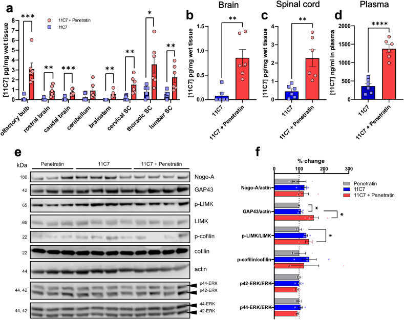
The intranasal pathway is a promising antibody delivery route for the treatment of neurological diseases, but the mechanisms mediating nose-to-brain/spinal cord transport are poorly understood. The aim of our study was to determine if the transport of antibodies can pharmacologically be modulated in the mouse CNS. The pharmacokinetics and distribution of recombinant antibodies were followed in brain and spinal cord homogenates and biofluids by ELISA and immunofluorescence. A non-CNS antigen-binding antibody (FG12) was used to monitor target-independent transport whereas 11C7 mAb, neutralizing the myelin-associated growth inhibitor Nogo-A, was applied to induce CNS target-dependent neuronal growth response. Fast axonal transport/neuronal activity were inhibited with Lidocaine pre-treatment on the olfactory mucosa. Antibody uptake was enhanced across the olfactory epithelium with the co-administration of the cell-penetrating peptide Penetratin. Growth signalling pathways were examined by Western blotting. FG12 was detected in the brain and spinal cord as early as 30 ​min after intranasal administration. After 1 ​h, the concentration of FG12 rapidly declined in all CNS areas and was back to baseline values at 24 ​h. Lidocaine prevented the early rise in FG12 concentration in the spinal cord. This effect was not observed in the brain. Penetratin allowed to maintain the elevation of FG12 and to activate 11C7-induced growth signalling in the spinal cord at 24 ​h. Our data suggest that the pharmacological modulation of transport mechanisms in the nose-to-CNS pathways may allow to control the therapeutic effects of antibodies in neurological diseases.

Keywords: Blood-brain barrier; Cell-penetrating peptide; Intranasal pathway; Neuronal plasticity; Nogo-A; Recombinant antibodies.


Conflict of interest statement

Declaration of competing interest AC has received speakers’/board honoraria from Actelion (Janssen/J&J), Almirall, Bayer, Biogen, Celgene (BMS), Genzyme, Merck KGaA (Darmstadt, Germany), Novartis, Roche, and Teva, all for hospital research funds. He received research support from Biogen, Genzyme, and UCB, the European Union, and the Swiss National Foundation. He serves as associate editor of the European Journal of Neurology, on the editorial board for Clinical and Translational Neuroscience and as topic editor for the Journal of International Medical Research.

Figures

Fig. 1
Fig. 1
Histological observation of FG12-HA distribution in the mouse nasal cavity. a Schematic presentation of intranasal administration of non-CNS antigen-binding antibody FG12-HA onto the olfactory mucosa of mice. b Distribution of FG12-HA in the nasal cavity 3 ​h post-administration. Cribriform plate (cr. pl.) (n ​= ​3). c, c’ Confocal images showing FG12-HA uptake at different locations of the olfactory epithelium cells 3 ​h post administration. FG12-HA was observed in the different layers of the olfactory epithelium, that are the fila olfactoria (f. olfac.), the sustentacular cell layer, the olfactory sensory neuron layer and the lamina propria. c’’ Close-up from c' showing FG12-HA in a sustentacular cell (star). Olfactory sensory neuron processes were also positive for FG12-HA (arrow). d FG12-HA was also found in the cell body (arrowhead) and neurites (arrow) of olfactory sensory neurons. e FG12-HA internalization was observed in beta-3-tubulin(+) olfactory sensory neurons. f Detection of FG12-HA in olfactory nerve bundles (ONB) but not in lymphatic vessels and blood vessels (BV) of the lamina propria (z) and the olfactory epithelium (x). Scale bar: 500 ​μm ​(b), 20 ​μm (c’), 10 ​μm (c’‘, d, e) 50 ​μm (f).
Fig. 2
Fig. 2
Distribution of FG12-HA in the CNS after intranasal administration. a Immunofluorescent detection of FG12-HA in the mouse brain (n ​= ​3/group). Colour coded immunofluorescence (Fire colour coding generated with the ImageJ/NIH software) shows staining rostrocaudal variations in brain regions. b-e Only mice treated with FG12-HA showed positive cells in the hippocampus (CA1) and in the cortex, compared with control mice left untreated. Close-ups in b and d allowed to distinguish FG12-HA immunoreactivity in individual cells. Close-ups from control mice in c and e exhibited very weak signal. Scale bars: 1 ​mm (a), 50 ​μm ​(b, c, d, e), 10 ​μm ​(b’, c’, d’, e’).
Fig. 3
Fig. 3
Pharmacokinetic analysis of FG12-HA. a ELISA detection of FG12-HA in CNS tissues at different time points following intranasal administration. PBS treatment was used as negative control treatment. For region-specific analyses, the CNS was dissected into olfactory bulb (OB, red), rostral brain (RCB, blue), caudal brain (CCB, green), cerebellum (CL, yellow), brainstem (BS, pink), cervical spinal cord (CSC, orange), thoracic spinal cord (TSC, turquoise) and lumbar spinal cord (LSC, grey) pieces. The number of animals per time point was 8 ​at 0.5 ​h, 4 ​at 1 ​h, 4 ​at 3 ​h, 8 ​at 6 ​h and 4 ​at 24 ​h. Statistical test: Multiple Mann-Whitney tests (∗p ​< ​0.05, ∗∗p ​< ​0.01, ∗∗∗p ​< ​0.001). b Time-course of FG12-HA variations calculated for the whole brain (OB, RCB, CCB, CL, BS), and spinal cord (CSC, TSC, LSC) tissues, compared to PBS-treated mice. c The level of FG12-HA was not significantly different between FG12-HA and PBS treatments in the CSF. d In the plasma, the concentration of FG12-HA plateaued from 1 ​h to 6 ​h. Statistical test: Kruskal-Wallis test with Dunn's correction for multiple comparison (∗p ​< ​0.05, ∗∗p ​< ​0.01, ∗∗∗p ​< ​0.001) a-d: Data are presented by means ​± ​SEM.
Fig. 4
Fig. 4
Lidocaine inhibits FG12-HA transport to the spinal cord. a Changes in FG12-HA concentrations 1 ​h post-administration. Ten min before antibody administration, Lidocaine was applied onto the olfactory mucosa to selectively block fast axonal transport/neuronal activity. In the control group, saline application preceded FG12-HA treatment. Statistical test: Multiple Mann-Whitney test (∗p ​< ​0.05, ∗∗p ​< ​0.01). b Lidocaine did not significantly affect FG12-HA concentration in the whole brain. c Lidocaine abolished FG12-HA transport to the spinal cord. d Plasma FG12-HA concentration was not modified by the intranasal administration of Lidocaine. Twelve mice were used in each treatment group. Bars represent means ​± ​SEM. b-d Statistical test: unpaired Mann-Whitney t-test (∗p ​< ​0.05, ∗∗p ​< ​0.01).
Fig. 5
Fig. 5
Enhanced antibody uptake with Penetratin sustains FG12-HA delivery in the brain and the spinal cord. a Co-administration of Penetratin and FG12-HA improved antibody uptake in the olfactory bulb at 1 ​h and in other brain and spinal cord regions at 24 ​h. Five mice were examined at 1 ​h and six mice at 24 ​h (dots represent independent mouse values). b At the whole brain and spinal cord levels, Penetratin only significantly increased FG12-HA at 24 ​h. c Penetratin increased the concentration of FG12-HA in the plasma at 1 ​h and 24 ​h. ​d FG12-HA was only detectable at 24 ​h in the CSF of mice receiving Penetratin. e When expressed in % of plasma levels, the concentration of FG12-HA was however not higher with Penetratin. a-d Histogram bars represent means ​± ​SEM. Statistical test: Multiple Mann-Whitney tests (∗p ​< ​0.05, ∗∗p ​< ​0.01∗∗∗p ​< ​0.001, ∗∗∗∗p ​< ​0.0001).
Fig. 6
Fig. 6
Penetratin-enhanced 11C7 uptake activates neuronal plasticity mechanisms in the spinal cord. a-d Penetratin strongly increased the level of 11C7 in the brain, spinal cord and plasma. Six mice were used per treatment group. e Western blot analysis of Nogo-A signalling in the spinal cord revealed pronounced changes with 11C7 ​+ ​Penetratin compared with 11C7 or Penetratin treatments given separately. Three-four mice were used for each treatment group (1 dot ​= ​1 mouse). Each sample comes from a different animal. f Quantitatively, the level of GAP43 was significantly higher in mice administered with 11C7 ​+ ​Penetratin than in those treated only with 11C7 or Penetratin. Only the phosphorylation state of LIMK, involved in Nogo-A signalling, was also statistically different. Protein signals shown in e were quantified by densitometry with the ImageJ software and normalized to the average values of Penetratin-treated mice in f. Statistical test: unpaired t-test (∗p ​< ​0.05). a-d, f Data are presented by means ​± ​SEM. a-d Statistical test: one way ANOVA (∗p ​< ​0.05).
figs1
figs1
figs2
figs2
figs3
figs3

References

    1. Shen J., Attar M. Antibody-Drug conjugate (ADC) research in ophthalmology--a review. Pharm Res. 2015;32(11):3572–3576. - PubMed
    1. Nisenbaum L., Martone R., Chen T., Rajagovindan R., Dent G., Beaver J., et al. CSF biomarker concordance with amyloid PET in Phase 3 studies of aducanumab. Alzheimer’s Dementia : the journal of the Alzheimer’s Association. 2023;19(8):3379–3388. - PubMed
    1. Perneczky R., Jessen F., Grimmer T., Levin J., Flöel A., Peters O., et al. Anti-amyloid antibody therapies in Alzheimer’s disease. Brain : J Neurol. 2023;146(3):842–849. - PubMed
    1. Winkelmann A., Loebermann M., Reisinger E.C., Hartung H.-P., Zettl U.K. Disease-modifying therapies and infectious risks in multiple sclerosis. 2016;12(4):217–233. - PubMed
    1. St-Amour I., Paré I., Alata W., Coulombe K., Ringuette-Goulet C., Drouin-Ouellet J., et al. Brain bioavailability of human intravenous immunoglobulin and its transport through the murine blood-brain barrier. J Cerebr Blood Flow Metabol. 2013;33(12):1983–1992. - PMC - PubMed