

Exploring Biochemical Characteristics of Pediatric Hyperdiploid Acute Lymphoblastic Leukemia by Raman Spectroscopy
- PMID: 40340365
- PMCID: PMC12096350
- DOI: 10.1021/acs.analchem.5c00410
Exploring Biochemical Characteristics of Pediatric Hyperdiploid Acute Lymphoblastic Leukemia by Raman Spectroscopy
Abstract
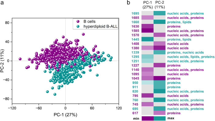
Hyperdiploid (HD) B-cell acute lymphoblastic leukemia (ALL) is widely recognized as the most common molecular subtype of leukemia, characterized by the presence of supernumerary chromosomes in the leukemic karyotype. While HD B-ALL is often associated with a favorable prognosis, an important subset of patients still experience relapse, reflecting the biological heterogeneity of this subtype. Current genomic and epigenetic research has shed light on the molecular complexity of HD B-ALL, yet rapid methods for capturing both the metabolic state and the chromosomal content of individual cells remain limited. Here, we introduce a novel Raman spectroscopy (RS)-based approach for the single-cell analysis of HD B-ALL. By detecting characteristic spectroscopic signatures of nucleic acids, proteins, and lipids, RS not only distinguishes malignant cells from normal B cells, but also discriminates between HD B-ALL and other molecular subtypes, including TCF3-PBX1, KMT2A-r, BCR-ABL1, and TEL-AML1. Notably, we developed a partial least-squares regression (PLS-R) model capable of accurately predicting chromosome number from each cell's Raman spectrum, thereby linking molecular fingerprints directly to genomic aberrations. This integrative spectroscopic strategy captures disease heterogeneity and informs therapeutic strategies. Taken together, our proof-of-concept findings highlight RS as a powerful, noninvasive tool for quantifying chromosomal alterations and metabolic phenotypes, adding crucial insights into the complex biology of HD B-ALL and paving the way for broader applications in precision medicine.
Figures


Similar articles
-
Raman classification of selected subtypes of acute lymphoblastic leukemia (ALL).Analyst. 2024 Jan 15;149(2):571-581. doi: 10.1039/d3an01708g. Analyst. 2024. PMID: 38099606
-
Advancing triage of acute lymphoblastic leukaemia subtypes diagnosis: label-free Raman spectroscopy for precise single-cell phenotyping and subtype classification.Analyst. 2024 Nov 4;149(22):5443-5454. doi: 10.1039/d4an00956h. Analyst. 2024. PMID: 39390798
-
MicroRNA characterize genetic diversity and drug resistance in pediatric acute lymphoblastic leukemia.Haematologica. 2011 May;96(5):703-11. doi: 10.3324/haematol.2010.026138. Epub 2011 Jan 17. Haematologica. 2011. PMID: 21242186 Free PMC article.
-
Pediatric acute lymphoblastic leukemia.Hematology Am Soc Hematol Educ Program. 2003:102-31. doi: 10.1182/asheducation-2003.1.102. Hematology Am Soc Hematol Educ Program. 2003. PMID: 14633779 Review.
-
Clinical implications of recurring chromosomal and associated molecular abnormalities in acute lymphoblastic leukemia.Semin Hematol. 2000 Oct;37(4):381-95. doi: 10.1016/s0037-1963(00)90018-0. Semin Hematol. 2000. PMID: 11071360 Review.
References
-
- Enshaei A., Vora A., Harrison C. J., Moppett J., Moorman A. V.. Defining Low-Risk High Hyperdiploidy in Patients with Paediatric Acute Lymphoblastic Leukaemia: A Retrospective Analysis of Data from the UKALL97/99 and UKALL2003 Clinical Trials. Lancet Haematol. 2021;8(11):e828–e839. doi: 10.1016/S2352-3026(21)00304-5. - DOI - PMC - PubMed
MeSH terms
LinkOut - more resources
Full Text Sources
Miscellaneous
