

Three-dimensional volumetric analysis of bone regeneration following jaw cyst enucleation with and without an autologous albumin gel-platelet-rich fibrin mixture (Alb-PRF): a randomized controlled clinical trial
- PMID: 40340767
- PMCID: PMC12060296
- DOI: 10.1186/s12903-025-06027-w
Three-dimensional volumetric analysis of bone regeneration following jaw cyst enucleation with and without an autologous albumin gel-platelet-rich fibrin mixture (Alb-PRF): a randomized controlled clinical trial
Abstract
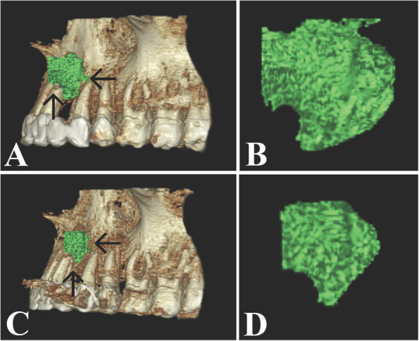
Introduction: The presence of an osseous cavity after cyst enucleation is a clinical challenge that needs to be considered. Using pure autologous concentrations of platelets, platelet-rich fibrin (PRF), as a graft material after cyst removal has shown promising effects. However, PRF has limitations in terms of durability, as it usually resorbs within 10-14 days, thus Mourão et al. introduced a new technique for PRF preparation to obtain an albumin gel-platelet-rich fibrin mixture (Alb-PRF), a new autologous material, that can remain stable for 4-6 months with the ability to regenerate bone. This research aimed to evaluate the effect of Alb-PRF on bone regeneration after jaw cyst enucleation via 3-dimensional (3D) volumetric analysis.
Methods: Twenty participants, with jaw cysts, were split into two groups. The Alb-PRF group included 10 individuals treated by enucleation and Alb-PRF application, and the control group included 10 individuals treated conventionally by enucleation without any additives. Cone beam computed tomography (CBCT) was conducted immediately following surgery (T1) and six months later (T2) to measure the volume of the residual bone cavity and the mean bone density of the regenerated bone using On-demand 3D viewer. Paired t test was used to compare the postoperative immediate results with the post-6-months results, whereas Student t test was used to compare the Alb-PRF group with the control group.
Results: At the 6-month follow-up, the volume of the residual bone cavity had declined and the bone density had increased significantly in both the Alb-PRF group and the control group (P1 < 0.001) compared with the immediate postoperative values. Although the changes in volume and density were greater in the Alb-PRF group than in the control group, there was no a noticeable difference between the two groups. (P = 0.821) and (P = 0.533), respectively.
Conclusion: There was no difference in bone regeneration between Alb-PRF and conventional blood clots after jaw cyst enucleation.
Trial registration: The trial was retrospectively registered at the Clinicaltrial.gov registry (Registration ID #NCT05658900). It was first submitted on 12/12/2022 and first posted on 21/12/2022.
Keywords: Albumin Gel-Platelet-Rich Fibrin Mixture; Bone Density; Bone Regeneration; Centrifugation; Cone Beam Computed Tomography; Jaw Cysts; Spontaneous Healing; Three-Dimensional Volumetric Analysis.
© 2025. The Author(s).
Conflict of interest statement
Declarations. Ethics approval and consent to participate: This research was approved by the Institutional Review Board of the Research Ethics Committee of the Faculty of Dentistry, Alexandria University, Egypt (International Number IORG0008839; Ethics Committee Number 0414–03/2022). These research activities followed the Declaration of Helsinki for human subjects. Each participant signed a formal written informed consent form before the operation. Consent for publication: Not applicable. Competing interests: The authors declare no competing interests.
Figures


Similar articles
-
Biological characterization of an injectable platelet-rich fibrin mixture consisting of autologous albumin gel and liquid platelet-rich fibrin (Alb-PRF).Platelets. 2021 Jan 2;32(1):74-81. doi: 10.1080/09537104.2020.1717455. Epub 2020 Jan 20. Platelets. 2021. PMID: 31959025
-
Increasing Bone Regeneration in Maxillary Sinus Augmentation Using Platelet-rich Fibrin: An Interventional Pre-Post Study.J Contemp Dent Pract. 2024 Sep 1;25(9):814-819. doi: 10.5005/jp-journals-10024-3747. J Contemp Dent Pract. 2024. PMID: 39791407
-
The autologous platelet rich fibrin: A novel approach in osseous regeneration after cystic enucleation: A pilot study.Indian J Dent Res. 2015 Nov-Dec;26(6):560-4. doi: 10.4103/0970-9290.176915. Indian J Dent Res. 2015. PMID: 26888231
-
Impact of Platelet-rich Plasma and Platelet-rich Fibrin in Mandibular Third Molar Extraction: A Systematic Review.J Contemp Dent Pract. 2024 Sep 1;25(9):904-910. doi: 10.5005/jp-journals-10024-3727. J Contemp Dent Pract. 2024. PMID: 39791420
-
Extended platelet-rich fibrin.Periodontol 2000. 2024 Feb;94(1):114-130. doi: 10.1111/prd.12537. Epub 2023 Nov 20. Periodontol 2000. 2024. PMID: 37986559
References
-
- Cawson R. et al. Oral Pathology and Oral Medicine. Vol. 53, Cawson’s Essentials of Oral Pathology and Oral Medicine. 2002. 287 p.
-
- Buchbender M, Neukam FW, Lutz R, Schmitt CM. Treatment of enucleated odontogenic jaw cysts: a systematic review. Oral Surg Oral Med Oral Pathol Oral Radiol. 2018;125(5):399–406. - PubMed
-
- Bodner L. Cystic lesions of the jaws in children. Int J Pediatr Otorhinolaryngol. 2002Jan 11;62(1):25–9. - PubMed
-
- Ettl T, Gosau M, Sader R, Reichert TE. Jaw cysts - Filling or no filling after enucleation? A review J Cranio-Maxillofacial Surg. 2012;40(6):485–93. - PubMed
Publication types
MeSH terms
Substances
Associated data
LinkOut - more resources
Full Text Sources
Medical
Miscellaneous
