. 2025 May 8;25(1):347.
doi: 10.1186/s12876-025-03937-5.

M64HCl, a focal adhesion kinase activator, promotes intestinal mucosal healing in rats

Affiliations

M64HCl, a focal adhesion kinase activator, promotes intestinal mucosal healing in rats

Guiming Liu et al. BMC Gastroenterol. .

Abstract

Background: Intestinal mucosal injury may arise from various factors. While many drugs target the causative factors, none directly stimulate mucosal wound healing. We found that the specific focal adhesion kinase (FAK) activator, M64HCl, promotes intestinal mucosal healing in mice. This study aims to further validate the therapeutic impact of M64HCl on intestinal mucosal repair in rats as a second species.

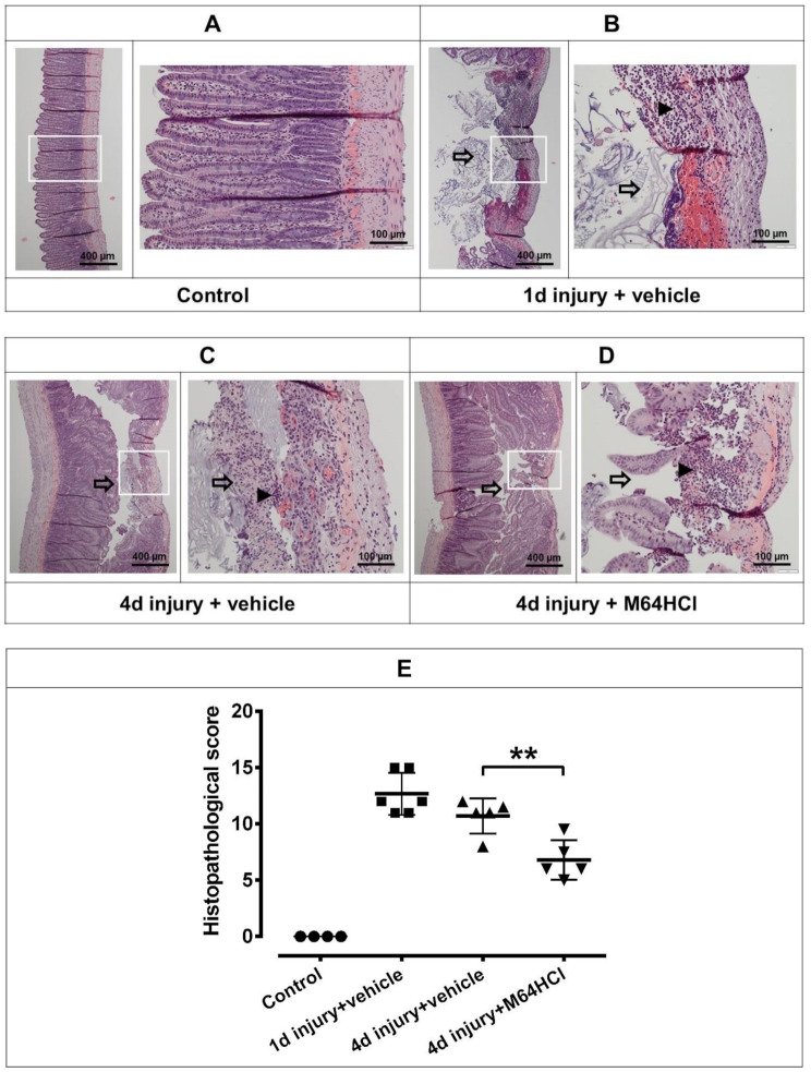
Methods: Wistar rats were assigned to one of four groups: normal control, 1-day injury + vehicle, 4-day injury + vehicle, or 4-day injury + M64HCl. Intestinal injury was induced by serosally applying 75% acetic acid. Immediately after injury, rats received either a continuous infusion of M64HCl (25 mg/kg/day) or its vehicle (saline). Four days post-injury, blood was drawn to measure M64HCl levels and assess liver and kidney function. The intestines were removed and opened, ulcer areas were photographed for size quantification, and tissues were fixed for histological and immunohistochemical analysis.

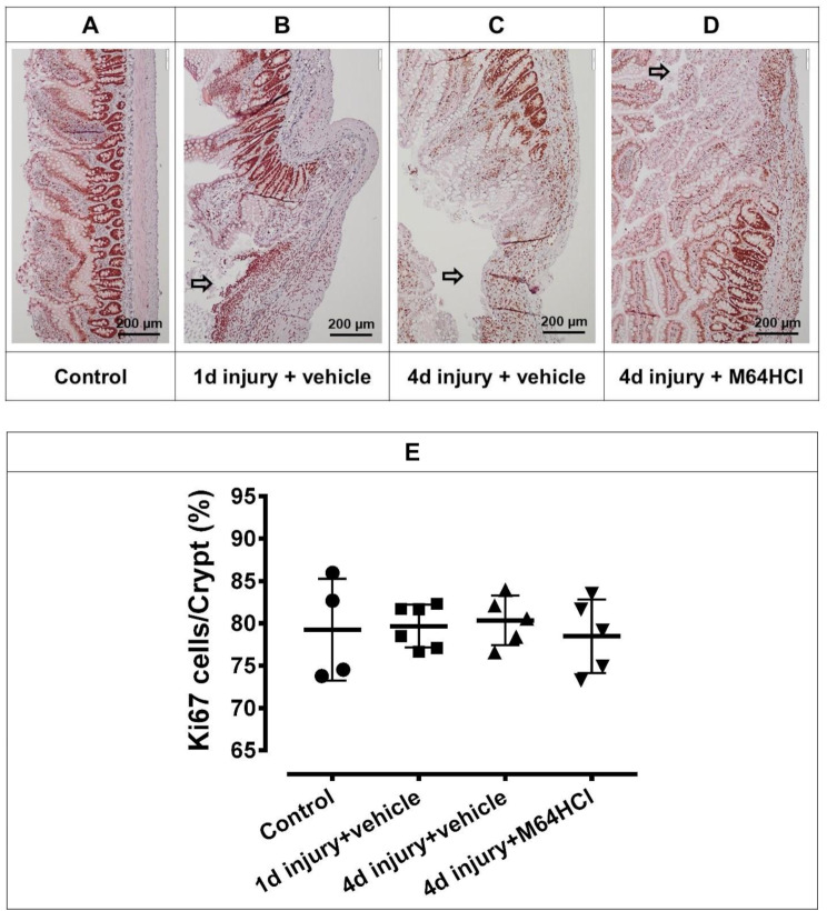
Results: M64HCl substantially reduced ulcer area on gross examination, while histological analysis showed alleviation of pathological changes with M64HCl treatment. Immunohistochemical analysis confirmed increased immunoreactivity for phosphorylated FAK in the epithelium adjacent to the injury in M64HCl-treated rats. However, there was no change in the percentage of Ki67-positive cells in each crypt at the edge of the ulcer area. Serum creatinine, ALT, and AST levels did not differ between the 4-day injury groups with or without M64HCl treatment.

Conclusions: M64HCl, a water-soluble FAK activator, promotes acetic acid-induced ulcer healing in rats and may be useful in treating gastrointestinal mucosal injury.

Keywords: Focal adhesion kinase; Mucosal healing; Small intestine, mucosal injury.


Conflict of interest statement

Declarations. Ethics approval and consent to participate: All experimental procedures involving animals were approved by the Institutional Animal Care and Use Committee (IACUC) of Northeast Ohio Medical University, with IACUC number 23-04-366. The experiments were conducted in accordance with relevant guidelines and regulations. Consent for publication: Not applicable. Competing interests: The University of North Dakota and the University of Minnesota have filed two patent applications on the use of small-molecule FAK activators to promote mucosal healing. MDB and VJG are listed as co-inventors, and one also includes RGM.

Figures

Fig. 1
Fig. 1
Administration of M64HCl accelerates the healing of acetic acid-induced intestinal ulcers in rats. Representative images show acetic acid-induced intestinal ulcers in a 1-day injury + vehicle rat (A), a 4-day injury + vehicle rat (B), and a 4-day injury + M64HCl rat (C). The black arrow indicates the ulcer area. Ulcer areas were quantified using ImageJ software. The ruler’s smallest unit is 1 mm. Statistical analysis of the acetic acid-induced intestinal ulcer areas (D) shows a significant reduction in the 4-day injury + M64HCl treatment group compared to the 4-day injury + vehicle group. The scatter plot graphs display individual data points, means, and standard deviation (SD) error bars
Fig. 2
Fig. 2
Histopathology of intestinal tissue. Intestinal tissues including ulcerated areas were collected from the normal control (A), 1-day injury + vehicle (B), 4-day injury + vehicle (C), and 4-day injury + M64HCl (D) groups and stained with hematoxylin and eosin (H&E). The left panels show low-magnification (40x) images of ulcerated areas and adjacent tissues, while the right panels display the same sections at higher magnification (200x), focusing on the ulcerated areas. The white square indicates where the high-magnification image was taken. The black arrow indicates the ulcer area. The black arrowheads indicate infiltration of inflammatory cells. Histopathological analysis revealed better healing in the 4-day injury + M64HCl group compared to the 4-day injury + vehicle group (E). The scatter plot graphs show individual data points along with mean values and SD error bars
Fig. 3
Fig. 3
Continuous sections used for H&E staining were also used to examine p-FAK expression through immunohistochemistry. This figure presents representative images of p-FAK immunohistochemical staining (brown) in the intestines of the normal control (A), 1-day injury + vehicle (B), 4-day injury + vehicle (C), and 4-day injury + M64HCl (D). p-FAK expression in the intestinal epithelial cells at the edge of the ulcer area is decreased in the 4-day injury + vehicle group, but returns to normal levels in the 4-day injury + M64HCl group (E). The left panels show low-magnification (40x) images, while the right panels display the same sections at higher magnification (200x). The white square indicates where the high-magnification image was taken. The black arrow indicates the ulcer area. The scatter plot graphs display individual data points, means, and SD error bars
Fig. 4
Fig. 4
Immunohistochemistry staining of Ki67 (n = 4–6). Representative images of Ki67 immunohistochemistry staining (brown color) in the intestines of the normal control (A), 1-day injury + vehicle (B), 4-day injury + vehicle (C) or 4-day injury + M64HCl (D) are shown in this figure. There was no significant difference in the percentage of Ki67-positive cells in each crypt at the edge of the ulcer area among all groups (E). The black arrow indicates the ulcer area. The scatter plot graphs show individual data points along with mean values and SD error bars

Similar articles

References

    1. Hu X, Yuan X, Zhang G, et al. The intestinal epithelial-macrophage-crypt stem cell axis plays a crucial role in regulating and maintaining intestinal homeostasis. Life Sci. 2024;344:122452. - PubMed
    1. Tai FWD, McAlindon ME. NSAIDs and the small bowel. Curr Opin Gastroenterol. 2018;34(3):175–82. - PubMed
    1. Goddard AF, James MW, McIntyre AS, Scott BB. Guidelines for the management of iron deficiency anaemia. Gut. 2011;60(10):1309–16. - PubMed
    1. Lee HS, Nam JH, Oh DJ, Moon YR, Lim YJ. Reduced risk of Gastrointestinal bleeding associated with eupatilin in aspirin plus acid suppressant users: nationwide population-based study. Korean J Intern Med. 2024;39(2):261–71. - PMC - PubMed
    1. Tian L, Huang C, Fu W, et al. Proton pump inhibitors May enhance the risk of digestive diseases by regulating intestinal microbiota. Front Pharmacol. 2023;14:1217306. - PMC - PubMed

Substances

LinkOut - more resources