. 2025 May 7;13(5):e010820.
doi: 10.1136/jitc-2024-010820.

Blocking LIF and PD-L1 enhances the antitumor efficacy of SBRT in murine PDAC models

Affiliations

Blocking LIF and PD-L1 enhances the antitumor efficacy of SBRT in murine PDAC models

Jian Ye et al. J Immunother Cancer. .

Abstract

Background: Recent preclinical and clinical data suggest that leukemia inhibitory factor (LIF) is a potential target for various tumor types including pancreatic ductal adenocarcinoma as LIF is involved in multiple protumor processes including cancer stem cell maintenance, epithelial-mesenchymal transition (EMT), immunosuppression, and chemo/radioresistance. Anti-LIF antibody therapy has demonstrated safety and tolerability but limited efficacy in phase 1 clinical trial in advanced solid tumors. This prompted us to explore combination therapies, suggesting that LIF blockade, when combined with standard-of-care chemotherapy, radiotherapy, and/or immunotherapy, could present a promising therapeutic strategy.

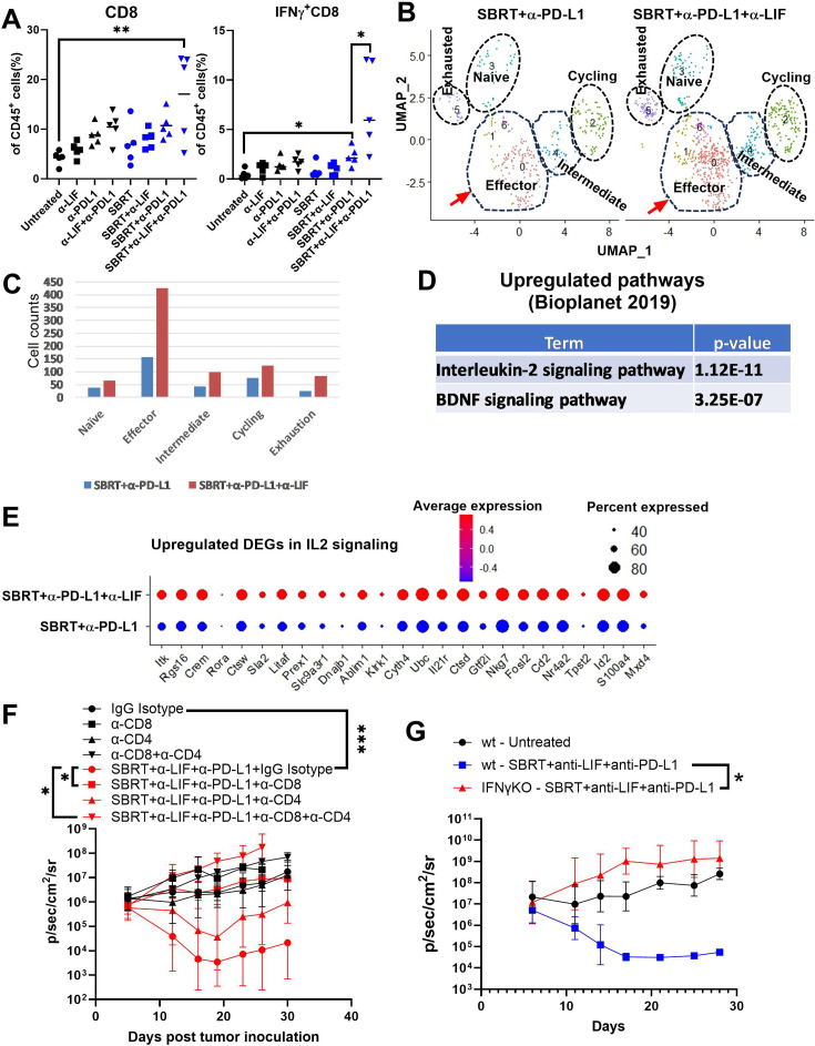
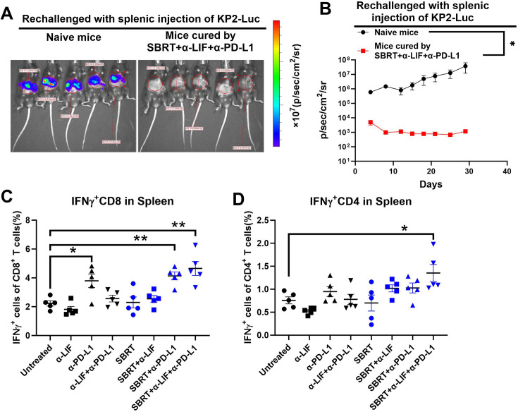
Methods: We evaluated the impact of combining systemic inhibition of LIF/programmed death-ligand 1 (PD-L1) with localized stereotactic body radiotherapy (SBRT) on tumor progression across multiple murine orthotopic pancreatic tumor models and examined systemic antitumor immunity using a hepatic rechallenge model. The antitumor immune response was characterized throughflow cytometry and Luminex assays. To identify differentially expressed genes and signaling pathways following treatment, we performed bulk RNA sequencing on pancreatic tumors. Additionally, single-cell RNA sequencing was conducted to further examine changes in tumor-infiltrating immune cells and their signaling pathways.

Results: We showed that simultaneous inhibition of LIF and PD-L1 significantly amplified the antitumor efficacy of SBRT, resulting in extended survival. The triple therapy (SBRT+anti-LIF+anti-PD-L1) generated an immunostimulatory tumor microenvironment, characterized by a proinflammatory shift in the cytokine/chemokine profile, increased infiltration of effector CD8+ T cells, and upregulated activation or maturation signals in tumor-infiltrating CD8+ T cells and macrophages. The beneficial effects of triple therapy were mostly abrogated by depletion of CD8+ T cells. In addition, triple therapy downregulated pathways related to tumor stemness, proliferation, and metabolism, and reduced EMT. Importantly, the combination of local SBRT treatment with systemic LIF and PD-L1 blockade resulted in long-term systemic antitumor memory.

Keywords: Gastrointestinal Cancer; Immunotherapy; Radiotherapy/radioimmunotherapy.


Conflict of interest statement

Competing interests: JE and NL hold AstraZeneca stock.

Figures

Figure 1
Figure 1. Blockade of LIF and PD-L1 significantly enhances the antitumor efficacy of SBRT in the murine orthotopic model of pancreatic cancer. (A) Schematic of the experimental design. C57BL/6J mice were injected with KCKO-Luc or KP2-Luc cells in the tail of the pancreas and treated with SBRT, anti-LIF, or anti-PD-L1 or combinations of two or three of these. Tumor growth was analyzed by IVIS twice a week. (B) and (E) Representative IVIS images from day 18 after KCKO-Luc (B) or KP2-Luc (E) implantation. (C) and (F) Tumor growth curves in KCKO-Luc (C) or KP2-Luc (F) were determined by IVIS imaging. Data shown are the geometric mean of IVIS value±SD from 10 to 15 mice/group. (D) and (G) Kaplan-Meier survival curves of mice bearing KCKO-Luc (D) or KP2-Luc (G) were observed. For KCKO-Luc model, untreated and SBRT groups (n=15 mice/group), other groups (n=10 mice/group); For KP2-Luc model, untreated (n=15 mice), other groups (n=10 mice/group). *, adjusted p<0.05, **, adjusted p<0.01, analyzed by one-way ANOVA with multiple comparison (adjusted by Bonferroni method) for growth curve; analyzed by log-rank (Mantel-Cox) test with multiple comparison (adjusted by Bonferroni method) for survival curve. ANOVA, analysis of variance; IVIS, in vivo imaging system; KCKO-Luc, luciferase-expressing KCKO; KP2-Luc; luciferase-expressing KP2 cells; LIF, leukemia inhibitory factor; PD-L1, programmed death-ligand 1; SBRT, stereotactic body radiation therapy.
Figure 2
Figure 2. Blockade of PD-L1 or LIF induced increased ICD in combination with SBRT in the orthotopic model of murine pancreatic cancer. Mice bearing KP2-Luc orthotopic tumors were treated with SBRT, anti-LIF or anti-PD-L1, and ICD was determined by immunohistochemistry (IHC) staining of cleaved caspase 3 (A), HMGB1 (B), and HSP70 (C). Results are expressed as the mean positivity±SEM from five mice per group and analyzed by one-way analysis of variance (ANOVA) with Dunnett post-test. *p<0.05, **p<0.01, compared with untreated group or monotherapy group. ICD, immunogenic cell death; KP2-Luc, luciferase-expressing KP2 cells; LIF, leukemia inhibitory factor; PD-L1, programmed death-ligand 1; SBRT, stereotactic body radiation therapy.
Figure 3
Figure 3. Triple therapy modulates intratumor cytokine/chemokine profiles and macrophage signals toward inflammatory phenotype. The change of intratumoral cytokines and chemokines after therapy was assessed using Luminex technology on day 14 post KCKO-Luc tumor implantation. (A) Fold changes of cytokines and chemokines in the tumor following treatment of SBRT/anti-LIF/anti-PD-L1. The levels of cytokines and chemokines of tumors from no treatment groups were set as 1, and data were fold changes as compared with the untreated group. (B) Principal components analysis (PCA) of cytokine/chemokines comparing untreated, SBRT+anti-PD-L1 and SBRT+anti-LIF+anti-PD-L1 groups. (C) IL-2, IL-12p70, IL-6, LIF, IL-10, MIP-1α, MIP-1β, and VEGF. Results are expressed as mean±SEM from five mice per group and analyzed by one-way ANOVA with Dunnett post-test. Significances are indicated by *p<0.05, **p<0.01, ***p<0.001, and ****p<0.0001 compared with untreated group. (D) and (E) Upregulated pathways in macrophages from scRNA-seq (D) and TNFa, BDNF, TLR and IL2 scores (E) shown in bridge plots. ANOVA, analysis of variance; IFNγ, interferon-gamma; IL, interleukin; KCKO-Luc, luciferase-expressing KCKO; LIF, leukemia inhibitory factor; PD-L1, programmed death-ligand 1; SBRT, stereotactic body radiation therapy; scRNA, single-cell RNA; TLR, Toll-like receptor; TSH, thyroid-stimulating hormone.
Figure 4
Figure 4. CD8+ T cells are critical for the antitumor effect in the orthotopic PDAC model. (A) Mice bearing orthotopic KP2-Luc pancreatic cancer were treated as in figure 1 and sacrificed on day 14. Tumor-infiltrating immune cells were determined by flow cytometry. Tumor-infiltrating CD8+ T cells and their expression of IFNγ were analyzed. Results are expressed as mean±SEM from five mice per group and analyzed by ANOVA with Dunnett post-test. Significance is indicated by *p<0.05, **p<0.01. (B–E) Tumor-infiltrating CD8+ T cells scRNA-seq analysis. (B) Reclustering of CD8 T cells shown in UMAP. (C) Cell counts in CD8 T cell subtypes across different treatments in pancreatic tumor were depicted, showing that triple therapy induced a relatively high amount of effector CD8 T cells in pancreatic tumor. (D) Upregulated pathways in CD8 T cells (triple therapy vs SBRT+anti-PD-L1 group). (E) DotPlot displaying upregulated DEGs in the IL2 signaling pathway in CD8 T cells from different treatments. (F) Tumor-bearing mice were treated with SBRT+anti-CD73+anti-PD-L1 as in figure 1. Mice were given isotype or antibodies against CD8, CD4 or a combination of both to deplete CD8+ T cells, CD4+ T cells, or both. Tumor growth was measured by IVIS. Data are represented as geometric mean±SD (n=5 for each group). *p<0.05, ***p<0.001. (G) Wild type or IFNγKO mice bearing KP2-Luc pancreatic cancer were treated as in figure 1. Tumor growth was measured by IVIS. Data are represented as geometric mean±SD (n=5 for each group). *p<0.05. ANOVA, analysis of variance; DEG, differentially expressed genes; IFNγ, interferon-gamma; IL, interleukin; IVIS, in vivo imaging system; KP2-Luc, luciferase-expressing KP2 cells; LIF, leukemia inhibitory factor; PDAC, pancreatic ductal adenocarcinoma; PD-L1, programmed death-ligand 1; SBRT, stereotactic body radiation therapy; scRNA-seq, single-cell RNA sequencing; triple therapy, SBRT+anti-PD-L1+anti LIF; UMAP, uniform manifold approximation and projection.
Figure 5
Figure 5. Triple therapy induces downregulation of stem-like transcription, cell cycle-related and metabolism-related pathways and reduction of EMT. (A–D) Mice bearing KCKO-Luc orthotopic pancreatic cancer were treated as in figure 1 and sacrificed on day 14. Tumor tissues were collected and processed for bulk RNA-seq. (A) Principal component analysis was performed showing the samples from triple therapy were relatively separated from other samples from other groups. (B) Heatmap showing the differentially expressed gene in triple therapy versus SBRT+anti-PD-L1 group. (C) Downregulated transcriptions (triple therapy vs SBRT+anti-PD-L1) in tumor stemness. (D) Downregulated pathways (triple therapy vs SBRT+anti-PD-L1) including cell growth (G2-M checkpoint), stemness (Myc, E2F), and metabolism (mTORC1, glycolysis, cholesterol homeostasis). (E–J) Tumor cells scRNA-seq analysis showing that triple therapy induced the reduction of mesenchymal tumor cells. (E) Subclusters of tumor cells across different treatments are shown in UMAP. (F) Cell counts in cell types across different treatments in pancreatic tumors were depicted, showing that triple therapy induced a decrease of mesenchymal cells and an increase of epithelial cells. (G) Downregulated pathways in stemness (Myc, epithelial mesenchymal transition) and metabolisms (Hypoxia, mTORC1, cholesterol homeostasis and glycolysis) when comparing triple therapy versus SBRT+anti-PD-L1. (H) Downregulated DEGs in EMT signal pathway shown in DotPlots. (I) Upregulated pathways when comparing triple therapy versus SBRT+anti-PD-L1. (J) Upregulated DEGs in apoptosis signal pathway shown in DotPlots. DEG, differentially expressed gene; EMT, epithelial to mesenchymal transition; KCKO-Luc, luciferase-expressing KCKO; LIF, leukemia inhibitory factor; PD-L1, programmed death-ligand 1; SBRT, stereotactic body radiation therapy; scRNA-seq, single-cell RNA sequencing; UMAP, uniform manifold approximation and projection.
Figure 6
Figure 6. Triple therapy induces systemic antitumor immune response in PDAC model. (A) and (B), mice that were cured by SBRT+anti-LIF+anti-PD-L1 in figure 1 were resistant to tumor rechallenge. Mice cured by triple therapy and age-matched naïve mice received 5×10⁵ KP2-Luc cells via hemisplenic injection. Tumor growth was monitored using IVIS imaging. (A) Representative of IVIS on day 7 post injection. (B) Growth curve of tumor liver metastasis (n=5 mice/group). *p<0.05, by multiple t-tests. (C) and (D) Triple therapy with SBRT, anti-PD-L1 and anti-LIF enhances activation of CD8 and CD4 cells in the spleen of murine pancreatic cancer orthotopic models. Mice bearing KP2-Luc orthotopic pancreatic cancer underwent treatment as described in figure 1 and were euthanized on day 14. Immune cells in the spleen were assessed by flow cytometry. Results are presented as mean±SEM from five mice per group and analyzed using ANOVA with Dunnett’s post-test. Significance is denoted by *p<0.05. ANOVA, analysis of variance; IFNγ, interferon-gamma; IVIS, in vivo imaging system; KP2-Luc, luciferase-expressing KP2 cells; LIF, leukemia inhibitory factor; PDAC, pancreatic ductal adenocarcinoma; PD-L1, programmed death-ligand 1; SBRT, stereotactic body radiation therapy.

References

    1. Rahib L, Smith BD, Aizenberg R, et al. Projecting cancer incidence and deaths to 2030: the unexpected burden of thyroid, liver, and pancreas cancers in the United States. Cancer Res. 2014;74:2913–21. doi: 10.1158/0008-5472.CAN-14-0155. - DOI - PubMed
    1. Siegel RL, Miller KD, Wagle NS, et al. Cancer statistics, 2023. CA Cancer J Clin. 2023;73:17–48. doi: 10.3322/caac.21763. - DOI - PubMed
    1. Doroshow DB, Bhalla S, Beasley MB, et al. PD-L1 as a biomarker of response to immune-checkpoint inhibitors. Nat Rev Clin Oncol. 2021;18:345–62. doi: 10.1038/s41571-021-00473-5. - DOI - PubMed
    1. Haslam A, Gill J, Prasad V. Estimation of the Percentage of US Patients With Cancer Who Are Eligible for Immune Checkpoint Inhibitor Drugs. JAMA Netw Open . 2020;3:e200423. doi: 10.1001/jamanetworkopen.2020.0423. - DOI - PMC - PubMed
    1. O’Reilly EM, Oh D-Y, Dhani N, et al. Durvalumab With or Without Tremelimumab for Patients With Metastatic Pancreatic Ductal Adenocarcinoma: A Phase 2 Randomized Clinical Trial. JAMA Oncol. 2019;5:1431–8. doi: 10.1001/jamaoncol.2019.1588. - DOI - PMC - PubMed

MeSH terms