. 2025 May 7;13(5):e009560.
doi: 10.1136/jitc-2024-009560.

Precision enhancement of CAR-NK cells through non-viral engineering and highly multiplexed base editing

Affiliations

Precision enhancement of CAR-NK cells through non-viral engineering and highly multiplexed base editing

Minjing Wang et al. J Immunother Cancer. .

Abstract

Background: Natural killer (NK) cells' unique ability to kill transformed cells expressing stress ligands or lacking major histocompatibility complexes (MHC) has prompted their development for immunotherapy. However, NK cells have demonstrated only moderate responses against cancer in clinical trials.

Methods: Advanced genome engineering may thus be used to unlock their full potential. Multiplex genome editing with CRISPR/Cas9 base editors (BEs) has been used to enhance T cell function and has already entered clinical trials but has not been reported in human NK cells. Here, we report the first application of BE in primary NK cells to achieve both loss-of-function and gain-of-function mutations.

Results: We observed highly efficient single and multiplex base editing, resulting in significantly enhanced NK cell function in vitro and in vivo. Next, we combined multiplex BE with non-viral TcBuster transposon-based integration to generate interleukin-15 armored CD19 chimeric antigen receptor (CAR)-NK cells with significantly improved functionality in a highly suppressive model of Burkitt's lymphoma both in vitro and in vivo.

Conclusions: The use of concomitant non-viral transposon engineering with multiplex base editing thus represents a highly versatile and efficient platform to generate CAR-NK products for cell-based immunotherapy and affords the flexibility to tailor multiple gene edits to maximize the effectiveness of the therapy for the cancer type being treated.

Keywords: Chimeric antigen receptor - CAR; Gene therapy; Hematologic Malignancies; Immunotherapy; Natural killer - NK.


Conflict of interest statement

Competing interests: MW, EJP, MGK, BM and BRW have filed patents covering the methods and approaches outlined in this work. All other authors declare they have no competing interests.

Figures

Figure 1
Figure 1. Highly efficient single gene KO in NK cells using BE. (A) Editing efficiency at genomic level quantified by A-to-G conversion of target base for each gene locus (n=3 independent NK cell donors). (B) Editing efficiency at protein level quantified by percentage of protein loss of each gene (n=3 independent NK cell donors). (C) Schema of killing assay to assess the functional improvement of AHR KO NK cells. (D) Ability of AHR KO versus Ctrl NK cells (with or without 14 days of L-Kynurenine pretreatment) to kill K562 cells at various E:T ratios as measured by luciferase luminescence assay. Assays run in triplicate in n=2 independent biological NK cell donors. (E) Statistical significance (p value) between each condition of AHR KO functional killing assay at E-to-T ratio of 1:2. (F) Schema of killing assay to assess the functional improvement of CISH KO NK cells. (G) Ability of CISH KO versus Ctrl NK cells to kill Molm-13 cells at various E:T ratios as measured by luciferase luminescence assay. Assays run in triplicate in n=3 independent biological NK cell donors. (H) Schema of killing assay to assess the functional improvement of TIGIT KO NK cells. (I) Ability of TIGIT KO versus Ctrl NK cells to kill Raji CD155hi cells at various E:T ratios as measured by luciferase luminescence assay. Assays run in triplicate in n=3 independent biological NK cell donors. (J) Schema of killing assay to assess the functional improvement of PDCD1 KO NK cells. (K) Ability of PDCD1 KO versus Ctrl NK cells to kill Raji PD-L1hi cells at various E:T ratios as measured by luciferase luminescence assay. Assays run in triplicate in n=3 independent biological NK cell donors. (L) Schema of ICS assay to assess the functional improvement of KLRG1 KO NK cells. (M) and (N) Cytokine production (M) and degranulation (N) ability of KLRG1 KO versus Ctrl NK cells against E-Cad+ Raji cells or E-Cad− Jurkat cells as measured by percentage of NK cells producing IFNγ and CD107a. Assays run in triplicate in n=3 independent biological NK cell donors. Data represented as mean±SD. P values calculated by two-way ANOVA test (n.s. p>0.05, *p≤0.05, **p≤0.01, ***p≤0.001, ****p≤0.0001). AHR, Aryl hydrocarbon receptor; ANOVA, analysis of variance; BE, base editor; CISH, cytokine-inducible SH2-containing protein; E:T, effector-to-target; ICS, intracellular cytokine staining; IFN‐γ, interferon-gamma; KLRG1, killer cell lectin-like receptor subfamily G member 1; KO, knockout; NK, natural killer; PDCD1, programmed cell death protein 1; TIGIT, T cell immunoreceptor with Ig and ITIM domains.
Figure 2
Figure 2. Highly efficient gain-of-function mutation in NK cells using BE. (A) Schema of how the S197P mutation renders NK cells non-cleavable by ADAM17 and results in enhanced ADCC cytotoxicity. (B) Schema of the recreation of S197P ncCD16a NK cells by a single base modification by BE. (C) Editing efficiency at genomic level quantified by A-to-G conversion of target base for CD16A (n=3 independent NK cell donors). (D) Editing efficiency at protein level quantified by CD16a retention on NK cell surface after PMA treatment (n=3 independent NK cell donors). (E) Cytokine production of S197P CD16a versus WT CD16a NK cells against CD20+Raji cells during ADCC (E:T ratio: 1:1). Plotted by folds increase of each cytokine with versus without anti-hCD20 mAb co-treatment. Assays run in duplicate in n=3 independent biological NK cell donors. (F) Ability of S197P CD16a versus WT CD16a NK cells to carry out ADCC against anti-hCD20 mAb treated CD20+Raji cells at various E:T ratios as measured by luciferase luminescence assay. Assays run in triplicate in n=3 independent biological NK cell donors. Data represented as mean±SD. P values calculated by two-way ANOVA test (*p≤0.05, **p≤0.01, ***p≤0.001, ****p≤0.0001). ADCC, antigen-dependent cellular cytotoxicity; ANOVA, analysis of variance; BE, base editor; CCA, cytosine-cytosine-adenine; E:T, effector-to-target; IFN‐γ, interferon-gamma; mAb, monoclonal antibody; mRNA, messenger RNA; NK, natural killer; PMA, phorbol 12-myristate 13-acetate; TCA, thymine-cytosine-adenine WT, wild type.
Figure 3
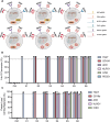
Figure 3. Highly efficient multiplex editing in NK cells using BE. (A) Schema of multiplex editing strategy. (B) Multiplex editing efficiency at genomic level quantified by A-to-G conversion of target base for each gene locus (n=2 independent NK cell donors). (C) Multiplex editing efficiency at protein level quantified by percentage of protein loss of each gene (n=2 independent NK cell donors). Data represented as mean±SD. AHR, Aryl hydrocarbon receptor; BE, base editor; CISH, cytokine-inducible SH2-containing protein; KLRG1, killer cell lectin-like receptor subfamily G member 1; NK, natural killer; PDCD1, programmed cell death protein 1; sgRNA, guide RNA; TIGIT, T cell immunoreceptor with Ig and ITIM domains.
Figure 4
Figure 4. Optimization of multiplex KO to maximize NK functionality against Rajihi/hi. (A) Schema of optimization strategy and all possible KO combinations included. (B) Editing efficiency at genomic level quantified by A-to-G conversion of target base for each gene locus (n=3 independent NK cell donors). (C) Editing efficiency at protein level quantified by percentage of protein loss of each gene (n=3 independent NK cell donors). (D) Ability of each KO combination to kill Rajihi/hi cells at various E:T ratios as measured by luciferase luminescence assay. Assays run in triplicate in n=2 independent biological NK cell donors. (E) Functional killing assay statistical significance (p value) between each KO combination at E to T ratio of 1:2 and 1:4. Data represented as mean±SD. P values calculated by two-way ANOVA test. ANOVA, analysis of variance; AHR, Aryl hydrocarbon receptor; CISH, cytokine-inducible SH2-containing protein; Ctrl, ABE8e mRNA only; E:T, effector-to-target; KO, knockout; mRNA, messenger RNA; NK, natural killer; PDCD1, programmed cell death protein 1; sgRNA, guide RNA; TIGIT, T cell immunoreceptor with Ig and ITIM domains; TP, TIGIT and PDCD1 KO; TPA, TIGIT, PDCD1, and AHR KO; TPAC, TIGIT, PDCD1, AHR, and CISH KO; TPC, TIGIT, PDCD1, and CISH KO.
Figure 5
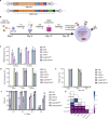
Figure 5. Simultaneous BE and non-viral transposon engineering exhibited enhanced NK cytotoxicity. (A) Schema of the designs of CD19 CAR constructs. (B) Schema of NK cell engineering timeline for simultaneous sgRNAs and CD19 CAR delivery. (C) Presorting and postsorting CAR integration rate quantified by percentage of RQR8 expression on NK cells (n=2 independent NK cell donors). (D) Postsorting editing efficiency at genomic level quantified by A-to-G conversion of target base for each gene locus (n=2 independent NK cell donors). (E) Postsorting editing efficiency at protein level quantified by percentage of protein loss of each gene (n=2 independent NK cell donors). (F) In vitro testing of the cytotoxicity of simultaneous BE and TcBuster engineered NK cells against Rajihi/hi cells at various E:T ratios as measured by luciferase luminescence assay. Assays run in triplicate in n=2 independent biological NK cell donors. (G) Killing assay statistical significance (p value) between each condition at E to T ratio of 1:4. Data represented as mean±SD. P values calculated by two-way ANOVA test. ANOVA, analysis of variance; BE, base editor; CAR, CD19 CAR RQR8; CAR15, CD19 CAR RQR8 IL-15; CAR/CTPKO, CD19 CAR RQR8 with CISH, TIGIT, and PDCD1 KO; CAR15/CTPKO, CD19 CAR RQR8 IL-15 with CISH, TIGIT, and PDCD1 KO; CISH, cytokine-inducible SH2-containing protein; Ctrl, ABE8e mRNA and CAR-expressing nanoplasmid only; CTPKO, CISH, TIGIT, and PDCD1 KO; E:T, effector-to-target; KO, knockout; NK, natural killer; PDCD1, programmed cell death protein 1; sgRNA, guide RNA; TIGIT, T cell immunoreceptor with Ig and ITIM domains; TPC, TIGIT, PDCD1, and CISH KO.
Figure 6
Figure 6. Multiplex edited CD19 CAR-NK cells are highly functional in vivo. (A) Schema of in vivo study design and timeline. (B) Luminescence (ROI) of individual tumor burden of mice bearing Rajihi/hi cells following treatment with PBS, Ctrl or engineered NK cells. Toxicity: mouse died due to suspected systemic toxicity, that is, rapid weight loss (>20%). (C) Tumor burden of each group on day 23, quantified by ROI (photons/sec) of each mouse (n=5). (D) Survival curve of each group shown in Kaplan-Meier curve (n=5). P values calculated by the Mantel-Cox test. Ctrl versus CAR15, **p≤0.01; Ctrl versus CAR15/CTPKO, *p≤0.05. (E) Cause of death of each animal in CAR15 and CAR15/CTPKO groups (n=5). BM/spleen: tumor ROI ≥ 1E8 with no obvious ovary and/or brain tumor; Ovary/brain: tumor ROI ≥ 1E8 with obvious ovary and/or brain tumor; Toxicity (No tumor): tumor ROI < 1E8. (F) Tumor burden of CAR15 and CAR15/CTPKO group at endpoint stratified by cause of death. (G) Quantifications of the number of NK cells in peripheral blood (measured by NK cell number per μL of blood), BM and spleen (measured by percentage of NK cells in BM or spleen) at end point. (H) Quantification of NK cell expansion in CAR15 and CAR15/TPCKO groups between day 25 and 30 (fold increase: day 30 NK cell count vs day 25 NK cell count). P values calculated by one-way ANOVA test (*p≤0.05, **p≤0.01, ***p≤0.001, ****p≤0.0001). Data represented as mean±SD. ANOVA, analysis of variance; BM, bone marrow; CAR, chimeric antigen receptor; IV, intravenous; NK, natural killer; PBS, phosphate-buffered saline.
Figure 7
Figure 7. Large-scale in vivo study of CAR15 and CAR15/CTPKO NK cells. (A) Luminescence (ROI) of individual tumor burden of mice bearing Rajihi/hi cells following treatment with PBS, Control or engineered NK cells. Toxicity: mouse died due to suspected systemic toxicity, that is, rapid weight loss (>20%) with less than 75 days of survival. (B) Tumor burden of each group on day 23, quantified by ROI (photons/sec) of each mouse (n=5). (C) Survival curve of each group shown in Kaplan-Meier curve (PBS and Ctrl, n=5; CAR15 and CAR15/CTPKO, n=10). P values calculated by Mantel-Cox test. Ctrl versus CAR15, **p≤0.01; Ctrl versus CAR15/CTPKO, ****p≤0.001. (D) Cause of death of each animal in CAR15 and CAR15/CTPKO groups (n=10) is defined based on the following criteria. BM/Spleen: tumor ROI ≥ 1E8 with no obvious ovary and/or brain tumor; Ovary/brain: tumor ROI ≥ 1E8 with obvious ovary and/or brain tumor; Toxicity (No tumor): tumor ROI < 1E8 w/ < 75 days of survival; Cleared: tumor ROI <1E8 and ≥75 days of survival. (E) Tumor burden of CAR15 and CAR15/CTPKO groups at endpoint stratified by cause of death. P values calculated by one-way ANOVA test (*p≤0.05, **p≤0.01, ****p≤0.0001). Data represented as mean±SD. ANOVA, analysis of variance; BM, bone marrow; CAR, chimeric antigen receptor; NK, natural killer; PBS, phosphate-buffered saline.

Update of

Similar articles

Cited by

References

    1. Ruggeri L, Capanni M, Urbani E, et al. Effectiveness of donor natural killer cell alloreactivity in mismatched hematopoietic transplants. Science. 2002;295:2097–100. doi: 10.1126/science.1068440. - DOI - PubMed
    1. Pan K, Farrukh H, Chittepu V, et al. CAR race to cancer immunotherapy: from CAR T, CAR NK to CAR macrophage therapy. J Exp Clin Cancer Res. 2022;41:119. doi: 10.1186/s13046-022-02327-z. - DOI - PMC - PubMed
    1. Suck G, Odendahl M, Nowakowska P, et al. NK-92: an “off-the-shelf therapeutic” for adoptive natural killer cell-based cancer immunotherapy. Cancer Immunol Immunother. 2016;65:485–92. doi: 10.1007/s00262-015-1761-x. - DOI - PMC - PubMed
    1. Miller JS, Lanier LL. Natural Killer Cells in Cancer Immunotherapy. Annu Rev Cancer Biol. 2019;3:77–103. doi: 10.1146/annurev-cancerbio-030518-055653. - DOI
    1. Judge SJ, Murphy WJ, Canter RJ. Characterizing the Dysfunctional NK Cell: Assessing the Clinical Relevance of Exhaustion, Anergy, and Senescence. Front Cell Infect Microbiol. 2020;10:49. doi: 10.3389/fcimb.2020.00049. - DOI - PMC - PubMed

Substances

LinkOut - more resources