Review
. 2025 May 9;10(1):149.
doi: 10.1038/s41392-025-02192-0.

The molecular mechanism and therapeutic landscape of copper and cuproptosis in cancer

Affiliations
Review

The molecular mechanism and therapeutic landscape of copper and cuproptosis in cancer

Ziyu Guo et al. Signal Transduct Target Ther. .

Abstract

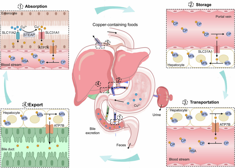
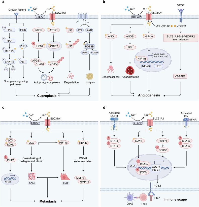
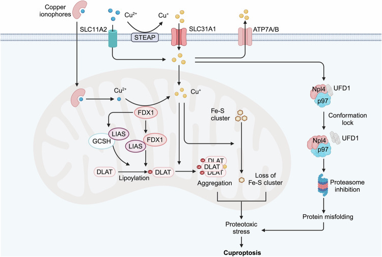
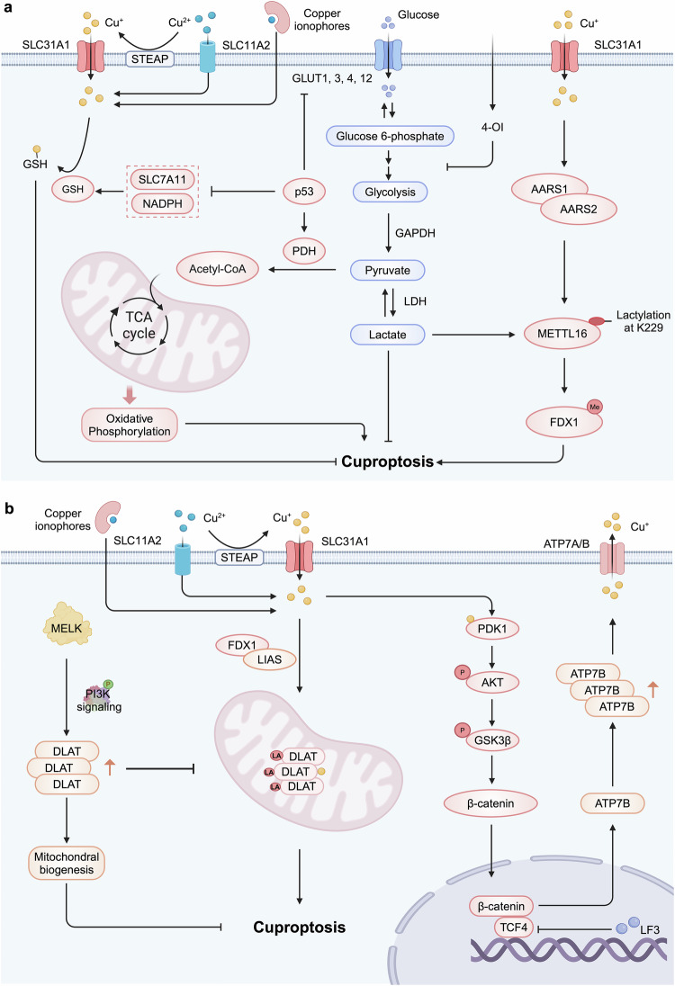
Copper, an essential micronutrient, plays significant roles in numerous biological functions. Recent studies have identified imbalances in copper homeostasis across various cancers, along with the emergence of cuproptosis, a novel copper-dependent form of cell death that is crucial for tumor suppression and therapeutic resistance. As a result, manipulating copper levels has garnered increasing interest as an innovative approach to cancer therapy. In this review, we first delineate copper homeostasis at both cellular and systemic levels, clarifying copper's protumorigenic and antitumorigenic functions in cancer. We then outline the key milestones and molecular mechanisms of cuproptosis, including both mitochondria-dependent and independent pathways. Next, we explore the roles of cuproptosis in cancer biology, as well as the interactions mediated by cuproptosis between cancer cells and the immune system. We also summarize emerging therapeutic opportunities targeting copper and discuss the clinical associations of cuproptosis-related genes. Finally, we examine potential biomarkers for cuproptosis and put forward the existing challenges and future prospects for leveraging cuproptosis in cancer therapy. Overall, this review enhances our understanding of the molecular mechanisms and therapeutic landscape of copper and cuproptosis in cancer, highlighting the potential of copper- or cuproptosis-based therapies for cancer treatment.


Conflict of interest statement

Competing interests: The authors declare no competing interests.

Figures

Fig. 1
Fig. 1
Systemic copper homeostasis. Systemic copper homeostasis involves intestinal absorption, hepatic storage, systemic transport, and biliary excretion. Dietary copper is predominantly absorbed in the small intestine, where copper (II) is reduced to copper (I) by members of the STEAP family. Copper (I) is then transported into enterocytes via SLC31A1, with a small amount entering through SLC11A2. Inside the intestinal epithelial cells, copper (I) binds to the copper chaperone and is transported to the basolateral side, where it is exported into the bloodstream via ATP7A. In the bloodstream, copper (I) binds to soluble copper chaperone proteins, primarily CP. Copper is transported to the liver through the portal vein, where hepatocytes uptake copper (I) from the bloodstream via SLC31A1. Within the hepatocytes, copper (I) can either be stored in MTs or re-enter the bloodstream through the ATP7B for distribution to other tissues. Excess copper is processed in the liver and excreted via bile, which is the primary route for copper elimination. Abbreviations: STEAP six-transmembrane epithelial antigen of the prostate, SLC31A1 solute carrier family 1 member 1, SLC11A2 solute carrier family 11 member 2, ATP7A/B ATPase copper transporter 7A and 7B, CP ceruloplasmin, MT metallothionein
Fig. 2
Fig. 2
Intracellular copper homeostasis. Within the cell, copper (I) can be sequestered by MTs and GSH, forming a dynamic copper pool, or it can bind to copper chaperone proteins, including CCS, COX17, CuL, and ATOX1, for transport to various organelles. CCS delivers copper (I) to the SOD1, facilitating the conversion of superoxide radicals into oxygen and thereby protecting the cell from oxidative stress. COX17 transports copper (I) to mitochondrial SCO1 and COX11, essential for COX assembly. CuL binds to copper in the cytosol and triggers copper transport into the mitochondria via SLC25A3. ATOX1 directs copper (I) to ATP7A/B in the TGN. When there is an excess of copper, ATP7A/B translocates to vesicular compartments and fuses with the plasma membrane to expel the excess copper. Also, CCS and ATOX1 are involved in transporting copper to the cell nucleus, which is crucial for activating various transcription factors. Abbreviations: MT metallothionein, GSH glutathione, CCS copper chaperone for superoxide dismutase, COX cytochrome oxidase, SCO1 synthesis of cytochrome c oxidase 1, CuL copper ligands, ATOX1 antioxidant protein 1, SOD1 superoxide dismutase 1, ATP7A/B ATPase copper transporter 7A and 7B, TGN trans-Golgi network
Fig. 3
Fig. 3
Mechanisms of copper in promoting tumor. a Copper binds to MEK1/2 and PDK1, activating oncogenic signaling pathways. It also activates ULK1/2 or enters the nucleus to induce the degradation of CRIP2, promoting autophagy. Additionally, copper interacts with the p53 protein, leading to its degradation. Copper also inhibits PDE3B, promoting lipolysis, which collectively contributes to cuproplasia. b Copper directly binds or activates angiogenic factors such as ANG and NO and interacts with HIF-1 to enhance NF-κB activity, promoting the expression of angiogenic mediators. Disulfide bonds formed between CTR1 and VEGFR2 activate VEGFR2 signaling, facilitating angiogenesis. c Copper promotes the expression of LOX/LOXL and HIF-1α, synergistically enhancing tumor metastasis through a positive feedback mechanism. Copper also binds to CD147 to promote its self-association, further enhancing metastasis. d Copper upregulates the expression of PD-L1 in cancer cells through multiple pathways, inhibiting T lymphocytes and inducing exhaustion, thereby facilitating immune escape. Created by BioRender. Abbreviations: ULK1/2 unc-51-like autophagy activating kinases 1 and 2, CRIP2 copper-binding protein cysteine-rich protein 2, PDE3B phosphodiesterase 3B, ANG angiogenin, NO nitric oxide, HIF-1 hypoxia-Inducible Factor-1, CTR1 copper transporter 1, VEGFR vascular endothelial growth factor receptor, LOX lysyl oxidase, LOXL lysyl oxidase-like protein, HIF-1α hypoxia-inducible factor-1α, PD-L1 programmed death-ligand 1
Fig. 4
Fig. 4
Mechanisms of copper-induced cell death. a Copper induces apoptosis primarily through the induction of ROS, DNA damage, and proteasome inhibition. Additionally, Copper induces TP53-dependent apoptosis by activating the transcription of TP53 target genes, while also triggering TP53-independent apoptosis through the inhibition of ribosomal synthesis and the induction of nucleolar stress. b Copper promotes pyroptosis by inducing ROS production and ER stress, resulting in NLRP3 inflammasome formation and membrane pore creation via GSDMD activation. c Copper toxicity activates the TLR4/NF-κB signaling pathway through oxidative stress, resulting in the phosphorylation and oligomerization of RIPK3 and MLKL, thereby triggering necroptosis. d Copper induces intracellular ROS through Fenton-like reactions and mitochondrial damage, leading to lipid peroxidation. It also binds to and induces the oligomerization of GPX4, promoting its autophagic degradation via the receptor TAXIBP1. e Copper initiates autophagy by activating AMPK, inhibiting mTOR, or directly binding to ULK1/2 kinases. Copper-mediated upregulation of autophagic genes and activation of the transcription factor TFEB contribute to the formation of autophagosomes and autolysosomes, which further promoting autophagy-dependent cell death. Created by BioRender. Abbreviations: ROS reactive oxygen species, ER endoplasmic reticulum, NLRP3 nod-like receptor family pyrin domain containing 3, GFDMD gasdermin D, TLR4 toll-like receptor 4, RIPK3 receptor-interacting protein kinase 3, MLKL mixed lineage kinase domain-like protein, GPX4 glutathione peroxidase 4, TAXIBP1 Tax1-binding protein 1, AMPK adenosine monophosphate-activated protein kinase, mTOR mammalian target of rapamycin, TFEB transcription factor EB
Fig. 5
Fig. 5
Major milestone of cuproptosis. The significance of copper ions has been recognized in 1928. Since 2022, research on cuproptosis and its regulatory mechanisms has surged. This timeline illustrates the evolution from the initial understanding of copper-induced cell death to the establishment of cuproptosis over the past few decades, providing insights into the major milestone surrounding cuproptosis and advancements in oncological research related to copper-associated cell death
Fig. 6
Fig. 6
Mechanism of cuproptosis. The mechanism of cuproptosis includes both mitochondrial-dependent and mitochondrial-independent pathways. Excess copper (II) enters the mitochondria, where it is reduced to the more toxic copper (I) by the mitochondrial protein FDX1. FDX1 also promotes protein lipoylation by directly binding to the LIAS and enhancing its interaction with the GCSH. Copper (I) binding induces the aggregation of DLAT and destabilizes Fe-S cluster proteins, triggering cellular stress responses that result in cuproptosis. Additionally, DSF/Cu mediates the aggregation and conformation lock of the Npl4-p97 protein in the cytoplasm, inhibiting the ubiquitin-proteasome degradation pathway, which contributes to proteotoxic stress and cuproptosis. Created by BioRender. Abbreviations: FDX1 ferredoxin 1, LIAS lipoic acid synthase, GCSH glycine cleavage system protein H, DLAT Dihydrolipoamide S-Acetyltransferase, Fe-S cluster iron-sulfur cluster, DSF Disulfiram, Npl4 an adaptor of the p97 segregase (also known as VCP)
Fig. 7
Fig. 7
Cancer-related pathways in cuproptosis. a Cuproptosis induction in tumor suppression. The tumor suppressor p53 inhibits glucose uptake and glycolysis, shifting metabolism towards the TCA cycle and oxidative phosphorylation, which sensitizes cells to cuproptosis. It also suppresses NADPH production by inhibiting G6PD and the pentose phosphate pathway, resulting in decreased GSH levels. Intratumoral copper enhances METTL16-K229 lactylation and activity through its interaction with AARS1 or AARS2, leading to increased FDX1 expression and cuproptosis induction. Additionally, the metabolite 4-OI alkylates cysteine residues in GAPDH, inhibiting its activity and suppressing aerobic glycolysis, thereby further promoting cuproptosis. b Cuproptosis evasion in tumor progression and therapeutic resistance. Increased MELK expression in tumors activates the PI3K/mTOR signaling pathway, leading to elevated DLAT expression and stabilized mitochondrial function, which enhances resistance to cuproptosis. Additionally, copper binds to PDK1, activating the downstream AKT-GSK3β-β-catenin pathway and promoting CSC characteristics. CSCs demonstrate heightened resistance to cuproptosis, as the β-catenin/TCF4 transcription complex binds to the ATP7B promoter, facilitating copper expulsion. Created by BioRender. Abbreviations: TCA tricarboxylic acid cycle, NADPH nicotinamide adenine dinucleotide phosphate, G6PD glucose-6-phosphate dehydrogenase, GSH glutathione, METTL16 methyltransferase-like 16, 4-OI 4-octyl itaconate, GAPDH glyceraldehyde-3-phosphate dehydrogenase, MELK maternal embryonic leucine zipper kinase. CSC cancer stem cell

References

    1. Guan, D. et al. Copper in cancer: from pathogenesis to therapy. Biomed. Pharmacother.163, 114791 (2023). - PubMed
    1. Garber, K. Cancer’s copper connections. Science349, 129 (2015). - PubMed
    1. Wang, Y. et al. Copper in colorectal cancer: from copper-related mechanisms to clinical cancer therapies. Clin. Transl. Med.14, e1724 (2024). - PMC - PubMed
    1. Festa, R. A. & Thiele, D. J. Copper: an essential metal in biology. Curr. Biol.21, R877–R883 (2011). - PMC - PubMed
    1. Linder, M. C. Copper homeostasis in mammals, with emphasis on secretion and excretion. a review. Int. J. Mol. Sci. 21, (2020). - PMC - PubMed