

Structural visualization of HECT-type E3 ligase Ufd4 accepting and transferring ubiquitin to form K29/K48-branched polyubiquitination
- PMID: 40341121
- PMCID: PMC12062229
- DOI: 10.1038/s41467-025-59569-6
Structural visualization of HECT-type E3 ligase Ufd4 accepting and transferring ubiquitin to form K29/K48-branched polyubiquitination
Abstract
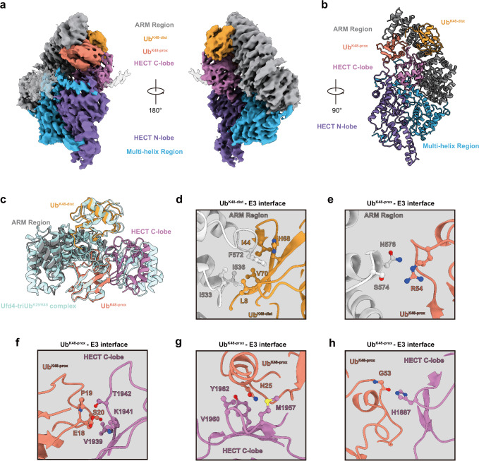
The K29/K48-linked ubiquitination generated by the cooperative catalysis of E3 ligase Ufd4 and Ubr1 is an enhanced protein degradation signal, in which Ufd4 is responsible for introducing K29-linked ubiquitination to K48-linked ubiquitin chains to augment polyubiquitination. How HECT-E3 ligase Ufd4 mediates the ubiquitination event remains unclear. Here, we biochemically determine that Ufd4 preferentially catalyses K29-linked ubiquitination on K48-linked ubiquitin chains to generate K29/K48-branched ubiquitin chains and capture structural snapshots of Ub transfer cascades for Ufd4-mediated ubiquitination. The N-terminal ARM region and HECT domain C-lobe of Ufd4 are identified and characterized as key structural elements that together recruit K48-linked diUb and orient Lys29 of its proximal Ub to the active cysteine of Ufd4 for K29-linked branched ubiquitination. These structures not only provide mechanistic insights into the architecture of the Ufd4 complex but also provide structural visualization of branched ubiquitin chain formation by a HECT-type E3 ligase.
© 2025. The Author(s).
Conflict of interest statement
Competing interests: The authors declare no competing interests.
Figures


Similar articles
-
The deubiquitinase TRABID stabilizes the K29/K48-specific E3 ubiquitin ligase HECTD1.J Biol Chem. 2021 Jan-Jun;296:100246. doi: 10.1074/jbc.RA120.015162. Epub 2021 Jan 8. J Biol Chem. 2021. PMID: 33853758 Free PMC article.
-
The N-end rule pathway is mediated by a complex of the RING-type Ubr1 and HECT-type Ufd4 ubiquitin ligases.Nat Cell Biol. 2010 Dec;12(12):1177-85. doi: 10.1038/ncb2121. Epub 2010 Nov 14. Nat Cell Biol. 2010. PMID: 21076411 Free PMC article.
-
Assembly and specific recognition of k29- and k33-linked polyubiquitin.Mol Cell. 2015 Apr 2;58(1):95-109. doi: 10.1016/j.molcel.2015.01.042. Epub 2015 Mar 5. Mol Cell. 2015. PMID: 25752577 Free PMC article.
-
Atypical Ubiquitination and Parkinson's Disease.Int J Mol Sci. 2022 Mar 28;23(7):3705. doi: 10.3390/ijms23073705. Int J Mol Sci. 2022. PMID: 35409068 Free PMC article. Review.
-
Emerging roles of the HECT E3 ubiquitin ligases in gastric cancer.Pathol Oncol Res. 2023 Feb 7;29:1610931. doi: 10.3389/pore.2023.1610931. eCollection 2023. Pathol Oncol Res. 2023. PMID: 36825281 Free PMC article. Review.
Cited by
-
TRIP12 structures reveal HECT E3 formation of K29 linkages and branched ubiquitin chains.Nat Struct Mol Biol. 2025 May 26. doi: 10.1038/s41594-025-01561-1. Online ahead of print. Nat Struct Mol Biol. 2025. PMID: 40419785
-
Protein semisynthesis reveals plasticity in HECT E3 ubiquitin ligase mechanisms.Nat Chem. 2024 Nov;16(11):1894-1905. doi: 10.1038/s41557-024-01576-z. Epub 2024 Jul 19. Nat Chem. 2024. PMID: 39030419
-
Structural basis for E4 enzyme Ufd2-catalyzed K48/K29 branched ubiquitin chains.Nat Chem Biol. 2025 Aug 15. doi: 10.1038/s41589-025-01985-2. Online ahead of print. Nat Chem Biol. 2025. PMID: 40817136
References
-
- Komander, D. & Rape, M. The ubiquitin code. Annu. Rev. Biochem.81, 203–229 (2012). - PubMed
-
- Husnjak, K. & Dikic, I. Ubiquitin-binding proteins: decoders of ubiquitin-mediated cellular functions. Annu. Rev. Biochem.81, 291–322 (2012). - PubMed
-
- Ding, Z. et al. Structural snapshots of 26S proteasome reveal tetraubiquitin-induced conformations. Mol. Cell73, 1150–1161.e6 (2019). - PubMed
-
- Kolla, S., Ye, M., Mark, K. G. & Rapé, M. Assembly and function of branched ubiquitin chains. Trends Biochem. Sci.47, 759–771 (2022). - PubMed
MeSH terms
Substances
LinkOut - more resources
Full Text Sources
