. 2025 May 9;10(1):150.
doi: 10.1038/s41392-025-02233-8.

Myeloid but not hepatocytic CD38 is a key driver for hepatic ischemia/reperfusion injury

Affiliations

Myeloid but not hepatocytic CD38 is a key driver for hepatic ischemia/reperfusion injury

Qi-Hang Zhao et al. Signal Transduct Target Ther. .

Abstract

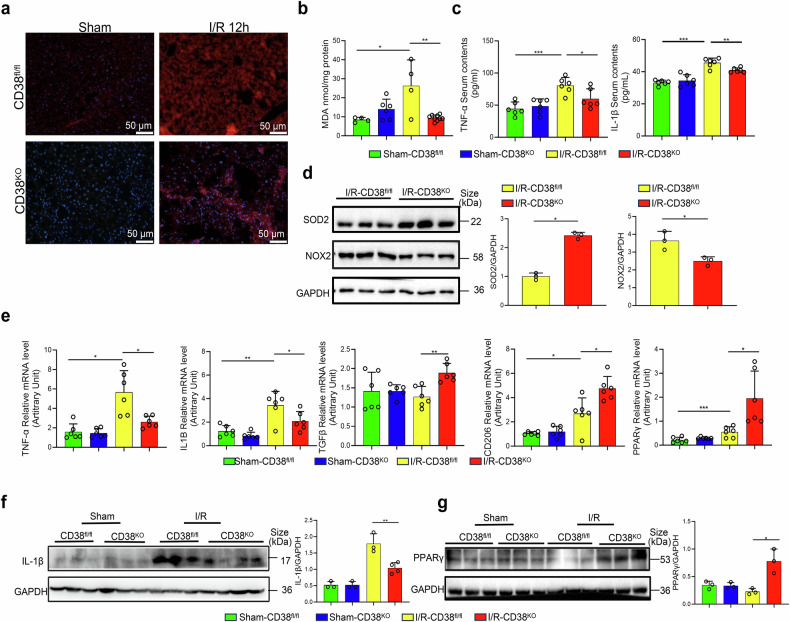
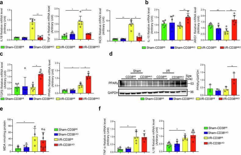
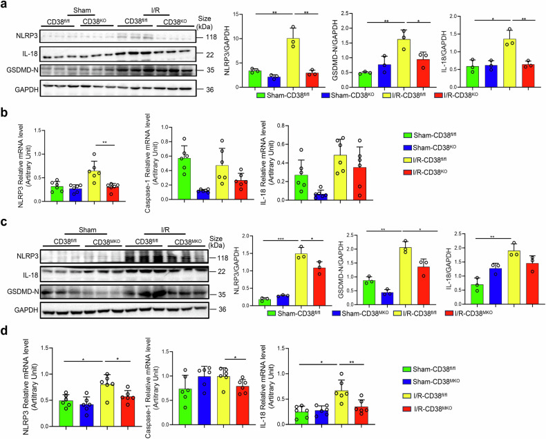
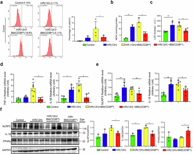
Hepatic ischemia-reperfusion injury (HIRI) is a critical condition that often occurs during liver transplantation and surgical liver resection. However, its mechanism has not been fully elucidated. Nicotinamide adenine dinucleotide (NAD+), functioning as a coenzyme or cofactor, is crucial for both redox and non-redox processes. In mammals, CD38 serves as the primary enzyme responsible for NAD+ degradation. In this study, we reported that the absence of CD38 markedly reduces HIRI in CD38 global knockout (CD38KO) and CD38 myeloid-specific knockout (CD38MKO) mice, but not in CD38 hepatocyte-specific knockout (CD38LKO) mice compared with the control (CD38fl/fl) mice by suppressing HIRI-induced hepatic oxidative stress, inflammatory responses, and pyroptosis. The findings were corroborated by a noticeable decrease in levels of alanine aminotransferase (ALT), aspartate transaminase (AST), and lactate dehydrogenase (LDH), along with reduced necrosis. Besides, we found that the expressions of SIRT1 and its downstream targets, p53 and PPARγ, were elevated in the liver tissues of CD38KO and CD38MKO mice compared to CD38fl/fl mice, while the acetylation levels of p53 were reduced. Furthermore, we demonstrated that myeloid CD38 deficiency not only promoted M2-type polarization and inhibited M1-type polarization of macrophages but also suppressed NLRP3-mediated pyroptosis by triggering NAD+/SIRT1 signaling in macrophages, resulting in the reduction of oxidative stress, inflammation, and pyroptosis in the liver, ultimately protecting against HIRI. This study highlights myeloid CD38 as a promising target for the prevention and treatment of HIRI clinically.


Conflict of interest statement

Competing interests: The authors declare no competing interests.

Figures

Fig. 1
Fig. 1
CD38 deficiency alleviated hepatic ischemia-reperfusion injury (HIRI) in mice. a CD38 expressions were determined by Western blotting and quantitative analysis in liver tissues after ischemia/reperfusion (I/R) injury. b The mRNA expressions of IL1B and IL-6 in liver tissues after I/R injury. c Representative fluorescence images of IL-1β/F4/80 and IL-1β/ASGR1 were taken from CD38KO mice subjected to HIRI, respectively. d, e Serum ALT and AST were measured in CD38KO and CD38MKO mice subjected to HIRI. f, g Representative H&E staining images and the quantitative analysis of liver ischemic necrosis were taken from CD38KO and CD38MKO mice subjected to HIRI, respectively. Data are shown as means ± SEM, *p < 0.05, **p < 0.01 and ***p < 0.001, n = 3–9 per group
Fig. 2
Fig. 2
Global CD38 deficiency inhibited oxidative stress and inflammation during HIRI. a Representative DHE staining images were taken from CD38KO and CD38fl/fl mice subjected to HIRI, respectively. b MDA contents were quantitatively measured in liver tissues from CD38KO and CD38fl/fl mice after I/R injury. c Serum TNF-α and IL-1β were detected by Elisa in CD38KO and CD38fl/fl mice after HIRI. d The protein expressions and the quantitative analysis of NOX2 and SOD2 were determined by western blot in liver tissues from CD38KO and CD38fl/fl mice after HIRI. e The mRNA expressions of TNF-α, IL1B, TGF-β, CD206, and PPARγ were determined by QPCR in CD38KO and CD38fl/fl mice after HIRI, respectively. f, g The expressions and the quantitative analysis of IL-1β and PPARγ were detected by Western blot in liver tissues in CD38KO and CD38fl/fl mice after HIRI, respectively. Data are shown as means ± SEM, *p < 0.05, **p < 0.01 and ***p < 0.001, n = 3–8 per group
Fig. 3
Fig. 3
Myeloid CD38 deficiency reduced hepatic ischemia-reperfusion-triggered inflammation. ac The mRNA expressions of IL1B, TNF-α, iNOS, IL-10, ARG1, TGF-β and PPARγ were analyzed by QPCR in CD38MKO and CD38fl/fl mice after HIRI. d The expressions and the quantitative analysis of PPARγ were detected by Western blot in liver tissues of CD38MKO and CD38fl/fl mice after HIRI. e MDA contents were measured in liver tissues of CD38LKO and CD38fl/fl mice after HIRI. f The mRNA expressions of TNF-α and IL1B were determined by QPCR in liver tissues of CD38LKO and CD38fl/fl mice after HIRI. Data are shown as means ± SEM, *p < 0.05, **p < 0.01 and ***p < 0.001, n = 3 ~ 9 per group
Fig. 4
Fig. 4
Global and myeloid deletion of CD38 ameliorated HIRI-induced pyroptosis in vivo. The protein expressions and the quantitative analysis of NLRP3, IL-18 and GSDMD-N and the mRNA expressions of NLRP3, Caspase-1 and IL-18 were examined by Western blot (a) and QPCR (b) in liver tissues of CD38KO and CD38fl/fl mice after HIRI, respectively. The protein expressions and the quantitative analysis of NLRP3, GSDMD-N and IL-18, and the mRNA expressions of NLRP3, Caspase-1 and IL-18 were determined by Western blot (c) and QPCR (d) in liver tissues from CD38MKO and CD38fl/fl mice after HIRI. Data are shown as means ± SEM, *p < 0.05, **p < 0.01and ***p < 0.001, n = 3–6 per group
Fig. 5
Fig. 5
Myeloid CD38 deficiency alleviated hepatocytic hypoxia/reoxygenation injury in a co-culture condition in vitro. a Representative flow cytometry plots of PI staining, along with the quantitative analysis, for hepatocytes from CD38fl/fl mice co-cultured with BMDMs from CD38KO mice after H/R injury. MDA contents (b), LDH activities (c), the mRNA expressions of IL-1B (d), TNF-α (d), NLRP3 (e) and IL-18 (e), and the protein expressions and the quantitative analysis (f) of NLRP3, IL-18 and PPARγ were examined in primary hepatocytes from CD38fl/fl mice co-cultured with BMDMs from CD38KO mice after H/R injury. Data are shown as means ± SEM, *p < 0.05, **p < 0.01 and ***p < 0.001, n = 3–6 per group
Fig. 6
Fig. 6
Global and myeloid deletion of CD38 upregulated SIRT1-p53 signaling pathway. The expressions and the quantitative analysis of SIRT1 (a, b), SIRT3 (a, b), p53 (c, d), and AC-p53 (c, d) were determined by Western blot in liver tissues from CD38KO, CD38MKO, and CD38fl/fl mice after HIRI. Data are shown as means ± SEM, *p < 0.05, **p < 0.01 and ***p < 0.001, n = 3 per group
Fig. 7
Fig. 7
Myeloid CD38 deficiency promoted macrophage M2 polarization through activating NAD+/SIRT1 pathway in vitro. The mRNA expressions of IL1B (a), CD206 (a), and the protein expressions and the quantitative analysis of SIRT1 (b) and SIRT3 (b) were determined by QPCR and Western blot in BMDMs from CD38fl/fl and CD38KO mice after LPS stimulation, respectively. c The mRNA expressions of IL1B and IL-6 were determined by QPCR in BMDMs from CD38fl/fl mice with the stimulation of hepatocytes-derived conditioned media after hypoxia/reoxygenation injury (HCMHR). The protein expressions and the quantitative analysis of SIRT1/SIRT3 (d), the NAD+ contents (e) and the infiltrations of the type 2 macrophages (CD206/Il-10, f, g), and type 1 macrophages (CD86/iNOS, f, g) were determined in BMDMs from CD38KO and CD38fl/fl mice with HCMHR stimulation. Data are shown as means ± SEM, *p < 0.05, **p < 0.01 and ***p < 0.001, n = 3–6 per group
Fig. 8
Fig. 8
Myeloid CD38 deficiency facilitated macrophage M2-type polarization through activating SIRT1-p53 and SIRT1-PPARγ pathway in vitro. The mRNA expressions of TNF-α (a), IL1B (b), IL-10 (c) and CD206 (d) were determined by QPCR in BMDMs from CD38KO and CD38fl/fl mice with the stimulation of the hepatocytes-derived conditioned media after hypoxia/reoxygenation injury (HCMHR) in the pretreatment of Compound C (AMPK inhibitor), PFT-α (P53 inhibitor) and T0070907(PPARγ inhibitor), respectively. Data are shown as means ± SEM, *p < 0.05, **p < 0.01 and ***p < 0.001, n = 3 per group. e The mechanism of myeloid-specific deletion of CD38 (CD38MKO) protecting against hepatic ischemia/reperfusion injury (HIRI): CD38MKO elevates the intracellular NAD+ levels in macrophages, and in turn, alleviates hepatic ischemia/reperfusion injury (HIRI)-induced inflammation and pyroptosis via activating SIRT1 signaling pathways in macrophages. On the one hand, SIRT1 promotes myeloid monocytes toward macrophage type 2 polarization through activating PPARγ signaling pathway in macrophages and inhibits monocytes toward macrophage type 1 polarization via activating p53 signaling, reducing HIRI-induced inflammation. On the other hand, SIRT1 also suppresses the release of pro-inflammatory factors such as IL1-β and IL18 through inactivating the canonical inflammasome-pyroptosis pathway of NLRP3-mediated caspase-1/GSDMD processing in macrophages. Image created with BioRender.com, with permission (agreement number: FG27ZL6X5O; citation to use: https://www.biorender.com/j12e628)

Similar articles

References

    1. Xiao, J. et al. Global liver disease burdens and research trends: Analysis from a Chinese perspective. J. Hepatol.71, 212–221 (2019). - PubMed
    1. Kadono, K. et al. SIRT1 regulates hepatocyte programmed cell death via GSDME - IL18 axis in human and mouse liver transplantation. Cell Death Dis.14, 762 (2023). - PMC - PubMed
    1. Hirao, H., Nakamura, K. & Kupiec-Weglinski, J. W. Liver ischaemia-reperfusion injury: a new understanding of the role of innate immunity. Nat. Rev. Gastroenterol. Hepatol.19, 239–256 (2022). - PubMed
    1. Ocuin, L. M. et al. Nilotinib protects the murine liver from ischemia/reperfusion injury. J. Hepatol.57, 766–773 (2012). - PMC - PubMed
    1. Li, F. et al. The protective effect of PNU-282987, a selective α7 nicotinic acetylcholine receptor agonist, on the hepatic ischemia-reperfusion injury is associated with the inhibition of high-mobility group box 1 protein expression and nuclear factor κB activation in mice. Shock39, 197–203 (2013). - PubMed

MeSH terms

LinkOut - more resources