. 2025 May 7;11(2):e005388.
doi: 10.1136/rmdopen-2024-005388.

Elevated serum levels of interleukin-18 discriminate Still's disease from other autoinflammatory conditions: results from the European ImmunAID cohort

Collaborators, Affiliations

Elevated serum levels of interleukin-18 discriminate Still's disease from other autoinflammatory conditions: results from the European ImmunAID cohort

Charlotte Girard-Guyonvarc'h et al. RMD Open. .

Abstract

Objectives: Systemic autoinflammatory diseases (SAIDs) represent a set of conditions with exaggerated innate immune responses. IL-1β and IL-18 are key cytokines involved in the pathogenesis of some SAID. We aimed to assess the diagnostic value of serum levels of IL-1β, IL-18, their respective inhibitors IL-1Ra and IL-18 binding protein (IL-18BP), and IFN-γ in SAID.

Methods: A cohort of patients with active SAID, including monogenic (mSAID) and genetically undiagnosed SAID (guSAID) from different European countries, with active disease at inclusion, was established. Serum levels of cytokines were measured by immunoassays.

Results: Sera from 53 mSAID, 220 guSAID and 49 controls without inflammatory disease were analysed. Serum levels of total and free IL-18 were significantly increased in Still's disease in comparison to most SAID and non-inflammatory controls. Levels of total IL-18 were also elevated in patients with familial Mediterranean fever to a comparable extent as in Still's disease. In contrast, free IL-18 levels were selectively higher in Still's disease. Receiver operating characteristic curve analysis showed that total IL-18 was the most sensitive and specific marker for the diagnosis of Still's disease (area under the curve=0.91). There was a positive correlation between IL-18 and ferritin. In 10 patients with Still's disease who had a second blood collection, we found a significant decrease in serum levels of free IL-18 after treatment.

Conclusions: Our results show that IL-18 can discriminate Still's disease from other SAID, and free IL-18 levels may be relevant to assess response to therapy in these patients.

Keywords: Biomarkers; Cytokines; Inflammation; Still's Disease, Adult-Onset.


Conflict of interest statement

Competing interests: CG has received research funds and has shares from AB2 Bio, Lausanne, Switzerland.

Figures
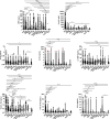
Figure 1
Figure 1. Levels of biomarkers of inflammation, cytokines and their inhibitors at first visit in the whole cohort. Individual measurements are represented as dots. For each disease group and non-inflammatory controls (NICs), results are shown as median (thick bars) and first and third quartiles (thin bars). On the graph showing serum levels of IL-1Ra, red dots represent patients treated with anakinra at the time of blood sampling. Comparisons between groups are performed using the Kruskal-Wallis test. Only significant differences are shown. *p<0.05, **p<0.01, ***p<0.001, ****p<0.0001. CAPS, cryopyrin-associated periodic syndrome; CRMO, Chronic Recurrent Multifocal Osteomyelitis; FMF, familial Mediterranean fever; IUO, inflammation of unknown origin; MKD, Mevalonate Kinase Disease; ND, neutrophilic dermatosis; RP, recurrent pericarditis; TRAPS, TNF receptor-associated periodic syndrome.
Figure 2
Figure 2. Correlations between cytokines and other markers of disease activity. Spearman correlation analyses were performed in the whole disease cohort (A) and in patients with Still’s disease only (B) between measured cytokines and their inhibitors and biological and clinical markers of disease activity collected at first visit (V1). Results are shown as heatmaps, with r coefficient represented as gradation of red (for positive correlation) and blue (for negative correlation). Strongest correlations, namely r values >0.4 or <−0.4 are shown in tables below, with corresponding p values. ALT, alanine transaminase; AST, aspartate transaminase; CRP, C reactive protein; DAA, disease activity assessment; ESR, erythrocyte sedimentation rate; Hb, haemoglobin; SAA, serum amyloid A.
Figure 3
Figure 3. ROC curves of total and free IL-18, IFN-γ, ferritin and CRP as diagnostic biomarkers of Still’s disease. The sensitivity and 100%-specificity of each biomarker to differentiate Still’s disease from other autoinflammatory diseases are shown. Areas under the curve (AUC) and p values are reported in the table for each biomarker. Cytokines, ferritin, CRP, SAA and AST levels at first visit (V1) were used for analyses. AST, aspartate aminotransferase; CRP, C reactive protein; ROC, receiver operating characteristic; SAA, serum amyloid A.
Figure 4
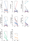
Figure 4. Levels of cytokines and their inhibitors at first and second visits in Still’s disease. Serum levels of IL-1β, IL-1Ra, IFN-γ, total IL-18, free IL-18 and IL-18BP of 10 patients with Still’s disease are individually reported at V1 and V2 and compared between both visits using the Mann-Whitney test (ns=p>0.05; **p<0.01 but >0.001) (A). Each symbol represents a single patient. Empty symbols and black lines represent patients without any treatment at V2. Blue symbols and lines represent patients treated with prednisone alone at V2. Red symbols and lines represent patients treated with anakinra alone at V2. Violet symbols and lines represent patients treated with prednisone and anakinra at V2. Teal blue squares and lines represent one patient receiving prednisone and tocilizumab at V2. Serum levels of free IL-18 are additionally reported according to disease activity at V2, with patients achieving complete remission in green, and patients in partial remission at V2 in orange (B). Again, the Mann-Whitney U test was used to compare free IL-18 levels between V1 and V2 (ns=p>0.05; **p<0.01 but >0.001).

References

    1. Georgin-Lavialle S, Ducharme-Benard S, Sarrabay G, et al. Systemic autoinflammatory diseases: Clinical state of the art. Best Pract Res Clin Rheumatol. 2020;34:101529. doi: 10.1016/j.berh.2020.101529. - DOI - PubMed
    1. Nigrovic PA, Lee PY, Hoffman HM. Monogenic autoinflammatory disorders: Conceptual overview, phenotype, and clinical approach. J Allergy Clin Immunol. 2020;146:925–37. doi: 10.1016/j.jaci.2020.08.017. - DOI - PMC - PubMed
    1. Park YH, Wood G, Kastner DL, et al. Pyrin inflammasome activation and RhoA signaling in the autoinflammatory diseases FMF and HIDS. Nat Immunol. 2016;17:914–21. doi: 10.1038/ni.3457. - DOI - PMC - PubMed
    1. Rathinam VAK, Fitzgerald KA. Inflammasome Complexes: Emerging Mechanisms and Effector Functions. Cell. 2016;165:792–800. doi: 10.1016/j.cell.2016.03.046. - DOI - PMC - PubMed
    1. Fautrel B, Mitrovic S, De Matteis A, et al. EULAR/PReS recommendations for the diagnosis and management of Still’s disease, comprising systemic juvenile idiopathic arthritis and adult-onset Still’s disease. Ann Rheum Dis. 2024;83:1614–27. doi: 10.1136/ard-2024-225851. - DOI - PMC - PubMed

LinkOut - more resources