

The characteristics of multimodal fundus imaging in AMN patients following COVID infection
- PMID: 40341186
- PMCID: PMC12062245
- DOI: 10.1038/s41598-025-99442-6
The characteristics of multimodal fundus imaging in AMN patients following COVID infection
Abstract
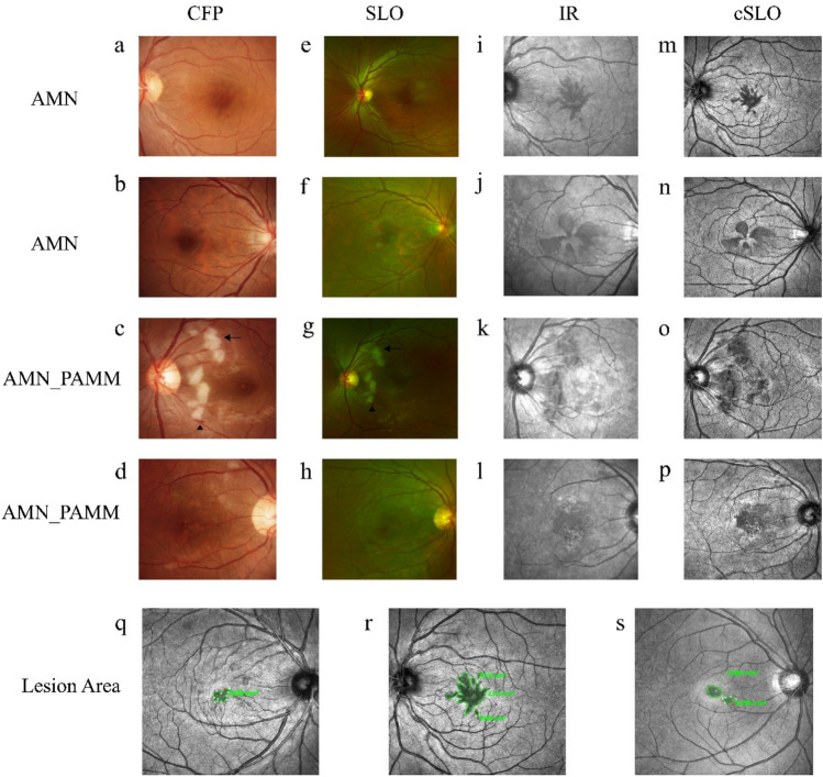
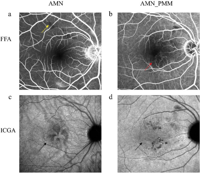
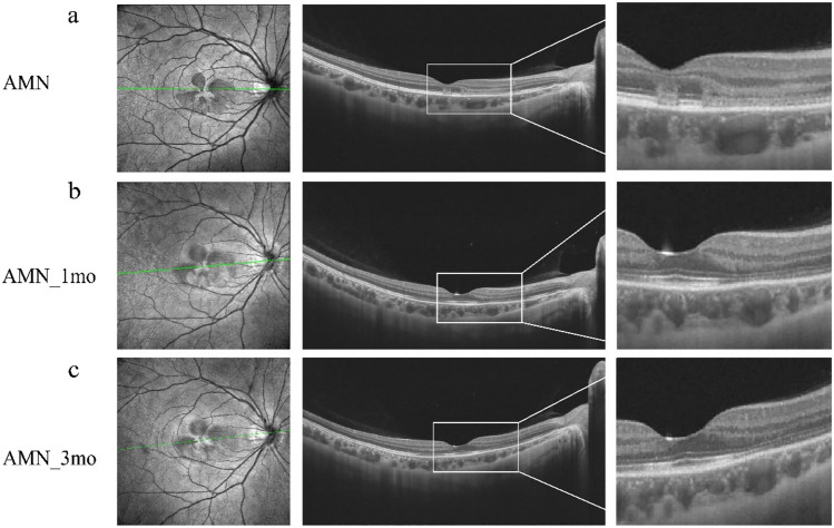
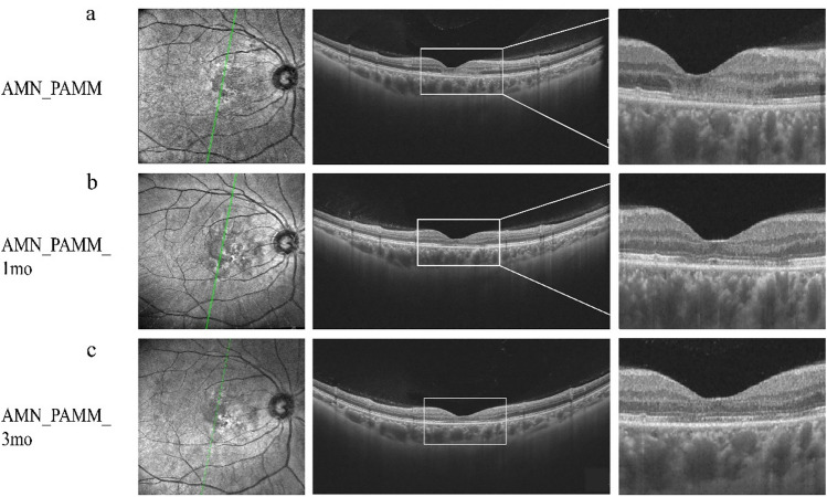
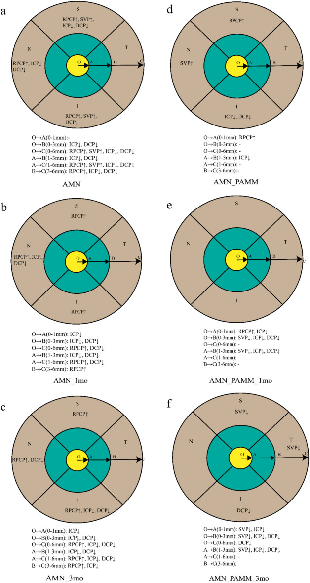
We investigated the features of multimodal fundus imaging in patients with both Coronavirus disease 2019 (COVID-19) and acute macular neuroretinopathy (AMN). This study included 15 patients with 29 eyes, all of whom underwent comprehensive fundus examinations and were followed for 3 months. Based on the diagnosis, patients were categorized into the AMN group and the AMN_PAMM group (AMN combined with paracentral acute middle maculopathy [PAMM]). At baseline, outer nuclear layer (ONL) thickness was not decreased in either group. However, a notable reduction in both outer retinal and full retinal thickness was observed in the AMN_PAMM group but not in the AMN group. Optical coherence tomography angiography (OCTA) demonstrated decreased vessel density (VD) in the intermediate capillary plexus (ICP) and deep capillary plexus (DCP), whereas the VD of the radial peripapillary capillary plexus (RPCP) and superficial vascular plexus (SVP) was increased in both groups. After 3 months of follow-up, ONL thickness and both outer and full retinal thickness were decreased in both groups. The VD of RPCP and SVP showed a significant decrease in the AMN_PAMM group. Visual acuity improvement was observed only in the AMN group, which may be attributed to the increase in choroid vascular index (CVI).
Keywords: Acute macular neuroretinopathy (AMN); Coronavirus disease 2019 (COVID-19); Optical coherence tomography angiography (OCTA); Paracentral acute middle maculopathy (PAMM).
© 2025. The Author(s).
Conflict of interest statement
Declarations. Competing interests: The authors declare no competing interests.
Figures
Similar articles
-
Association of Acute Macular Neuroretinopathy or Paracentral Acute Middle Maculopathy with Sickle Cell Disease.Ophthalmol Retina. 2021 Nov;5(11):1146-1155. doi: 10.1016/j.oret.2021.01.003. Epub 2021 Jan 19. Ophthalmol Retina. 2021. PMID: 33476854
-
RETINAL MICROVASCULATURE ALTERATION IN PARACENTRAL ACUTE MIDDLE MACULOPATHY AND ACUTE MACULAR NEURORETINOPATHY: A QUANTITATIVE OPTICAL COHERENCE TOMOGRAPHY ANGIOGRAPHY STUDY.Retin Cases Brief Rep. 2020 Fall;14(4):343-351. doi: 10.1097/ICB.0000000000000709. Retin Cases Brief Rep. 2020. PMID: 29443808
-
Projection-Resolved OCT Angiography of Microvascular Changes in Paracentral Acute Middle Maculopathy and Acute Macular Neuroretinopathy.Invest Ophthalmol Vis Sci. 2018 Jun 1;59(7):2913-2922. doi: 10.1167/iovs.18-24112. Invest Ophthalmol Vis Sci. 2018. PMID: 30025133 Free PMC article.
-
Clinical and Diagnostic Findings of Acute Macular Neuroretinopathy and Paracentral Acute Middle Maculopathy in the COVID-19 Era.Ophthalmologica. 2023;246(3-4):181-191. doi: 10.1159/000533530. Epub 2023 Aug 11. Ophthalmologica. 2023. PMID: 37573773 Free PMC article. Review.
-
Paracentral acute middle maculopathy spectral-domain optical coherence tomography feature of deep capillary ischemia.Curr Opin Ophthalmol. 2014 May;25(3):207-12. doi: 10.1097/ICU.0000000000000045. Curr Opin Ophthalmol. 2014. PMID: 24614148 Review.
References
-
- Negri, E. M. et al. Ultrastructural characterization of alveolar microvascular damage in severe COVID-19 respiratory failure. J. Appl. Physiol.135, 950–955. 10.1152/japplphysiol.00424.2023 (2023). - PubMed
-
- Zhang, Y. & Stewart, J. M. Retinal and choroidal manifestations of COVID-19. Curr. Opin. Ophthalmol.32, 536–540. 10.1097/ICU.0000000000000801 (2021). - PubMed
MeSH terms
Grants and funding
LinkOut - more resources
Full Text Sources
Medical
Miscellaneous
