. 2025 Jul 16;31(8):1139-1153.
doi: 10.1261/rna.080342.124.

Structural and functional characterization of the SLA' structure at the 3' terminus of the Zika virus negative-strand intermediate

Affiliations

Structural and functional characterization of the SLA' structure at the 3' terminus of the Zika virus negative-strand intermediate

Quinn H Abram et al. RNA. .

Abstract

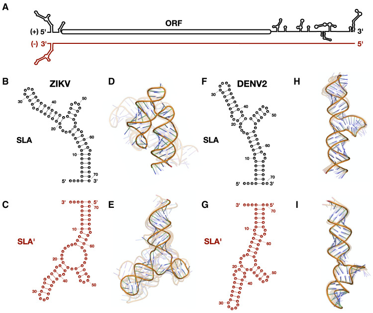
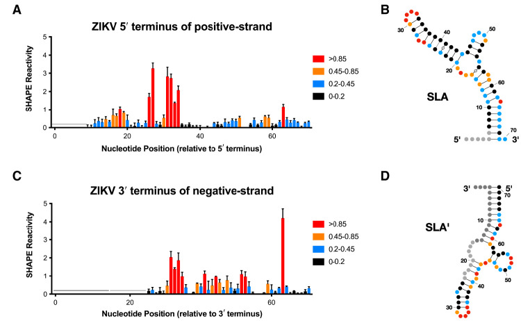
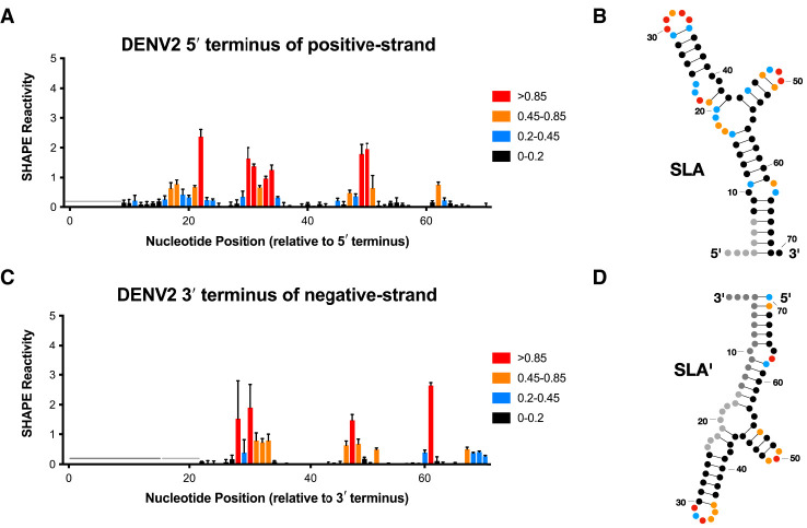
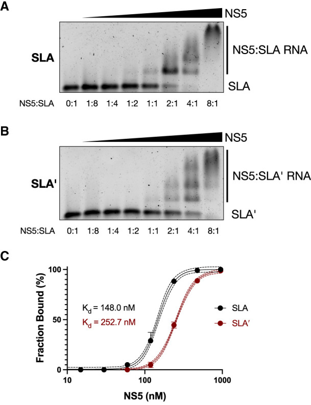
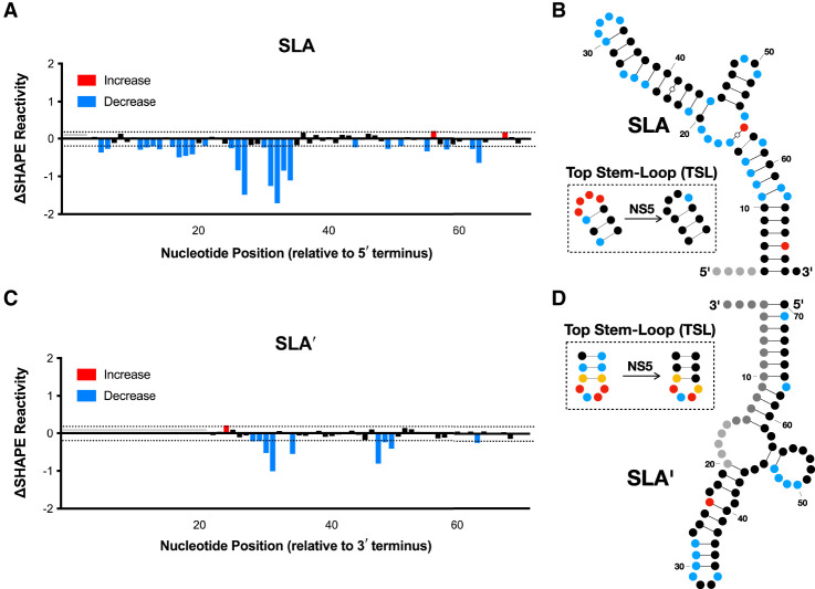
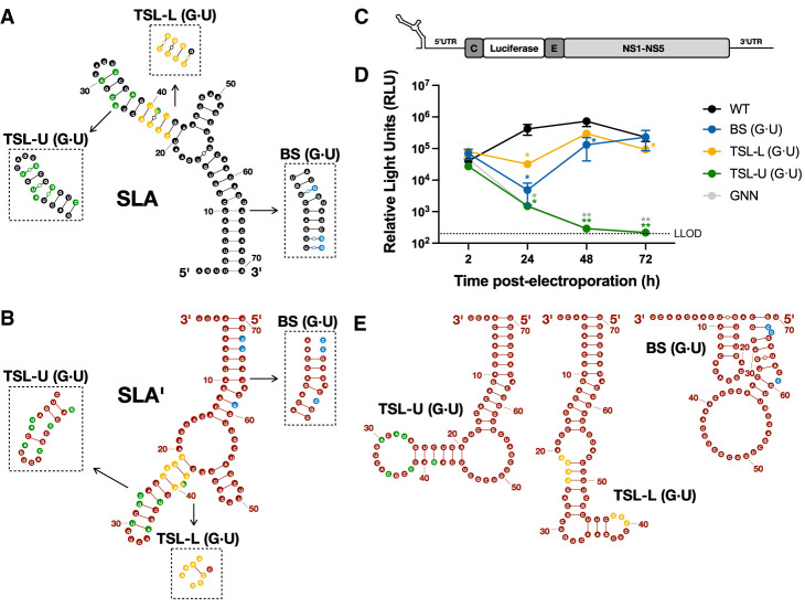
Flavivirus infections, including those of Dengue virus (DENV) and Zika virus (ZIKV), result in a high disease burden globally, yet many aspects of their viral life cycle remain poorly understood. For example, while some features of the mechanism of negative-strand RNA synthesis are known, relatively little is known about the initiation of positive-strand RNA synthesis in the flavivirus life cycle. Viral RNA replication is initiated via the recruitment of the viral NS5 RNA-dependent RNA polymerase (RdRp) to stem-loop A (SLA) at the 5' terminus of positive-strand genomic RNA. Subsequent genome cyclization is thought to facilitate loading of NS5 onto the 3' terminus of the genomic RNA to initiate negative-strand RNA synthesis. Conversely, it is not clear whether RNA structures in the negative-strand replicative intermediate similarly recruit NS5 to promote positive-strand RNA synthesis, providing specificity to this process. Herein, we characterized the secondary structure of the 3' terminus of the negative-strand replicative intermediate in ZIKV and DENV1-4 in silico and in vitro. We observed that the 3' terminus of the negative strand is capable of forming a secondary structure which mirrors SLA, which we term SLA'. While we demonstrate that SLA' forms in vitro and is capable of interacting with NS5, introduction of G·U wobble base pairs that disrupt SLA', while keeping SLA largely intact, suggest that SLA' is not necessary for viral RNA replication. As such, this work suggests that in contrast to related viruses, the positive-strand promoter is unlikely to be provided by specific structure(s) at the 3' terminus of the negative-strand replicative intermediate.

Keywords: SHAPE; Zika virus; flavivirus; negative-strand replicative intermediate; nonstructural protein 5 (NS5); stem–loop A (SLA).


Figures

FIGURE 1.
FIGURE 1.
Zika virus (ZIKV) and Dengue virus 2 (DENV2) are predicted to form SLA′ at the 3′ terminus of their negative strands. (A) Cartoon diagram of the ZIKV positive-strand genomic RNA (black) and its complementary negative-strand intermediate (cayenne). Stem–loop A (SLA) is shown at the 5′ terminus of the positive strand, while the predicted SLA′ structure is shown at the 3′ terminus of the negative strand. Predicted secondary and tertiary structures of the first 70 nt of the 5′ terminus of the positive strand and the last 70 nt of the 3′ terminus of the negative strand of (BE) ZIKV and (FI) DENV2, respectively. Tick marks represent 10 nt intervals. The highest confidence tertiary structure prediction is fully opaque, while the next four highest-ranked predictions are translucent.
FIGURE 2.
FIGURE 2.
In vitro selective 2′ hydroxyl acylation analyzed by primer extension (SHAPE) analysis of the 3′ terminus of the negative strand in Zika virus (ZIKV) is consistent with SLA′ formation. (A) Normalized SHAPE reactivities of the first 70 nt at the 5′ terminus of the positive strand of ZIKV. Data are shown as the normalized SHAPE reactivity from four biological replicates, and error bars represent the SEM. Nucleotides with very low (≤0.2), low (0.2–0.4, blue), intermediate (0.4–0.85, orange), and high (≥0.85, red) SHAPE reactivity are indicated. Nucleotides 1–6 were omitted due to high background reactivity (light gray). (B) Prediction of the lowest free energy structure formed by the first 70 nt of the ZIKV positive strand as constrained by the normalized SHAPE reactivity data from (A). Tick marks represent 10 nt intervals. (C) Normalized SHAPE reactivities of the last 70 nt at the 3′ terminus of the negative strand of ZIKV. Data are shown as the normalized SHAPE reactivity from four biological replicates, and error bars represent the SEM. Nucleotides 1–15 were bound by the primer (dark gray), and nucleotides 16–23 and 157–163 were omitted due to high background reactivity (light gray). (D) Prediction of the lowest free energy structure formed by the last 70 nt of the ZIKV negative strand as constrained by the normalized SHAPE reactivity data from (C).
FIGURE 3.
FIGURE 3.
In vitro SHAPE analysis of the 3′ end of the negative strand in DENV2 is also consistent with SLA′ formation. (A) Normalized SHAPE reactivities of the first 70 nt at the 5′ terminus of the positive strand of DENV2. Data are shown as the normalized SHAPE reactivity from four biological replicates, and error bars represent the SEM. Nucleotides with very low (≤0.2), low (0.2–0.4, blue), intermediate (0.4–0.85, orange), and high (≥0.85, red) SHAPE reactivity are indicated. Nucleotides 1–7 were omitted due to high background reactivity (light gray). (B) Prediction of the lowest free energy structure formed by the first 70 nt of the DENV2 positive strand as constrained by the normalized SHAPE reactivity data from (A). Tick marks represent 10 nt intervals. (C) Normalized SHAPE reactivities of the last 70 nt at the 3′ terminus of the negative strand of DENV2. Data are shown as the normalized SHAPE reactivity from four biological replicates, and error bars represent the SEM. Nucleotides 1–15 were bound by the primer (dark gray), and nucleotides 16–24 and 157–163 were omitted due to high background reactivity (light gray). (D) Prediction of the lowest free energy structure formed by the last 70 nt of the DENV2 negative strand as constrained by the normalized SHAPE reactivity data from (C).
FIGURE 4.
FIGURE 4.
ZIKV NS5 binds to both SLA- and SLA′-containing RNAs. Electrophoretic mobility shift assays (EMSAs) were performed using ZIKV NS5 and (A) the first 163 nt of the ZIKV positive strand, or (B) the last 163 nt of the ZIKV negative strand. For each RNA, 2.5 pmol (150 ng) of RNA was incubated with increasing (twofold) amounts of NS5 ranging from 0 to 20 pmol (0–2 µg) and then analyzed via agarose gel electrophoresis. Molar ratios of NS5:RNA are indicated. Data shown are representative of four independent replicates. (C) Binding curves of NS5 to SLA- and SLA′-containing RNAs based on the bound RNA fraction from the EMSAs in A and B. Data for each RNA were fit to a sigmoidal four-parameter logistic curve model, with R2 values of R2 = 0.9962 and R2 = 0.9987 for SLA and SLA′, respectively. The dashed lines indicate 95% confidence intervals.
FIGURE 5.
FIGURE 5.
NS5 binding results in a change in SHAPE reactivity in the top loop of SLA and SLA′. ΔSHAPE reactivity plots for (A), the first 70 nt of the 5′ terminus of the positive strand and (C), the last 70 nt of the 3′ terminus of the negative-strand in the presence or absence of ZIKV NS5 (i.e., ΔSHAPE = NS5 + RNA reactivity − RNA only reactivity). Data are shown as the normalized change in SHAPE reactivity upon NS5 binding at each nucleotide position from four biological replicates, and error bars represent the SEM. Significant (>magnitude of the average ΔSHAPE reactivity) increases (red) and decreases (blue) in SHAPE reactivity upon NS5 binding are indicated. The baseline significance is indicated with a dashed line. Nucleotides 1–3 in A and 16–20 in C were omitted due to high background reactivity (light gray), while nucleotides 1–15 in C were bound by the primer (dark gray). (B,D) ΔSHAPE reactivity data from (A,C) mapped onto the predicted structures of (B) SLA and (D) SLA′. Tick marks represent 10 nt intervals. Insets represent the normalized SHAPE reactivities of the respective top loop regions in the absence and presence of NS5. Nucleotides with very low (≤0.2), low (0.2–0.4, blue), intermediate (0.4–0.85, orange), and high (≥0.85, red) normalized SHAPE reactivity are indicated in each inset.
FIGURE 6.
FIGURE 6.
SLA′ is not necessary for ZIKV RNA replication. Cartoon diagrams of WT (A) SLA and (B) SLA′ with insets for the three sets of mutations that introduce G·U wobble base pairs into the lower and upper portion of the TSL (TSL-L [G·U], yellow and TSL-U [G·U], green), and base stem (BS [G·U], blue). (C) Schematic representation of the ZIKV subgenomic replicon system, which contains the Renilla luciferase (RLuc) gene in place of the majority of the ZIKV structural proteins. (D) Full-length capped WT, TSL-L (G·U), TSL-U (G·U), or BS (G·U) ZIKV subgenomic RLuc reporter replicon RNAs were electroporated into BHK-21 cells. Luciferase activity was measured at 2–72 h post-electroporation. The lower limit of detection is indicated (LLOD). Data are representative of three independent biological replicates with three technical replicates, and error bars represent standard deviation (SD) of the mean. Statistical significance was determined by multiple Student's t-tests, (*) P ≤ 0.1, (**) P ≤ 0.05. (E) Most stable predicted secondary structure of the last 70 nt at the 3′ terminus of the negative strand for the TSL-U (G·U), TSL-L (G·U), and BS (G·U) mutants, as constrained by normalized SHAPE reactivity data.
Quinn H. Abram
Quinn H. Abram

Similar articles

References

    1. Abram QH, Landry BN, Wang AB, Kothe RF, Hauch HCH, Sagan SM. 2024. The myriad roles of RNA structure in the flavivirus life cycle. RNA Biol 21: 14–30. 10.1080/15476286.2024.2357857 - DOI - PMC - PubMed
    1. Abramson J, Adler J, Dunger J, Evans R, Green T, Pritzel A, Ronneberger O, Willmore L, Ballard AJ, Bambrick J, et al. 2024. Accurate structure prediction of biomolecular interactions with AlphaFold 3. Nature 630: 493–500. 10.1038/s41586-024-07487-w - DOI - PMC - PubMed
    1. Alvarez DE, Lodeiro MF, Luduena SJ, Pietrasanta LI, Gamarnik AV. 2005. Long-range RNA-RNA interactions circularize the dengue virus genome. J Virol 79: 6631–6643. 10.1128/JVI.79.11.6631-6643.2005 - DOI - PMC - PubMed
    1. Barnard TR, Abram QH, Lin QF, Wang AB, Sagan SM. 2021. Molecular determinants of flavivirus virion assembly. Trends Biochem Sci 46: 378–390. 10.1016/j.tibs.2020.12.007 - DOI - PubMed
    1. Barnard TR, Landry BN, Wang AB, Sagan SM. 2023. Zika virus NS3 and NS5 proteins determine strain-dependent differences in dsRNA accumulation in a host cell type-dependent manner. J Gen Virol 104. 10.1099/jgv.0.001855 - DOI - PubMed

MeSH terms