. 2025 May 1;66(5):21.
doi: 10.1167/iovs.66.5.21.

Muscone Attenuates Uveitis Through the PI3K/AKT Signaling Pathway

Affiliations

Muscone Attenuates Uveitis Through the PI3K/AKT Signaling Pathway

Xianyang Liu et al. Invest Ophthalmol Vis Sci. .

Abstract

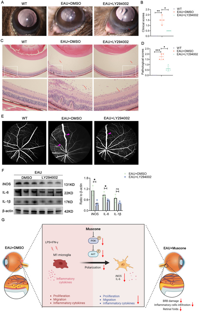
Purpose: Uveitis is an immune-mediated ocular disorder that poses a significant threat to vision, particularly among young and middle-aged adults. The treatment of uveitis is complicated by the presence of the blood-retinal barrier (BRB), which restricts the passage of large molecular drugs into the eye, thus limiting effective therapeutic options. The primary objective of this study is to identify a novel therapeutic agent capable of treating uveitis and explore its underlying mechanism.

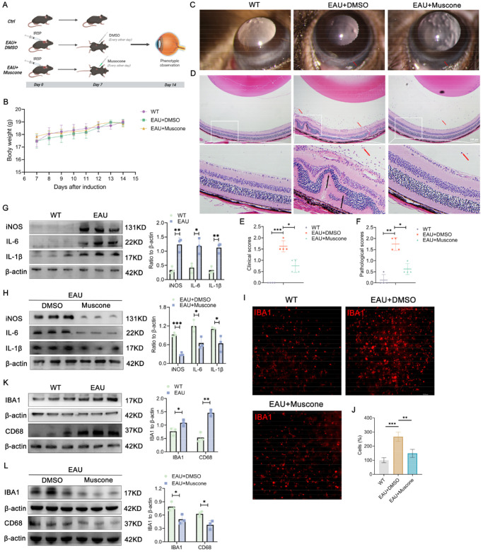
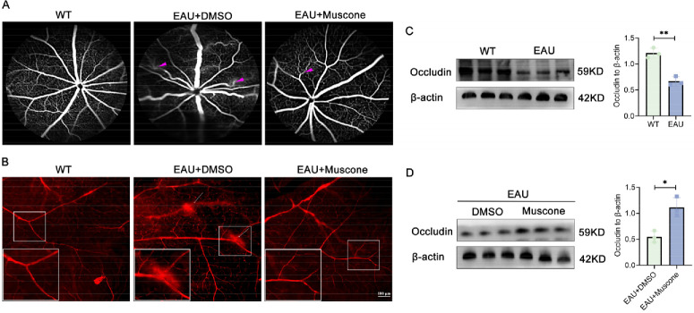
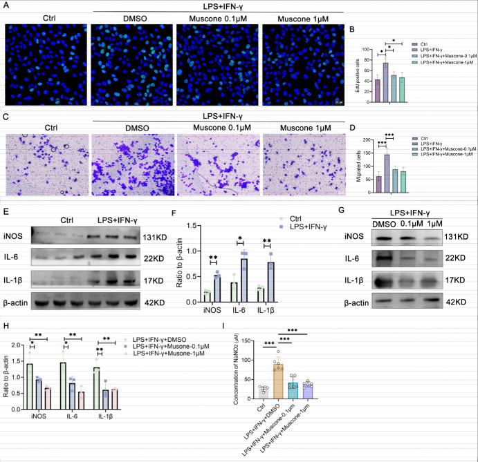
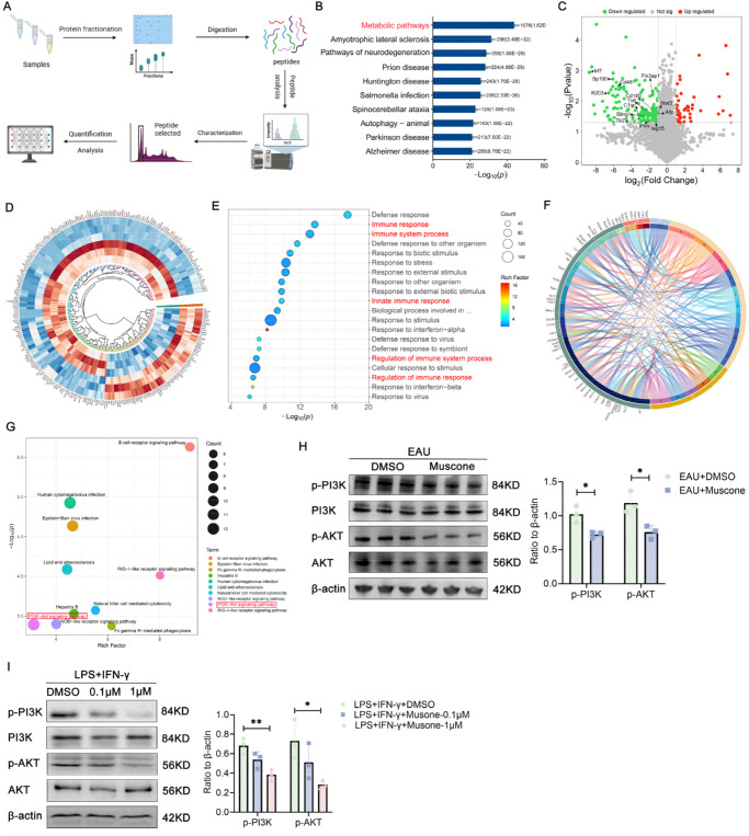
Methods: In this study, we used a mouse model of experimental autoimmune uveitis (EAU) induced by interphotoreceptor retinoid-binding protein (IRBP) and lipopolysaccharide (LPS) and interferon-gamma (IFN-γ)-induced inflammatory BV2 cells. Evans blue and fundus fluorescein angiography (FFA) experiments were performed to evaluate the destruction of BRB. Silt lamp and hematoxylin and eosin (H&E) staining were conducted to evaluate the inflammatory response. In vivo proteomics and Western blot were carried to investigate the underlying mechanisms.

Results: Our study reveals that Muscone significantly alleviates EAU and restores the integrity of BRB. Moreover, Muscone treatment markedly downregulated inflammatory factors within the retinas and BV2 cells. In vivo proteomic combined with liquid chromatography-mass spectrometry (LC-MS) has elucidated that Muscone exerts its anti-inflammatory effects by modulating the PI3K-AKT signaling pathway. Moreover, by using LY294002 to specifically inhibit PI3K, we observed a marked decrease in inflammatory phenotype and BRB destruction of EAU.

Conclusions: In summary, this study establishes the protective efficacy of Muscone against the progression of EAU and provides insights into the molecular mechanisms responsible for its therapeutic action.


Conflict of interest statement

Disclosure: X. Liu, None; H. Zuo, None; C. Wu, None; N. Li, None; Q. Zhou, None; F. Cao, None; B. Chu, None; S. Zeng, None; H. Feng, None; Y. Wang, None; F. Lei, None; K. Hu, None; S. Hou, None

Figures

Figure 1.
Figure 1.
Muscone effectively mitigates retinal inflammation in mice with EAU. (A) Schematic depiction of the post-immunization treatment procedure with DMSO or Muscone in mice. (B) Comparative body weight tracking across different treatment groups including control mice, EAU mice with DMSO, and EAU mice with Muscone. (C, E) Clinical scores of control mice, EAU mice with DMSO, and those treated with Muscone (mean ± SD; n = 4/group; *P < 0.05, ***P < 0.001; Mann-Whitney U test; red arrow = ciliary congestion). (D, F) Pathological scores in the same groups mentioned above (mean ± SD; n = 4/group; *P < 0.05, **P < 0.01; Mann-Whitney U test; red arrow = inflammatory cells, black arrow = retinal folds). (G) Protein levels and quantitative chart of iNOS, IL-6, and IL-1β in retinal tissues between control and EAU mice (mean ± SD; n = 3/group; *P < 0.05, **P < 0.01, unpaired Student's t-test). (H) Protein levels and quantification of iNOS, IL-6, and IL-1β in retinal tissues between EAU mice and EAU mice with Muscone (mean ± SD; n = 3/group; *P < 0.05, ***P < 0.001, unpaired Student's t-test). (I, J) Immunofluorescence visualization of the microglial marker IBA1 in retinas from control, EAU, and EAU with Muscone groups (mean ± SD; n = 3/group; ***P < 0.001, **P < 0.01, 1-way ANOVA). (K) Protein levels and quantification of IBA1 and CD68 in control and EAU mice (mean ± SD; n = 3/group; *P < 0.05, **P < 0.01, unpaired Student's t-test). (L) Protein levels and quantification of IBA1 and CD68 between EAU mice and EAU mice with Muscone (mean ± SD; n = 3/group; *P < 0.05, unpaired Student's t-test).
Figure 2.
Figure 2.
Muscone protects the integrity of the BRB. (A) Representative images of control mice, EAU mice, and EAU mice treated with Muscone following intraperitoneal injection of FFA (purple triangle = blood vessel curvature and leakage). (B) Evans blue staining images for the three groups mentioned above (white arrow = retinal vascular leakage). (C) Quantitative analysis of Occludin protein levels in the retinas of control and EAU mice (mean ± SD; n = 3/group; **P < 0.01, unpaired Student's t-test). (D) Comparative quantitative analysis of Occludin protein levels in the retinas of EAU mice with and without Muscone treatment (mean ± SD; n = 3/group; *P < 0.05, unpaired Student's t-test).
Figure 3.
Figure 3.
Muscone exerts an inhibitory effect in activated BV2 cells. (A, B) EdU555 assay for the control, LPS + IFN-γ treated, and LPS + IFN-γ treated with 0.1 µM or 1 µM Muscone (mean ± SD; n = 3/group; *P < 0.05, ***P < 0.001, 1-way ANOVA). (C, D) Transwell assay and quantitative analysis for the four groups described (mean ± SD; n = 4/group; ***P < 0.001, 1-way ANOVA). (E, F) Protein levels and quantitative analysis of iNOS, IL-6, and IL-1β in BV2 cells with or without LPS + IFN-γ stimulation (mean ± SD; n = 3/group; *P < 0.05, **P < 0.01, unpaired Student's t-test). (G, H) Protein levels and quantification of iNOS, IL-6, and IL-1β protein levels in LPS + IFN-γ stimulated BV2 cells treated with DMSO, 0.1 µM or 1 µM Muscone (mean ± SD; n = 3/group; **P < 0.01, unpaired Student's t-test). (I) Measurement of NaNO2 concentration in the control, LPS + IFN-γ, LPS + IFN-γ + 0.1 µM Musocne, LPS + IFN-γ + 1 µM Muscone groups (mean ± SD; n = 5/group; ***P < 0.001, 1-way ANOVA).
Figure 4.
Figure 4.
Muscone exhibits anti-inflammatory properties by modulating the PI3K/AKT signaling pathway. (A) Overview of the DIA experimental workflow. (B) KEGG pathway analysis of total proteins identified by LC-MS. (C) Volcano plot derived from DIA proteomics data. (D) Circular heatmap of significantly differential proteins. (E) Top 20 GO terms of the data. (F) Chord diagram illustrating the interconnectivity between differential proteins and their respective pathways. (G) Top 10 KEGG pathways. (H) Quantitative analysis of PI3K, p-PI3K, AKT, and p-AKT protein levels in the retinas of EAU mice with and without Muscone treatment (mean ± SD; n = 3/group; *P < 0.05, unpaired Student's t-test). (I) Analysis of PI3K, p-PI3K, AKT, and p-AKT protein levels in LPS + IFN-γ treated BV2 cells with DMSO, 0.1 µM Muscone, and 1 µM Muscone (mean ± SD; n = 3/group; *P < 0.05, **P < 0.01, unpaired Student's t-test).
Figure 5.
Figure 5.
A PI3K-selective inhibitor effectively reduces inflammation in EAU. (A, B) Clinical scores of control mice, EAU mice with DMSO, EAU mice intravitreal injection with LY294002 (mean ± SD; n = 4/group; *P < 0.05, ***P < 0.001; Mann-Whitney U test; red arrow = ciliary congestion). (C, D) Pathological scores in the same groups mentioned above (mean ± SD; n = 4/group; *P < 0.05, **P < 0.01; Mann-Whitney U test; red arrow = inflammatory cells, black arrow = retinal folds). (E) Representative images from three groups intraperitoneal FFA injection (purple triangle = blood vessel curvature and leakage). (F) The protein level and quantification of iNOS, IL-6, and IL-1β in the retinas of EAU mice with LY294002 (mean ± SD; n = 3/group; ns > 0.05, *P < 0.05, unpaired Student's t-test). (G) The function and mechanism of Muscone in EAU.

Similar articles

References

    1. Joltikov KA, Lobo-Chan AM.. Epidemiology and risk factors in non-infectious uveitis: a systematic review. Front Med (Lausanne). 2021; 8: 695904. - PMC - PubMed
    1. Burek-Michalska A, Turno-Kręcicka A.. Adalimumab in the treatment of non-infectious uveitis. Adv Clin Exp Med. 2020; 29(10): 1231–1236. - PubMed
    1. Bertrand PJ, Jamilloux Y, Ecochard R, et al. .. Uveitis: autoimmunity… and beyond. Autoimmun Rev. 2019; 18(9): 102351. - PubMed
    1. Triggianese P, Fatica M, Caso F, et al. .. Rheumatologist's perspective on non-infectious uveitis: patterns from tertiary referral rheumatologic clinics in Italy. Int J Mol Sci. 2023; 24(11): 9690. - PMC - PubMed
    1. Liu X, Meng J, Liao X, et al. .. A de novo missense mutation in MPP2 confers an increased risk of Vogt-Koyanagi-Harada disease as shown by trio-based whole-exome sequencing. Cell Mol Immunol. 2023; 20(11): 1379–1392. - PMC - PubMed

Substances