. 2025 May 1;66(5):20.
doi: 10.1167/iovs.66.5.20.

LncRNA HOTAIR Interaction With WTAP Promotes m6A Methyltransferase Complex Assembly and Posterior Capsule Opacification Formation by Increasing THBS1

Affiliations

LncRNA HOTAIR Interaction With WTAP Promotes m6A Methyltransferase Complex Assembly and Posterior Capsule Opacification Formation by Increasing THBS1

Xi Chen et al. Invest Ophthalmol Vis Sci. .

Abstract

Purpose: To explore the role of long non-coding RNAs (lncRNAs) and N6-methyladenosine (m6A) in posterior capsule opacification (PCO) and their underlying mechanisms.

Methods: The localization of lncRNAs and proteins was analyzed using fluorescence in situ hybridization and immunofluorescence staining. RNA m6A quantification, RNA immunoprecipitation, co-immunoprecipitation, MeRIP-seq, MeRIP-qPCR, western blotting, wound healing, and Transwell assays were applied to elucidate the underlying mechanisms.

Results: The levels of lncRNA HOX transcript antisense intergenic RNA (HOTAIR) and m6A methylation increased significantly during epithelial-mesenchymal transition (EMT) in lens epithelial cells (LECs). HOTAIR promoted EMT and m6A methyltransferase activity but had no effect on methyltransferase activity and was not modified by m6A. Nevertheless, HOTAIR interacted with WT1-associated protein (WTAP), a key m6A writer protein, facilitating WTAP-mediated recruitment of METTL3-METTL14 heterodimers and enhancing m6A modification. The HOTAIR/WTAP complex elevated m6A levels, thrombospondin 1 (THBS1) expression, and EMT in LECs.

Conclusions: LncRNA HOTAIR enhances the assembly of the WTAP/METTL3/METTL14 complex and promotes EMT in LECs by upregulating m6A modification and THBS1 expression. Targeting the HOTAIR/WTAP/THBS1 pathway may prevent or treat PCO.


Conflict of interest statement

Disclosure: X. Chen, None; C. Li, None; J. Li, None; Z. Guo, None; S. Zhang, None; C. Guo, None; H. Yan, None

Figures

Figure 1.
Figure 1.
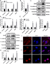
lncRNA HOTAIR expression and localization in the PCO model. (A, B) mRNA levels of lncRNA HOTAIR, vimentin, N-cadherin, E-cadherin, and ZEB1 in LECs of the NS group and 5 days post-surgery were determined using qRT-PCR analysis. (C, D) Protein levels of EMT markers in LECs of the NS group and 5 days post-surgery were determined using western blotting analysis. (E, F) lncRNA HOTAIR, vimentin, N-cadherin, E-cadherin, and ZEB1 mRNA levels after a 48-hour treatment of LECs with TGF-β2 (10 ng/mL). (G, H) Protein levels of EMT markers in TGF-β2 (10 ng/mL, 48 hours)–induced LECs. (I) Nuclear localization of lncRNA HOTAIR in HLE-B3 cells by RNA-FISH assay. Scale bar: 20 µm. Data are presented as mean ± SEM (n = 3). Only P < 0.05 was considered significant. *P < 0.05, **P < 0.01, ***P < 0.001 (Student's t-test); ns, not significant.
Figure 2.
Figure 2.
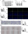
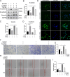
LncRNA HOTAIR overexpression promotes EMT, cell viability, proliferation, and migration of TGF-l –treated LECs. (A, B) After transfection of the HLE-B3 cells with HOTAIR overexpression or NC lentivirus followed by treatment with 10 ng/mL TGF-β2 for 48 hours, western blotting indicated vimentin, N-cadherin, and E-cadherin protein levels. (C) The viability of HLE-B3 cells was examined using the CCK-8 assay. (D, E) The proliferation of HLE-B3 cells was examined using EdU analysis. Scale bar: 200 µm. (F, G) Transwell migration results expressed as the number of migrated cells. Scale bar: 200 µm. (H, I) Wound healing assay showed cell migration at 0 and 48 hours. Scale bar: 100 µm. Data are presented as mean ± SEM (n = 3). Only P < 0.05 was considered significant. *P < 0.05, **P < 0.01, ***P < 0.001 (one-way ANOVA); ns, not significant.
Figure 3.
Figure 3.
LncRNA HOTAIR knockdown inhibits TGF-bi–induced EMT, cell viability, proliferation, and migration of LECs. (A, B) After transfection of the HLE-B3 cells with si-HOTAIR or si-NC followed by treatment with 10 ng/mL TGF-β2 for 48 hours, western blotting indicated EMT marker protein levels. (C) The viability of HLE-B3 cells was examined using the CCK-8 assay. (D, E) The proliferation of HLE-B3 cells was examined using EdU analysis. Scale bar: 200 µm. (F, G) Transwell migration results expressed as the number of migrated cells. Scale bar: 200 µm. (H, I) Wound healing assay showed cell migration at 0 and 48 hours. Scale bar: 100 µm. Data are presented as mean ± SEM (n = 3). Only P < 0.05 was considered significant. *P < 0.05, **P < 0.01, ***P < 0.001 (one-way ANOVA); ns, not significant.
Figure 4.
Figure 4.
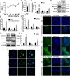
Total m6A levels and expression and localization of WTAP, METTL3, and METTL14 in the PCO model. (A) RNA m6A quantification assay showed total m6A modification levels in LECs from the NS and 1, 2, 3, 4, and 5 days after cataract surgery. (B) Total m6A modification level in TGF-β2 (10 ng/mL)–treated LECs after 48 hours. (CE) mRNA and protein levels of WTAP, METTL3, and METTL14 in LECs from the NS group and 5 days post-surgery determined using qRT-PCR and western blotting analysis. (FH) mRNA and protein levels of WTAP, METTL3, and METTL14 in TGF-β2 (10 ng/mL)–treated LECs after 48 hours. (I) IF staining assay indicated nuclear localization of WTAP, METTL3, and METTL14 (green) in HLE-B3 cells, with nuclei stained with DAPI (blue). Scale bar: 40 µm. (JL) IF staining assay indicated the expression levels of WTAP, METTL3, and METTL14 (green) in lens capsules of the NS group and 5 days post-surgery, with nuclei stained with DAPI (blue). Scale bar: 50 µm. Data are presented as mean ± SEM (n = 3). Only P < 0.05 was considered significant. *P < 0.05, **P < 0.01, ***P < 0.001 (Student's t-test); ns, not significant.
Figure 5.
Figure 5.
Interaction between lncRNA HOTAIR and WTAP protein and the effect of HOTAIR on the WTAP/METTL3/METTL14 complex and total m6A level in EMT of LECs. (A, B) RIP and qRT-PCR showed the interaction between HOTAIR and WTAP. (C, D) HOTAIR expression levels were examined using qRT-PCR in HLE-B3 cells with overexpression and knockdown of WTAP. (E, H) Total m6A level in HOTAIR overexpression and knockdown LECs after TGF-β2 treatment (10 ng/mL for 48 hours). (F, G, I, J) Western blotting indicated WTAP, METTL3, and METTL14 protein levels in HOTAIR overexpression and knockdown LECs after TGF-β2 treatment (10 ng/mL for 48 hours). (K) Dual RNA-FISH and IF indicated the colocalization of HOTAIR and WTAP in TGF-β2–induced HLE-B3 cells with low or overexpression of HOTAIR. Scale bar: 30 µm. (L) Co-IP and western blotting results showed the binding efficiency of WTAP with METTL3 and METTL14 in LECs transfected with si-NC or si- HOTAIR, and co-transfected with NC or WTAP overexpression lentivirus. Data are presented as mean ± SEM (n = 3). Only P < 0.05 was considered significant. *P < 0.05, **P < 0.01, ***P < 0.001 (Student's t-test and one-way ANOVA); ns, not significant.
Figure 6.
Figure 6.
Knockdown of WTAP inhibits the enhanced EMT, cell proliferation and migration of LECs induced by overexpression of lncRNA HOTAIR. (AD) Western blotting results showed vimentin, N-cadherin, and E-cadherin protein levels in LECs with NC or HOTAIR overexpression lentivirus and co-transfected with si-NC or si-WTAP. (E, F) Cell proliferation was tested using EdU assay. (G) Cell viability was detected using CCK-8 assay. (H, I) Transwell migration assay showed the number of migrated cells. Scale bar: 200 µm. (J, K) Wound healing assay showed migrating cells. Scale bar: 100 µm. Data are presented as mean ± SEM (n = 3). Only P < 0.05 was considered significant. *P < 0.05, **P < 0.01, ***P < 0.001 (one-way ANOVA).
Figure 7.
Figure 7.
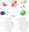
Identification of the lncRNA HOTAIR/WTAP complex targets via RNA-seq and MeRIP-seq. (A) DEGs identified by RNA-seq in NC and HOTAIR overexpression HLE-B3 cells with TGF-β2 treatment. (B) Heatmap showed consistency between NC and HOTAIR overexpression groups. (C) Predominant consensus motif GGAC. (D) The number of m6A peaks in NC and HOTAIR overexpression groups. (E) Density distribution of m6A peaks across mRNA transcripts. (F) Distribution of different m6A peaks in NC and HOTAIR overexpression groups. (G, H) GO and KEGG enrichment analysis of the genes of differential peaks.
Figure 8.
Figure 8.
THBS1 serves as the downstream marker of the HOTAIR/WTAP complex in LECs EMT. (A) m6A levels of lncRNA HOTAIR did not change in the control group or TGF-β2 treatment group. (BF) RIP-qPCR revealed the relative m6A modification levels of five mRNA candidates (LZTS1, THBS1, FOSB, KLF6, and FZD5) in TGF-β2–induced HLE-B3 cells stably transfected with NC or HOTAIR overexpression lentivirus. (G) The levels of five mRNA candidates in TGF-β2–induced HLE-B3 cells stably transfected with NC or HOTAIR overexpression lentivirus were detected using qRT-PCR assay. Data are presented as mean ± SEM (n = 3). Only P < 0.05 was considered significant. *P < 0.05, **P < 0.01, ***P < 0.001 (Student's t-test and one-way ANOVA); ns, not significant.
Figure 9.
Figure 9.
HOTAIR/WTAP complex enhances the expression of THBS1 in an m6A-dependent manner in the EMT of LECs. (AF) Western blotting showed the expression levels of LZTS1, THBS1, FOSB, KLF6, and FZD5 in TGF-β2–induced HLE-B3 with stable NC or HOTAIR overexpression lentivirus transfection. (G, H) Western blotting showed THBS1 protein levels in TGF-β2–induced WTAP overexpression HLE-B3 cells. (IK) After TGF-β2–induced HLE-B3 cells were transfected with si-NC or si-HOTAIR and then co-transfected with NC or WTAP overexpression lentivirus, RIP-qPCR showed the relative m6A modification levels of THBS1 mRNA. Data are presented as mean ± SEM (n = 3). Only P < 0.05 was considered significant. *P < 0.05, **P < 0.01, ***P < 0.001 (one-way ANOVA); ns, not significant.
Figure 10.
Figure 10.
The expression of THBS1 in TGF-β2–induced cells and mouse cataract surgery models of PCO. (AC, G) The expression levels of THBS1 in LECs from the NS group and 5 days post-surgery were determined using qRT-PCR, western blotting, and immunofluorescence staining. Scale bar: 50 µm. (DF, H) The expression levels of THBS1 in LECs after TGF-β2 (10 ng/mL) treatment for 48 hours. Scale bar: 100 µm. Data are presented as mean ± SEM (n = 3). Only P < 0.05 was considered significant. *P < 0.05, **P < 0.01, ***P < 0.001, ****P < 0.0001 (Student's t-test); ns, not significant.
Figure 11.
Figure 11.
HOTAIR/WTAP complex promotes EMT of LECs by enhancing THBS1 expression. (AD) Western blotting analysis showed the expression levels of EMT markers after TGF-β2–induced HLE-B3 cells were transfected with NC or HOTAIR overexpression followed by co-transfection with si-NC or si-THBS1. (E) IF staining showed the vimentin level. Scale bar: 100 µm. (F, G) Transwell assay showed migrating cells in TGF-β2-induced HOTAIR overexpression HLE-B3 cells with THBS1 low expression. Scale bar: 200 µm. (H, I) Wound healing assay showed migrating cells in TGF-β2–induced HOTAIR overexpression HLE-B3 cells with THBS1 low expression. Scale bar: 100 µm. Data are presented as mean ± SEM (n = 3). Only P < 0.05 was considered significant. *P < 0.05, **P < 0.01, ***P < 0.001, ****P < 0.0001 (one-way ANOVA); ns, not significant.
Figure 12.
Figure 12.
The mechanistic scheme of this study. lncRNA HOTAIR/WTAP/THBS1 axis regulates EMT of LECs in PCO.

Similar articles

References

    1. Liu YC, Wilkins M, Kim T, Malyugin B, Mehta J. Cataracts. Lancet. 2017; 390(10094): 600–612. - PubMed
    1. Konopinska J, Mlynarczyk M, Dmuchowska DA, Obuchowska I. Posterior capsule opacification: a review of experimental studies. J Clin Med. 2021; 10(13): 2847. - PMC - PubMed
    1. Milazzo S, Grenot M, Benzerroug M.. Posterior capsule opacification. J Fr Ophtalmol. 2014; 37(10): 825–830. - PubMed
    1. Sen P, Kshetrapal M, Shah C, Mohan A, Jain E, Sen A. Posterior capsule opacification rate after phacoemulsification in pediatric cataract: hydrophilic versus hydrophobic intraocular lenses. J Cataract Refract Surg. 2019; 45(10): 1380–1385. - PubMed
    1. Cinar E, Yuce B, Aslan F, Erbakan G. Influence of Nd:YAG laser capsulotomy on toric intraocular lens rotation and change in cylinder power. J Cataract Refract Surg. 2024; 50(1): 43–50. - PubMed

MeSH terms

LinkOut - more resources