. 2025 May 8;16(1):4300.
doi: 10.1038/s41467-025-59105-6.

Rational design of 19F NMR labelling sites to probe protein structure and interactions

Affiliations

Rational design of 19F NMR labelling sites to probe protein structure and interactions

Julian O Streit et al. Nat Commun. .

Abstract

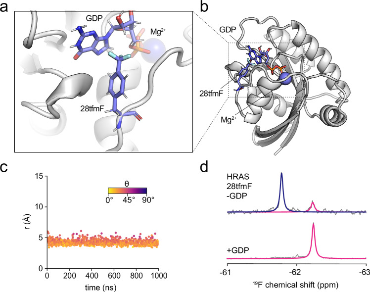
Proteins are investigated in increasingly more complex biological systems, where 19F NMR is proving highly advantageous due to its high gyromagnetic ratio and background-free spectra. Its application has, however, been hindered by limited chemical shift dispersions and an incomprehensive relationship between chemical shifts and protein structure. Here, we exploit the sensitivity of 19F chemical shifts to ring currents by designing labels with direct contact to a native or engineered aromatic ring. Fifty protein variants predicted by AlphaFold and molecular dynamics simulations show 80-90% success rates and direct correlations of their experimental chemical shifts with the magnitude of the engineered ring current. Our method consequently improves the chemical shift dispersion and through simple 1D experiments enables structural analyses of alternative conformational states, including ribosome-bound folding intermediates, and in-cell measurements of protein-protein interactions and thermodynamics. Our strategy thus provides a simple and sensitive tool to extract residue contact restraints from chemical shifts for previously intractable systems.


Conflict of interest statement

Competing interests: The authors declare no competing interests.

Figures

Fig. 1
Fig. 1. Aromatic ring currents dominate the secondary chemical shift of a fluorinated protein.
a Chemical structure of 4-trifluoromethyl-L-phenylalanine (tfmF). b Crystal structure of FLN5 (PDB 1QFH) with residues used for tfmF labelling highlighted (colour scheme corresponds to legend shown in panel c). c 19F NMR spectra of folded FLN5 (top) and unfolded FLN5 (A3A3 mutant,, middle) labelled at various positions as highlighted in panel (b). The bottom spectra show wild-type (675Phe) and mutant (Phe675Ala) FLN5 655tfmF. All spectra were recorded at 298 K and 500 MHz. d Close-up view of Tyr655 and Phe675 in the crystal structure of FLN5. e Schematic illustration of a trifluoromethyl (CF3) group interacting with an aromatic benzene ring, defined by the distance, r, between the CF3 and ring centres of mass and the angle, θ, between the vector normal to the ring plane and the vector between the CF3 and ring centres of mass. f All-atom MD simulation of FLN5 655tfmF showing an interaction between the CF3 group of 655tfmF and the aromatic ring of Phe675 quantified using r and θ.
Fig. 2
Fig. 2. Rational de novo design of ring current shifts across different structural motifs.
a Flow chart of the design method. b (Left) Structural model of FLN5 726tfmF/746Phe, highlighting interaction between 726tfmF and 746Phe across two β-strands. (Middle) Distance (r) and angle (θ) between the CF3 group of 726tfmF and aromatic ring of 746Phe observed in a representative all-atom MD simulation. (Right) 19F NMR spectra of FLN5 726tfmF with and without 746Phe recorded at 298 K and 500 MHz. c (Left) Structural model of HRAS 157tfmF/153His, highlighting an interaction between 157tfmF and 153His within an α-helix. (Middle) Distance (r) and angle (θ) between the CF3 group of 157tfmF and aromatic ring of 153His observed in a representative all-atom MD simulation. (Right) 19F NMR spectra of HRAS 157tfmF with and without 153His recorded at 298 K and 500 MHz. d Scatter plots (each data point represents a protein variant) correlating the distances (coloured by the fraction of time (MD) spent or models (ColabFold/AF3) in the plane of the ring defined as θ > 54.6°) predicted by ColabFold (left), AF3 (middle) and MD simulations (ff15ipq force field, right) with secondary 19F chemical shifts for variants of FLN5, FLN4, I27, FLNa21, FLNa21/migfilin complex, HemK, and HRAS. The error bars represent one s.d. over the five predicted models for ColabFold and AF3, and the s.e.m. obtained from three independent simulations for MD. PPV = positive predictive value; NPV = negative predictive value. Positive secondary chemical shift > 0.2 ppm in magnitude. The vertical lines represent the distance cut-off values for perpendicular and in-plane interactions (lower and higher distance, respectively). All variants included solvent-exposed residues on the protein surface. e Correlations between predicted geometric factors (1-3cos2θ)/r3) and secondary 19F chemical shifts, the corresponding Pearson correlation coefficients (rP) and intercepts for lines of best fit. Errors in all figures represent the s.e.m. from three independent replicates unless otherwise stated.
Fig. 3
Fig. 3. Engineered ring current shifts enable detection and characterisation of alternative protein conformational states.
a Crystal structure (left) of the HemK NTD (residues 1–73, PDB 1T43) and the predicted interaction between 38tfmF and Phe42 observed by MD (right). b Distance (r) and angle (θ) between the CF3 group of 38tfmF and the aromatic ring of Phe42 observed in a representative all-atom MD simulation. c 19F NMR spectra of HemK NTD 38tfmF variants: Mut (harbours I26V/R34K/Q46R mutations) and the Phe42Ala (below), recorded at 298 K and 500 MHz. d Backbone (Cα) root mean square fluctuation (RMSF) analysis for HemK NTD observed in long-timescale all-atom MD simulations (average ± s.e.m. from six independent simulations of 20 μs). The locations of the mutations are indicated by stars, and diamonds represent the tfmF labelling site and aromatic ring location within helix h3. e Representative structures of wild-type and mutant HemK obtained from all-atom MD simulations. f Structural ensembles of natively folded (N) FLN5 (left) obtained from an all-atom MD simulation using the ff15ipq force field and the FLN5Δ6 folding intermediate (I). The Tyr655 and Phe675 sidechains are shown as sticks for both ensembles. g 19F NMR spectrum of FLN5Δ6 655tfmF recorded at 283 K and 500 MHz, showing unfolded (U), intermediate (I) and native (N) conformations at equilibrium. h Probability distributions of r and the geometric factor (1-3cos2θ)/r3) calculated for the FLN5 N and I state and average values. i Structural model of a folded FLN5 nascent chain tethered to the ribosome. j Structural models of FLN5 655tfmF/675Phe and 726tfmF/746Phe highlighting the A/B and F/G strand pairs, respectively. k 19F NMR spectra of FLN5+47 RNCs with the 655tfmF/675Phe and 726tfmF/746Phe labelling pairs, recorded at 298 K and 500 MHz. For FLN5+47 655tfmF/675Phe two folding intermediates (I1 and I2) have been identified previously.
Fig. 4
Fig. 4. Probing protein-ligand interactions.
a, b Structural model of HRAS 28tfmF bound to GDP obtained from all-atom MD simulations. c Distance (r) and angle (θ) between the CF3 group of 28tfmF and aromatic rings of GDP observed in a representative all-atom MD simulation. d 19F NMR spectrum of HRAS 28tfmF purified in the absence (top) and presence (bottom) of Mg2+/GDP recorded at 298 K and 500 MHz.
Fig. 5
Fig. 5. Direct and residue-specific detection of protein-protein interactions in cells.
a Structural model of the FLNa21 (2274tfmF) – migfilin (WT) complex obtained from all-atom MD simulations. b Distance (r) and angle (θ) between the CF3 group of 2274tfmF and the aromatic ring of Phe14 observed in a representative all-atom MD simulation. c Structural model of the FLNa21 (2274tfmF) – migfilin (Ser12His) complex obtained from all-atom MD simulations. d Distance (r) and angle (θ) between the CF3 group of 2274tfmF and the aromatic ring of 12His observed in a representative all-atom MD simulation. e 19F NMR spectrum of purified FLNa21 2274tfmF alone and with 0.5−1.5 molar equivalents (eq) of migfilin WT and 12His. f In-cell 19F NMR spectrum of FLNa21 2274tfmF and co-expressed with WT and 12H migfilin. Free amino acid (tfmF) is also detected as part of the growth medium. All spectra were recorded at 298 K and 500 MHz.
Fig. 6
Fig. 6. Quantitative protein thermodynamics measured inside living cells.
a In-cell (the arrow highlights free tfmF as part of the growth medium), b in-lysate, and (c) purified 19F NMR spectra of FLN5 655tfmF 672Ala at various temperatures recorded at 500 MHz showing an unfolded and folded population. The arrow indicates the position of the free tfmF peak. d Temperature-dependence of the folding equilibrium constant (Keq) in all conditions. Data were fit to a modified Gibbs-Helmholtz equation (Eq. 1). Data points were calculated from fitted peak integrals from an NMR spectrum of a single biological sample averaged across 256 (purified, lysate) or 512 (in-cell) technical repeats, and errors (standard error) propagated from bootstrapping (200 iterations) of line shape fits. e Thermodynamic parameters of protein folding. Values were obtained from fits to the temperature-dependence data in panel (d) (from one biological sample), and with errors representing the 95% confidence interval.

Similar articles

References

    1. Boeszoermenyi, A. et al. Aromatic (19)F-(13)C TROSY: a background-free approach to probe biomolecular structure, function, and dynamics. Nat. Methods16, 333–340 (2019). - PMC - PubMed
    1. Huang, Y. et al. Use of paramagnetic (19)F NMR to monitor domain movement in a glutamate transporter homolog. Nat. Chem. Biol.16, 1006–1012 (2020). - PMC - PubMed
    1. Huang, Y. et al. Environmentally ultrasensitive fluorine probe to resolve protein conformational ensembles by (19)F NMR and cryo-eM. J. Am. Chem. Soc.145, 8583–8592 (2023). - PMC - PubMed
    1. Liu, J. J., Horst, R., Katritch, V., Stevens, R. C. & Wuthrich, K. Biased signaling pathways in beta2-adrenergic receptor characterized by 19F-NMR. Science335, 1106–1110 (2012). - PMC - PubMed
    1. Pan, B., Liu, D., Yang, L. & Wuthrich, K. GPCR large-amplitude dynamics by (19)F-NMR of aprepitant bound to the neurokinin 1 receptor. Proc. Natl. Acad. Sci. USA119, e2122682119 (2022). - PMC - PubMed

LinkOut - more resources