. 2025 Jun;44(12):3468-3491.
doi: 10.1038/s44318-025-00451-y. Epub 2025 May 8.

Control of replication and gene expression by ADP-ribosylation of DNA in Mycobacterium tuberculosis

Affiliations

Control of replication and gene expression by ADP-ribosylation of DNA in Mycobacterium tuberculosis

Rachel E Butler et al. EMBO J. 2025 Jun.

Erratum in

Abstract

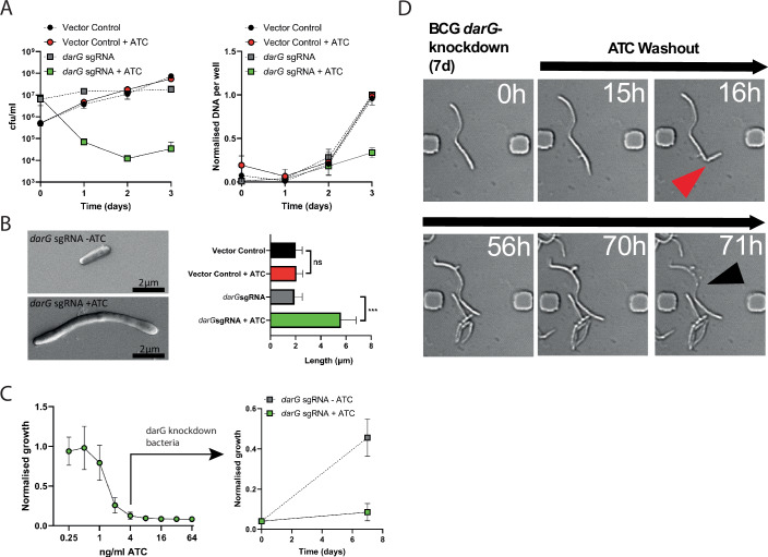
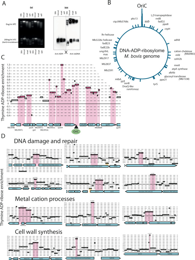
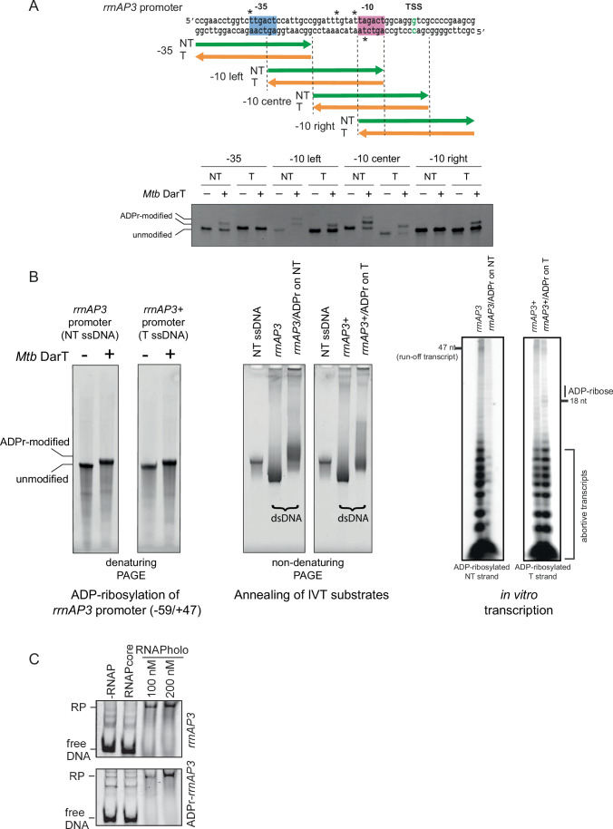
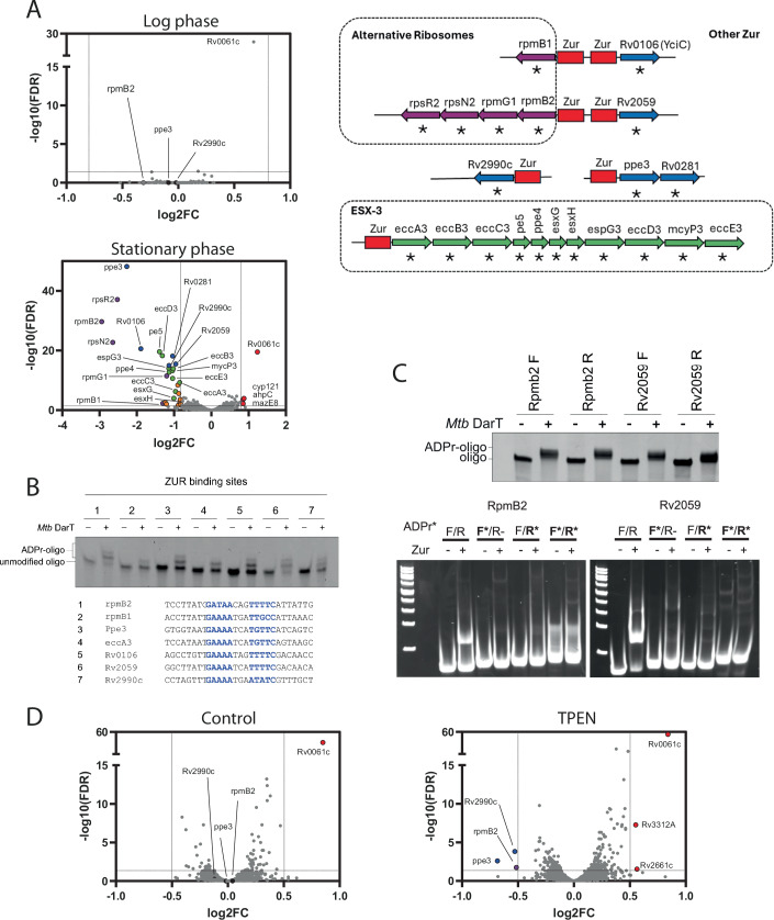
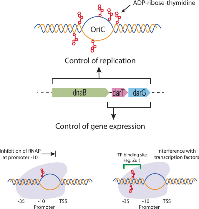
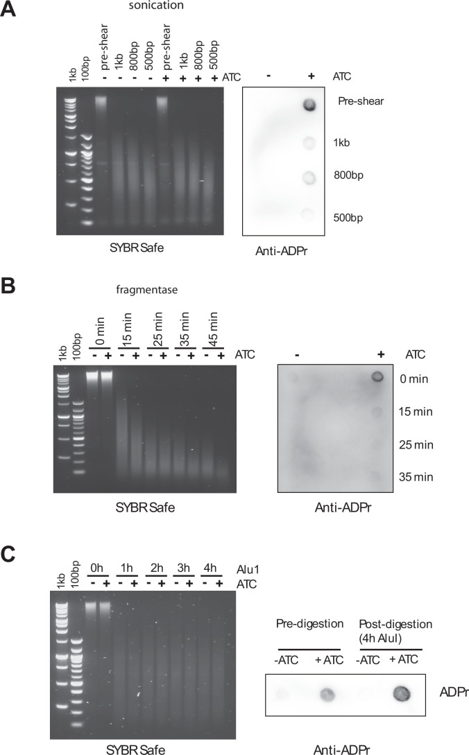
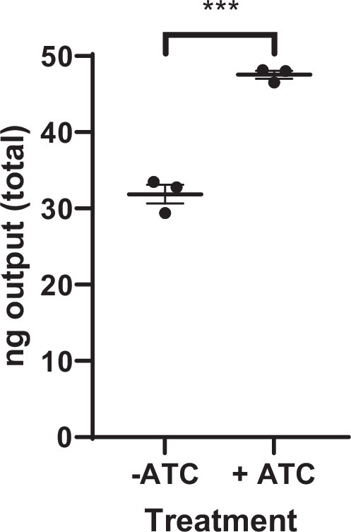
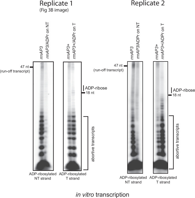
Mycobacterium tuberculosis maintains long-term infections characterised by the need to regulate growth and adapt to contrasting in vivo environments. Here we show that M. tuberculosis complex bacteria utilise reversible ADP-ribosylation of single-stranded DNA as a mechanism to coordinate stationary phase growth with transcriptional adaptation. The DNA modification is controlled by DarT, an ADP-ribosyltransferase, which adds ADP-ribose to thymidine, and DarG, which enzymatically removes this base modification. Using darG-knockdown M. bovis BCG, we map the first DNA ADP-ribosylome from any organism. We show that inhibition of replication by DarT is reversible and accompanied by extensive ADP-ribosylation at the origin of replication (OriC). In addition, we observe ADP-ribosylation across the genome and demonstrate that ADP-ribose-thymidine alters the transcriptional activity of M. tuberculosis RNA polymerase. Furthermore, we demonstrate that during stationary phase, DarT-dependent ADP-ribosylation of M. tuberculosis DNA is required to optimally induce expression of the Zur regulon, including the ESX-3 secretion system and multiple alternative ribosome proteins. Thus, ADP-ribosylation of DNA can provide a mechanistic link through every aspect of DNA biology from replication to transcription to translation.

Keywords: ADP-ribosylation; ADPr-Seq; DNA Modification; PARP; Transcription Regulation.


Conflict of interest statement

Disclosure and competing interests statement. The authors declare no competing interests.

Figures

Figure 1
Figure 1. Reversible control of mycobacterial replication by the DarT/DarG ADP-ribosylation system.
(A) Left panel: Viability of M. bovis BCG is reduced when DarT activity is deregulated by CRISPRi knockdown of the “antitoxin” DarG ADP-ribosylglycohydrolase, induced by addition of anhydrotetracycline (ATC) at 200 ng/ml to M. bovis BCG darG-sgRNA. Right panel: DNA synthesis is inhibited by deregulation of DarT. Data are mean ± SD, n = 3 independent cultures. (B) Scanning electron micrograph showing filamentous appearance of BCG darG-knockdown mycobacteria with unregulated DarT ADP-ribosylation. Average cell length increased threefold in BCG darG-knockdown. Data are mean ± SD, N = 3 independent experiments, ~350 cell measurements per strain/treatment, ***P = 0.0008 by one-way ANOVA with Sidak’s multiple comparison test. (C) Level of growth inhibition is controllable by titration of ATC-induced knockdown of DarG and reversible by washout of ATC. Right panel shows M. bovis BCG darG-sgRNA inhibited for growth with 4 ng/ml ATC, washed and then restimulated with ATC or carrier alone. Data are mean ± SD, n = 3 independent cultures, and are representative of three independent experiments. (D) Captured images from video of a darG-sgRNA (growth inhibited) M. bovis BCG after washout of ATC showing resumption of cell division (red arrow), and eventual necrosis of one daughter cell (black arrow). The full video and selected stills are available in Movie EV3 and Fig. EV3. Source data are available online for this figure.
Figure 2
Figure 2. The DNA ADP-ribosylome of M. bovis BCG.
(A) DarT-dependent ADP-ribosylation of DNA in M. bovis BCG darG-sgRNA was stimulated by 200 ng/ml ATC-induced knockdown of darG. DNA from ATC-induced and uninduced cells was Southern blotted and probed with antibodies against ADP-ribosylated DNA, indicating widespread DNA modification in DarG-knockdown cells. Anti-dsDNA blotting as control for DNA transfer. (B) ADPr-Seq of DNA from darG-knockdown M. bovis BCG. Alu1-digested DNA from ATC+ and ATC- M. bovis BCG darG-sgRNA was sequenced before and after immune-affinity capture of ADP-ribosylated DNA fragments. Significant enrichment of ADP-ribosylated DNA fragments was revealed by generalised linear regression of read counts to estimate the relative enrichment of modified fragments in the induced DarG-knockdown mycobacteria compared to control uninduced bacteria. The density of ADP-ribosylated DNA (FDR ≤0.05, n = 3)) is plotted across the M. bovis genome. (C) ADP-ribosylation of the chromosome origin of replication (OriC), including the AT-rich region and surrounding genes. Significantly enriched (ADP-ribosylated) DNA fragments determined by negative binomial generalised linear regression of read counts to estimate the relative enrichment of ADPr fragments in the induced DarG-knockdown mycobacteria versus uninduced mycobacteria (FDR ≤0.05, n = 3) are shown in pink. (D) ADP-ribosylated DNA was distributed across the genome in genes associated with functional roles including DNA damage and repair, metal ion transport and biology and cell wall synthesis. Examples of ADP-ribose-enriched fragments are shown (FDR ≤0.05, n = 3). Source data are available online for this figure.
Figure 3
Figure 3. The modulatory effects of ADP-ribosylation of ssDNA on RNA polymerase and transcription.
(A) DarT from M. tuberculosis ADP-ribosylates the rrnAP3 promoter in vitro. Overlapping oligonucleotides covering the promoter region on both template (T) and non-template (NT) strands were reacted with M. tuberculosis DarT and β-NAD+, and assessed by PAGE to reveal ADP-ribose modifications. The strict TNT sequence requirement for DarT allowed inference of ADP-ribosylated thymidines (indicated by *) with preference for the non-template strand adjacent in the −10 region. (B) Transcriptional activity of RNA polymerase (RNAP) holoenzyme is inhibited by ADP-ribosylation of DNA. The NT strand of the rrnAP3 promoter region (−59 to +47) was ADP-ribosylated with DarT enzyme. The T strand of the rrnAP3+ promoter (carrying an g to t base change at +23) was ADP-ribosylated. ADP-ribose modified NT strand rrnAP3 was annealed with the unmodified T strand to make rrnAP3/ADPr template DNA for in vitro transcription (IVT). ADP-ribose modified T strand of rrnAP3+ was annealed with the unmodified NT strand to make rrnAP3 + /ADPr template DNA for IVT. IVT was performed with the M. tuberculosis RNAP holoenzyme on rrnAP3/ADPr template and rrnAP3 + /ADPr template alongside templates carrying no ADP-ribose modifications. This showed that ADP-ribosylation of the NT strand of rrnAP3 inhibited the formation of both abortive transcripts as well as run-off transcripts, whereas ADP-ribosylation on the template strand of rrnAP3+ did not inhibit transcription although there was some indication for formation of stalled elongation complex near the introduced ADP-ribosylation site. This IVT assay is shown alongside a replicate experiment in Fig. EV6. (C) EMSA showing RNAP holoenzyme binding to the rrnAP3 promoter irrespective of ADP-ribose modification. Source data are available online for this figure.
Figure 4
Figure 4. Regulation of stationary phase gene expression by DarT-dependent ADP-ribosylation of DNA in M. tuberculosis.
(A) RNA-Seq of wild-type M. tuberculosis GC1237 versus M. tuberculosis ΔdarTdarG in logarithmic phase growth at 6 days (upper panel) and stationary phase growth at 20 days (lower panel). Genes with significantly altered transcription between wild-type and ΔdarTdarG-knockout strain (FDR <0.05) and a log2 fold change (log2FC) of >0.8 (n = 4) are coloured, and functional category is indicated in the colour-coordinated genomic organisation diagram (right panel, *significantly altered transcription, Zur-binding sites in red). (B) DarT-dependent ADP-ribosylation of Zur-binding site DNA sequences from the promoters of genes differentially regulated in stationary phase M. tuberculosis ΔdarTdarG. Zur-binding motifs are highlighted in blue. (C) DarT-dependent ADP-ribosylation of either DNA strand in Zur-binding sites from the divergent promoter between rpmB2 and Rv2059 inhibited Zur binding to DNA. (D) RNA-Seq of wild-type M. tuberculosis GC1237 versus M. tuberculosis ΔdarTdarG in zinc-replete media (left panel) and under zinc deficiency by exposure to 1.2 µM TPEN (right panel). Genes with significantly altered transcription between wild-type and ΔdarTdarG-knockout strain (FDR <0.05) and a log2FC of >0.5 are coloured. n = 4. Source data are available online for this figure.
Figure 5
Figure 5. Coordination of DNA replication and transcription by DarT-dependent ADP-ribosylation of thymidine in M. tuberculosis complex bacteria.
DarT ADP-ribosyltransferase and DarG ribosylglycohydrolase (transcriptionally linked to the replicative helicase DnaB) reversibly modify thymidine at the chromosome origin of replication (OriC) to regulate DNA replication. In coordination, ADP-ribosylation regulates gene expression by modification of thymidine at the promoter to inhibit escape of RNAP from the promoter or alter the binding of transcription factors.
Figure EV1
Figure EV1. In cellulo ADP-ribosylation of gDNA inhibits DNA synthesis.
In cellulo ADP-ribosylation of gDNA in M. bovis BCG was induced in late log phase by CRISPRi of the antitoxin darG with 200 ng/ml ATC (or 0 ng/ml ATC uninduced control). After 24 h, the cultures were diluted to stimulate replication, and the ATC concentration replenished as indicated. After 3 days, optical density (OD600 nm; A), and colony forming units (cfu; B)) were assessed. Genomic DNA (C) was isolated from the cultures by phenol:chloroform:isoamyl extraction and quantified as detailed in “Methods” section. Data are mean +/− SD for n = 3 replicate cultures; n.s = not significant, **P = 0.0096. ****P = 0.00002 by unpaired two-tailed T test. Source data are available online for this figure.
Figure EV2
Figure EV2. Time-lapse stills from Movies EV1 and EV2.
An experiment schematic is included in the panel. BCG darG-sgRNA and BCG carrying an empty vector were pretreated for 48 h with 200 ng/ml ATC, then loaded into a microfluidic device. The bacteria were imaged every hour for 59 h, with continued ATC treatment. BCG-vector control bacilli divide over 59 h (AC), whereas BCG darG-sgRNA stop dividing and elongate (DF). An example is circled in each treatment. Source data are available online for this figure.
Figure EV3
Figure EV3. Time-lapse stills from Fig. 1D and Movie EV3.
An experiment schematic is included in the panel. BCG darG-sgRNA were minimally inhibited with ATC in a microtitre plate for 7 days, and loaded into a microfluidic device (A). After 12 h, ATC was washed out of the device (B). The bacterium marked with a white arrow was followed over time (C). After 16 h in fresh medium, division occurs at one pole (DF). After 58 in fresh medium, branching occurs at the other pole (G) but is followed by swelling (H) and necrosis (I).
Figure EV4
Figure EV4. Survival of the E6F6A ADPr epitope after gDNA fragmentation.
In cellulo ADP-ribosylation of gDNA was induced in BCG by CRISPRi knockdown of the antitoxin darG with 200 ng/ml ATC (or 0 ng/ml ATC control) for 48 h. gDNA was extracted and fragmented by (A) sonication (S220 Focussed Ultrasonicator, Covaris); (B) NEBNext Fragmentase Enzymes (FR); (C) AluI digestion. Fragmentation was analysed by agarose gel electrophoresis (left panels). Equal quantities of DNA were bound to a Zetaprobe membrane and epitope stability assessed by dot-blot using the E6F6A antibody against ADP-ribose. The ADPr-Seq technique requires the generation of gDNA fragments that are affinity purified using the anti-ADPr antibody E6F6A, which are then sequenced by NGS. This approach required the ADPr epitope to remain on the DNA following fragmentation. We tried three approaches to generating gDNA fragments. Typically sonication is used to generate 500 bp fragments for NGS studies, however this led to the loss of the ADPr epitope (A). Random fragmentation using NEBNext dsDNA Fragmentase also led to loss of the ADPr epitope, and unequal fragment sizes were generated between control gDNA (-ATC) and ADPr-gDNA ( + ATC) (B). Digestion with AluI maintained the epitope post fragmentation, and resulted in a similar fragmentation pattern in control gDNA (-ATC) and ADPr-gDNA ( + ATC) (C).
Figure EV5
Figure EV5. Yield of DNA fragments recovered by immunoprecipitation with anti-ADPr antibody.
DNA from darG-silenced ( + ATC) and uninduced control BCG (-ATC) was fragmented with AluI and immunoprecipitated using the E6F6A anti-ADPr antibody. The yield of DNA from each precipitation was measured using the dsDNA Quantifluor system (Promega). Error bars are mean +/− SEM, ***P = 0.0003 by unpaired T test.
Figure EV6
Figure EV6. Replicate in vitro transcription assay showing effects of ADP-ribosylation of ssDNA on transcription by M. tuberculosis RNA polymerase.
IVT was performed with the M. tuberculosis RNAP holoenzyme on rrnAP3/ADPr template (ADP-ribose on non-template NT strand) and rrnAP3 + /ADPr template (ADP-ribose on template T strand) alongside substrates carrying no ADP-ribose modifications. This showed that ADP-ribosylation of the NT strand of rrnAP3 inhibited the formation of both abortive transcripts as well as run-off transcripts, whereas ADP-ribosylation on the template strand of rrnAP3+ did not inhibit transcription although there was some indication for formation of stalled elongation complex near the introduced ADP-ribosylation site. For direct comparison, Fig. 3B image (replicate 1) is shown alongside replicate 2.

Similar articles

References

    1. Banerjee R, Rudra P, Prajapati RK, Sengupta S, Mukhopadhyay J (2014) Optimization of recombinant Mycobacterium tuberculosis RNA polymerase expression and purification. Tuberculosis 94:397–404 - PubMed
    1. Barth VC, Chauhan U, Zeng JM, Su XY, Zheng HY, Husson RN, Woychik NA (2021) Mycobacterium tuberculosis VapC4 toxin engages small ORFs to initiate an integrated oxidative and copper stress response. Proc Natl Acad Sci USA 118:e2022136118 - PMC - PubMed
    1. Barth VC, Zeng JM, Vvedenskaya IO, Ouyang M, Husson RN, Woychik NA (2019) Toxin-mediated ribosome stalling reprograms the Mycobacterium tuberculosis proteome. Nat Commun 10:3035 - PMC - PubMed
    1. Beste DJ, Espasa M, Bonde B, Kierzek AM, Stewart GR, McFadden J (2009) The genetic requirements for fast and slow growth in mycobacteria. PLoS ONE 4:e5349 - PMC - PubMed
    1. Beste DJV, Laing E, Bonde B, Avignone-Rossa C, Bushell ME, McFadden JJ (2007) Transcriptomic analysis identifies growth rate modulation as a component of the adaptation of mycobacteria to survival inside the macrophage. J Bacteriol 189:3969–3976 - PMC - PubMed

MeSH terms

LinkOut - more resources