. 2025 May 8;8(1):712.
doi: 10.1038/s42003-025-08138-0.

Structural and functional insights into the nuclear role of Parkinson's disease-associated α-synuclein as a histone chaperone

Affiliations

Structural and functional insights into the nuclear role of Parkinson's disease-associated α-synuclein as a histone chaperone

Sneha Jos et al. Commun Biol. .

Abstract

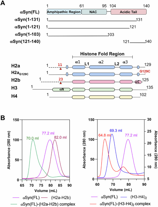
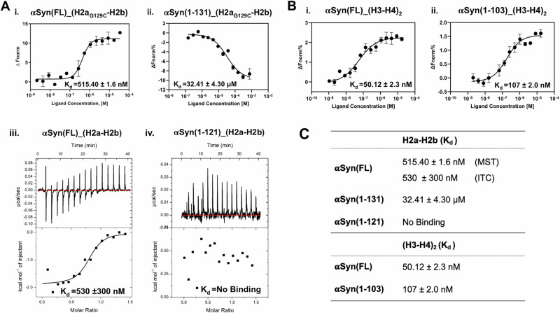
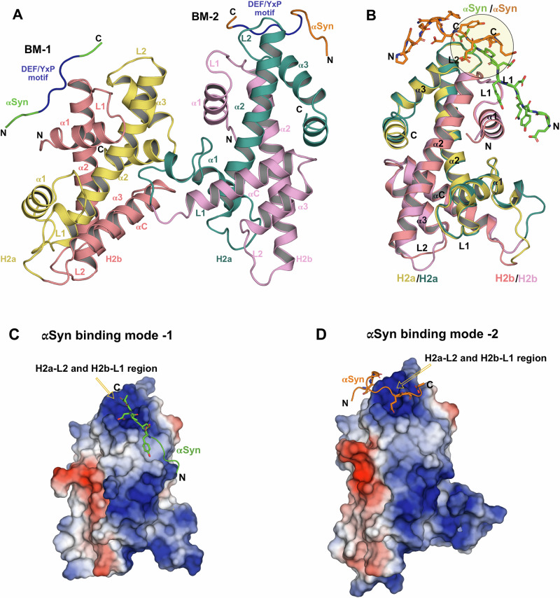
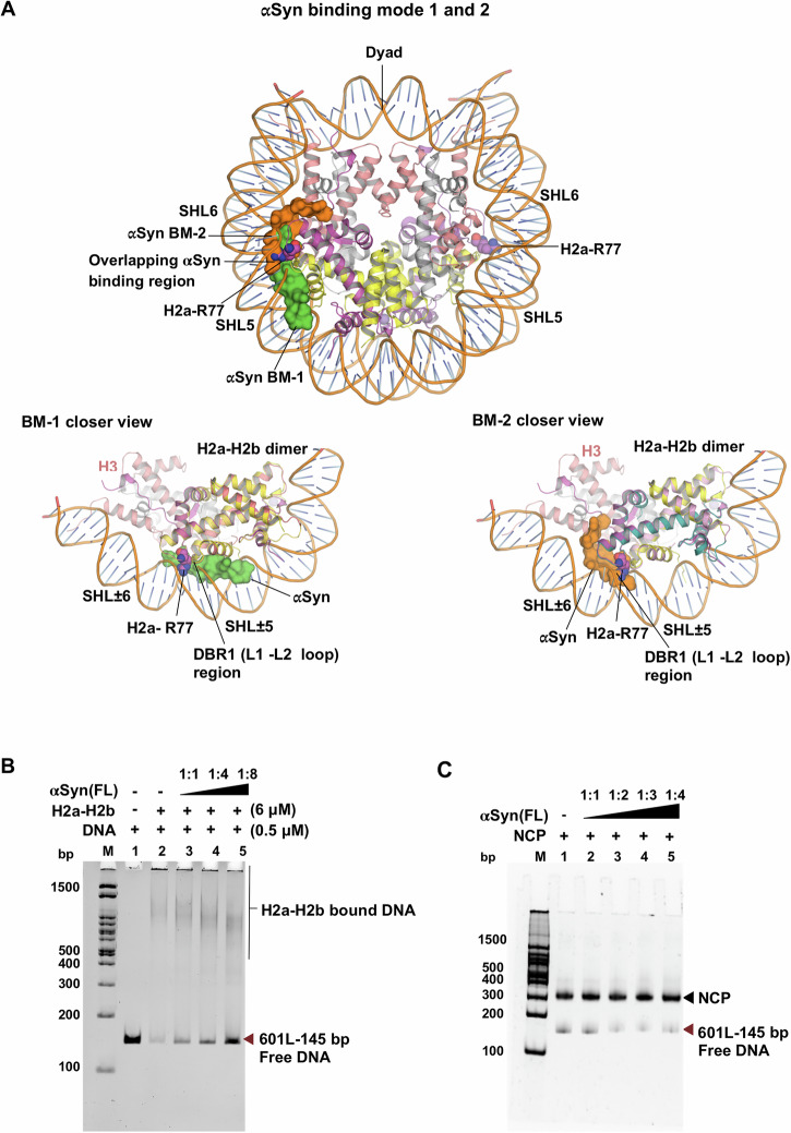
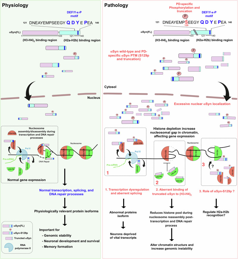
α-Synuclein (αSyn) plays a critical role in the pathogenesis of 'Synucleinopathies'. Although increased nuclear αSyn localization induces neurotoxicity, its definitive physiological role remains elusive. Previous studies on nuclear αSyn are limited to its interactions with individual histones and dsDNA, leaving a significant gap in understanding its interactions with assembled histone H2a-H2b dimer and (H3-H4)2 tetramer, as well as its role in chromatin regulation. Here, we demonstrate that αSyn binds specifically to both H2a-H2b and (H3-H4)2 with high affinity. Truncation studies reveal that αSyn(1-103) region interacts with (H3-H4)2, while the acidic (121-140) C-terminal end is crucial for H2a-H2b binding and contains a conserved DEF/YxP motif present in other dimer-binding histone chaperones. High-resolution structure of αSyn(121-140) with H2a-H2b complex reveals that αSyn adopts two binding modes (BM-1 and BM-2). Nonetheless, the αSyn C-terminal end in both modes overlap but runs in opposite orientations, specifically interacting with the H2a-L2 and H2b-L1 loop regions of the dimer and cap the H2a-R78 residue. Mutational analysis confirms that αSyn-Y136 and P138 residues, part of the DEF/YxP motif, together with H2a-R78, are critical for αSyn-(H2a-H2b) interaction. The chaperoning assay supports αSyn's function as a histone chaperone, suggesting the potential role of αSyn in the nucleosome assembly/disassembly process.


Conflict of interest statement

Competing interests: The authors declare no competing interests.

Figures

Fig. 1
Fig. 1. αSyn forms complex with assembled histone H2a-H2b and (H3-H4)2.
A Schematic representation of αSyn and histone constructs used in this study. The red arrow indicates the H2a and H2b N-terminal tail truncation boundary, while the G129C mutation introduced in histone H2a is marked in red. B Size-exclusion chromatography shows αSyn(FL) forms a complex with (H2a-H2b) dimer and (H3-H4)2 tetramer.
Fig. 2
Fig. 2. Biophysical studies of αSyn with H2a-H2b and (H3-H4)2.
A MST analysis of αSyn(FL) (i) and αSyn(1-131) (ii) with fluorescently labeled H2aG129C-H2b dimer. iii-iv. ITC analysis of the αSyn(FL) and αSyn(1-121) with H2a-H2b dimer, respectively. B MST analysis of the αSyn(FL) (i) and αSyn(1-103) (ii) with fluorescently labeled (H3-H4)2 tetramer. Error bars represent SD (N = 3). The Kd values are displayed in individual panels and summarized in table (C).
Fig. 3
Fig. 3. Crosslinking assay, HSQC NMR spectra, and sequence analysis.
A SDS-PAGE gel of DSS and EDC mediated cross-linking experiment (Lane M- protein marker along with the indicated protein concentrations at the top). i. αSyn(FL) with both (H2a-H2b) and (H2a-H2b)TL, ii. αSyn(1-131) and αSyn(1-121) with (H2a-H2b)TL, and iii. αSyn(121-140) with (H2a-H2b)TL. The cross-linking assays were performed in the presence of 150 mM NaCl. B HSQC NMR spectra of 15N-labeled αSyn(FL) in the absence (blue) and presence (red) of (H2a-H2b) dimer (1:1 ratio) showed significant chemical shift perturbations for αSyn C-terminal (120-140) residues. C Sequence alignment of the human αSyn-dimer recognition region (121-140) with Swc5 in yeast (top) and multiple sequence alignment with other species (bottom). The DEF/YxP motif is indicated by (*) on top of the sequence analysis.
Fig. 4
Fig. 4. αSyn with ScH2a-H2b complex structure.
A The overall structure of αSyn(121-140)-ScH2a-H2b complex is shown in cartoon representation. The asymmetric unit contains two molecules, and αSyn has different binding modes for each unit of ScH2a-H2b. The DEF/YxP motif is indicated in blue. B Superimposition of αSyn(121-140)-ScH2a-H2b complex within asymmetric unit shows that αSyn(136-140) overlaps and runs in opposing directions and is highlighted in a circle. C, D The electrostatic interface between αSyn(121-140) and ScH2a-H2b. ScH2a-H2b is shown in the surface model and colored according to its electrostatic potential, and αSyn is shown in stick representation.
Fig. 5
Fig. 5. The interface between αSyn with H2a-H2b complex.
A, B αSyn binding mode-1 and 2; close-up view of residues involved in the αSyn-ScH2a-H2b interface interactions are shown in stick representation. C Binding analysis of αSyn(FL) and αSyn mutants (Y136A, P138A, Y136A-P138A) with H2aG129C-H2b dimer and H2a(R78A)G129C-H2b dimer using MST (i-vii). The Kd values are summarized in the table. D, E Superimposition of αSyn BM-1 and BM-2 with other known dimer-specific chaperone structures. αSyn (BM-1, green), αSyn (BM-2, orange), Anp32e (PDB: 4CAY, pink), YL1 (PDB: 5CHL, red), Swc5 (PDB: 6KBB, purple), Chz1 (PDB: 6AE8, yellow), Spt16 (PDB: 4WNN, cyan), YL1 (PDB: 5FUG, brown), Spt16 (PDB: 8I17, blue) and H2a-H2b/H2a.Z-H2b dimer (gray). In BM-1, the position of Y136 and E135 is conserved with other histone chaperones, whereas in BM-2, the position of E137 is conserved, indicated in a dotted circle.
Fig. 6
Fig. 6. αSyn(121-140) competes with the DNA binding region of H2a-H2b.
A Superimposition of αSyn(121-140)-ScH2a-H2b complex structure with the NCP structure (PDB ID: 3X1S). The H2a-L2 loop R77 residue anchored to a minor groove of the DNA at SHL ± 5.5 in the nucleosome shown in spheres. The αSyn(136-140) region in both binding modes (BM-1 and BM-2) overlaps, exclusively binds to DBR1 (H2a-L2 and H2b-L1 loop), and caps conserved H2a-R77 residue. In BM-1, αSyn peptide clashed with the DNA-binding site of H2a-H2b in the nucleosome. In BM-2, αSyn peptide clashed with both DNA-binding sites of H2a-H2b and competes for the H3 binding site. B Native PAGE analysis of histone chaperoning assay shows that αSyn(FL) competes with 145 bp Widom 601L DNA nucleosome positioning sequence for binding to H2a-H2b dimer. Lane M: 100 bp DNA ladder. Lane 1: DNA alone. Lane 2: DNA + H2a-H2b dimer. Lanes 3-5: Increasing H2a-H2b: αSyn(FL) ratios (1:1, 1:4, 1:8). C Electrophoretic mobility shift assay (EMSA) with NCP and αSyn(FL) shows no mobility shift, indicating no binding. Lane M: 100 bp DNA ladder. Lane 1: NCP alone. Lanes 2-5: Increasing NCP:αSyn(FL) ratios (1:1, 1:2, 1:3, 1:4).
Fig. 7
Fig. 7. Model of nuclear αSyn role in physio-pathological conditions.
Under physiological conditions, the nuclear-localized αSyn possibly regulates nucleosome assembly/disassembly during transcription and DNA repair, which is essential for normal gene expression. Conversely, excessive nuclear αSyn localization depletes the histone pool, increasing the nucleosomal gap and adversely affecting gene expression under pathological conditions.

Similar articles

References

    1. Goedert, M. Alpha-synuclein and neurodegenerative diseases. Nat. Rev. Neurosci.2, 492–501 (2001). - PubMed
    1. Maroteaux, L., Campanelli, J. T. & Scheller, R. H. Synuclein: a neuron-specific protein localized to the nucleus and presynaptic nerve terminal. J. Neurosci.8, 2804–2815 (1988). - PMC - PubMed
    1. Spillantini, M. G. et al. Alpha-synuclein in Lewy bodies. Nature388, 839–840 (1997). - PubMed
    1. Lashuel, H. A. Do Lewy bodies contain alpha-synuclein fibrils? and Does it matter? A brief history and critical analysis of recent reports. Neurobiol. Dis.141, 104876 (2020). - PubMed
    1. Goers, J. et al. Nuclear localization of alpha-synuclein and its interaction with histones. Biochemistry42, 8465–8471 (2003). - PubMed