Review
. 2025 Jul;56(1):103.
doi: 10.3892/ijmm.2025.5544. Epub 2025 May 9.

Molecular mechanisms of programmed cell death and potential targeted pharmacotherapy in ischemic stroke (Review)

Affiliations
Review

Molecular mechanisms of programmed cell death and potential targeted pharmacotherapy in ischemic stroke (Review)

Wan-Li Duan et al. Int J Mol Med. 2025 Jul.

Abstract

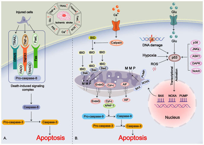
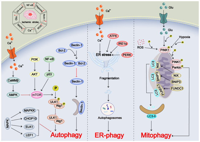
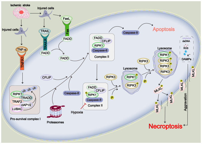
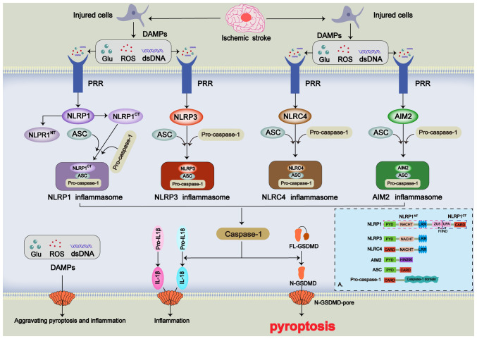
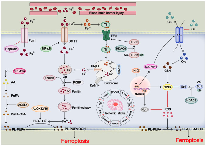
Stroke poses a threat to the elderly, being the second leading cause of death and the third leading cause of disability worldwide. Ischemic stroke (IS), resulting from arterial occlusion, accounts for ~85% of all strokes. The pathophysiological processes involved in IS are intricate and complex. Currently, tissue plasminogen activator (tPA) is the only Food and Drug Administration‑approved drug for the treatment of IS. However, due to its limited administration window and the risk of symptomatic hemorrhage, tPA is applicable to only ~10% of patients with stroke. Additionally, the reperfusion process associated with thrombolytic therapy can further exacerbate damage to brain tissue. Therefore, a thorough understanding of the molecular mechanisms underlying IS‑induced injury and the identification of potential protective agents is critical for effective IS treatment. Over the past few decades, advances have been made in exploring potential protective drugs for IS. The present review summarizes the specific mechanisms of various forms of programmed cell death (PCD) induced by IS and highlights potential protective drugs targeting different PCD pathways investigated over the last decade. The present review provides a theoretical foundation for basic research and insights for the development of pharmacotherapy for IS.

Keywords: IS; PCD; molecular mechanisms; pharmacotherapy; protective drugs.


Conflict of interest statement

The authors declare that they have no competing interests.

Figures

Figure 1
Figure 1
Potential mechanisms of ischemic stroke-induced apoptosis. (A) Extrinsic apoptotic pathway. TRAIL, TNF-α and FasL are released from injured cells and activate extrinsic apoptotic pathways via binding to 'death receptors' on the cell membrane. (B) Intrinsic apoptotic pathway. Hypoxia, elevated Ca2+ levels, ROS, Glu and upregulated P53 levels can activate the intrinsic apoptotic pathways. AIF, apoptosis-inducing factor; ASK1, apoptosis signal-regulated kinase-1; BID, BH3-interacting domain; Cyt-c, cytochrome c; DAPK, death-associated protein kinase; DR4/5, death receptor 4; EndoG, endonuclease G; FADD, fas-associated protein with death domain; Glu, glutamate; MMP, mitochondrial membrane permeabilization; NOXA, NADPH oxidase activator; PUMP, p53-upregulated modulator of apoptosis; ROS, reactive oxygen species; tBID, truncated BID; TNFR1, tumor necrosis factor receptor 1; TRADD, TNFRSF1A associated via death domain; TRAIL, TNF-related apoptosis-inducing ligand.
Figure 2
Figure 2
Potential mechanisms of IS-induced autophagy. Excessive autophagy as well as the deficiency of autophagy can promote IS-induced cell injury. Ca2+ overload, upregulation of MAPK, high expression levels of NF-κB and upregulation of Beclin-1 cause disruption of autophagic flux in IS. Ca2+ overload can cause ER stress and promote ER-phagy. Hypoxia, ROS and extracellular Glu excess caused by IS promote intracellular mitophagy. AMPK, 5'-AMP-activated protein kinase; ATF6, activating transcription factor 6; Atg1, autophagy-related 1; BNIP3, Bcl-2/adenovirus E1B 19 kDa protein-interacting protein 3; CAMMβ, calmodulin-dependent kinase β; ELK1, ETS transcription factor ELK1; ER, endoplasmic reticulum; FUNDC1, FUN14 domain containing 1; Glu, glutamate; IRE1α, inositol-requiring enzyme 1α; IS, ischemic stroke; LEF1, lymphoid enhancer-binding factor 1; NIX, Nip3-like protein X; P, phosphorylated; PERK, protein kinase R-like endoplasmic reticulum kinase; PINK1, PTEN-induced putative kinase 1; ROS, reactive oxygen species; ULK1, UNC-51-like kinase 1.
Figure 3
Figure 3
Potential mechanisms of IS-induced necroptosis. TNF-α, TRAIL and FasL released from injured cells can form a pro-survival complex upon binding to their receptors. Additionally, hypoxia caused by IS inhibits the function of caspase-8, and thus, activates the necroptosis pathway that follows RIPK1/RIPK3/MLKL signaling. Following the onset of necroptosis, intracellular DAMPs are released, further promoting necroptosis. CFLIP, cellular FLICE-like inhibitory protein; cIAP1/2, cellular inhibitor of apoptosis protein 1/2; DAMPs, damage associated molecular patterns; dsDNA, double-stranded DNA; FADD, fas-associated protein with death domain; IS, ischemic stroke; LUBAC, linear ubiquitin chain assembly complex; MLKL, mixed lineage kinase domain-like protein; P, phosphorylated; RIPK1, receptor interacting serine/threonine kinase 1; RIPK3, receptor interacting serine/threonine kinase 3; ROS, reactive oxygen species; TNFR1, tumor necrosis factor receptor 1; TRADD, TNFRSF1A associated via death domain; TRAF2, TNF receptor associated factor 2; TRAIL, TNF-related apoptosis-inducing ligand; TRAILR, TNF-related apoptosis-inducing ligand receptor; ub, ubiquitin.
Figure 4
Figure 4
Potential mechanisms of IS-induced pyroptosis. During IS pathology, DAMPs released from injured cells can bind to PRRs on the cell membrane and activate downstream inflammasomes. PRRs include NLRs and Toll-like receptors, and inflammasomes include NLRP1 inflammasomes, NLRP3 inflammasomes, NLRC4 inflammasomes and AIM2 inflammasomes. Activation of inflammasomes promotes the formation of N-GSDMD and causes pyroptosis. After pyroptosis occurs, intracellular DAMPs are released and further aggravate pyroptosis and the inflammatory response. (A) Schematic diagram of protein structural domains. AIM2, absent in melanoma 2; ASC, apoptosis-associated speck-like protein containing a CARD; CARD, caspase recruitment domain; CT, C-terminal; DAMPs, damage associated molecular patterns; dsDNA, double-stranded DNA; FIIND, function to find; FL-GSDMD, full-length gasdermin D; Glu, glutamate; HIN200, hematopoietic interferon-inducible nuclear protein with a 200-amino acid repeat; IS, ischemic stroke; LRR, leucine-rich repeat; N-GSDMD, N-terminal gasdermin D; NLR, NOD-like receptor; NLRC4, NLR family CARD domain containing 4; NLRP, NLR family pyrin domain containing; NT, N-terminal; PRR, pattern recognition receptor; PYD, pyrin domain; ROS, reactive oxygen species; UPA, ubiquitin protease associated domain; ZU5, ZO-1 and Unc5-like domain 5.
Figure 5
Figure 5
Potential mechanisms of IS-induced ferroptosis. During IS pathology, high expression levels of cPLA2α, ACSL4 and ALOX12/15 cause disruption of cellular lipid metabolism, promote the formation of PL-PUFA-OOH and cause ferroptosis. Fe2+ overload, high expression levels of hepcidin, activation of NF-κB and HIF-1α, and high expression levels of HDAC9 cause disruption of cellular ferrometabolism, thus contributing to ferroptosis. High expression levels of HDAC9, low expression levels of GPX4 and high levels of Glu inhibit ferroptosis resistance, thereby promoting the onset of cellular ferroptosis. AA, arachidonic acid; AC, acetyl; ACSL4, acyl-CoA synthetase long-chain family member 4; ALOX12/15, arachidonic acid lipoxygenase 12/15; cPLA2α, cytosolic phospholipase A2; Cys, cysteine; DMT1, divalent metal transporter 1; Fpn1, ferroportin 1; Glu, glutamate; GPX4, glutathione peroxidase 4; GSH, glutathione; HDAC9, histone deacetylase 9; HIF-1α, hypoxia-inducible factor-1α; Ho-1, heme oxygenase 1; IS, ischemic stroke; Lip; Nrf2, nuclear factor erythroid 2-related factor 2; PCBP1, poly (rC)-binding protein 1; PL-PUFA, phospholipid-polyunsaturated fatty acids; PL-PUFA-OOH, phospholipid-polyunsaturated fatty acids-hydroperoxide; PuFA, polyunsaturated fatty acid; ROS, reactive oxygen species; SLC7A11, solute carrier family 7 member 11; Sp1, specificity protein 1; STEAP3, six-transmembrane epithelial antigen of the prostate 3; Tf, transferrin; TfR1, transferrin receptor 1; ub, ubiquitin; Zip8/14, zinc transporter 8/14.

Similar articles

Cited by

References

    1. Katan M, Luft A. Global burden of stroke. Semin Neurol. 2018;38:208–211. doi: 10.1055/s-0038-1649503. - DOI - PubMed
    1. Benjamin EJ, Virani SS, Callaway CW, Chamberlain AM, Chang AR, Cheng S, Chiuve SE, Cushman M, Delling FN, Deo R, et al. Heart disease and stroke statistics-2018 update: A report from the American heart association. Circulation. 2018;137:e67–e492. doi: 10.1161/CIR.0000000000000558. - DOI - PubMed
    1. Tao T, Liu M, Chen M, Wang C, Xu T, Jiang Y, Guo Y, Zhang JH. Natural medicine in neuroprotection for ischemic stroke: Challenges and prospective. Pharmacol Ther. 2020;216:107695. doi: 10.1016/j.pharmthera.2020.107695. - DOI - PubMed
    1. Datta A, Sarmah D, Mounica L, Kaur H, Kesharwani R, Verma G, Veeresh P, Kotian V, Kalia K, Borah A, et al. Cell death pathways in ischemic stroke and targeted pharmacotherapy. Transl Stroke Res. 2020;11:1185–1202. doi: 10.1007/s12975-020-00806-z. - DOI - PubMed
    1. Guo J, Tuo QZ, Lei P. Iron, ferroptosis, and ischemic stroke. J Neurochem. 2023;165:487–520. doi: 10.1111/jnc.15807. - DOI - PubMed

MeSH terms

Substances