Review
. 2025 May;48(5):404-425.
doi: 10.1007/s12272-025-01545-1. Epub 2025 May 8.

Targeting vascular endothelial growth receptor-2 (VEGFR-2): structural biology, functional insights, and therapeutic resistance

Affiliations
Review

Targeting vascular endothelial growth receptor-2 (VEGFR-2): structural biology, functional insights, and therapeutic resistance

Fahad Hassan Shah et al. Arch Pharm Res. 2025 May.

Abstract

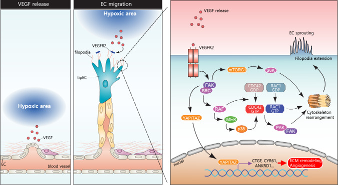
Angiogenesis, the process of new blood vessel formation, is a fundamental physiological process implicated in several pathological disorders. The vascular endothelial growth factors (VEGFs) and their receptors (VEGFRs) are crucial for angiogenesis and vasculogenesis. Among them, the tyrosine kinase receptor VEGFR-2 is primarily expressed in endothelial cells (ECs). These cells regulate various physiological responses, including differentiation, cell proliferation, migration, and survival, by binding to VEGF mitogens. Vascular Endothelial Growth Factor Receptor 2 (VEGFR-2) is a key regulator of this process, making it a prime target for therapeutic intervention. Several drugs targeting VEGFR-2 have been approved and are currently utilized to halt the pathological axis of VEGF-VEGFR. This review will focus on the recent developments in the molecular structure and function of VEGFR-2, the molecular mechanism of VEGFR-2 activation, and its downstream signaling pathway. It will also discuss therapies and experimental drugs approved to inhibit the function of VEGFR-2 and the resistance mechanism.

Keywords: Angiogenesis; Pathology; Resistance; Signaling; VEGF; VEGFR-2.


Conflict of interest statement

Declarations. Conflict of interest: The authors declare no competing financial interests.

Figures

Fig. 1
Fig. 1
The molecular illustration of human VEGFR-2 structure. The VEGFR-2 structure is comprised of a signal peptide, an extracellular domain containing seven immunoglobulin-like subdomains (IgD1-7), transmembrane-, juxtamembrane domain (JMD), a catalytic tyrosine kinase domain composed of ATP binding (ADB)-, kinase insert domain (KID) and phosphotransferase domain (PTD) as well as C-terminal domain. Among these domains, key tyrosine residues are phosphorylated upon binding of VEGF to VEGFR2, such as Tyr801 implicated in VEGFR2 activation. Tyr951, Tyr1504, and Tyr1059 located within KID and PTD, are involved in ECs proliferation, migration, and tube formation. The residues present in the c-terminal domain (Tyr1175 and Tyr1214) activate SHB-SCK-PI3 K, PLCγ and NCK signaling pathways which are essential for angiogenesis
Fig. 2
Fig. 2
The molecular mechanism regulated by VEGFR-2 mediated angiogenesis. Upon binding of VEGF mitogens to the VEGFR-2 receptor, it orchestrates dimerization and phosphorylation of (tyrosine Y1175) residue of the c-terminal domain that activates phospholipase C gamma (PLCγ1). PLCγ1 mediates the activation of phosphatidylinositol 4,5-bisphosphate (PIP2) that stimulates inositol trisphosphate (IP3) and diacylglycerol (DAG). Inositol trisphosphate (IP3) fosters calcium ions production whereas diacylglycerol (DAG) activates protein kinase C which in turn stimulates sphingosine kinase (SPK). SPK activates protein kinase D (PKD) and Raf protein. PKD interacts with histone deacetylases, heat shock protein 27 (HSP27), and cAMP-response-element binding protein (CREB) that aids in the process of angiogenesis whereas RAF modulates MEK and ERK to direct the endothelial cells'differentiation, and proliferation, and survival. The dimerization and phosphorylation event attracts the binding of TSAd that modulates a cascade of activation of different molecular factors such as SRC, PI3 K, PIP2, AKT, PKD1, and PKD2 that directs protein synthesis, metabolism, and cell growth and survival
Fig. 3
Fig. 3
Molecular Depiction of VEGFR-2 role in Vascular Permeability of Endothelial cells. The binding of VEGF mitogen on VEGFR-2 receptor facilitates dimerization and phosphorylation of Tyrosine 801 (Y801) activates protein kinase C (PKC) that stimulates PI3 K and AKT pathway that results in the production of nitric oxide species (NO species) which in turn activate several other molecular factors such as PLCγ, PIP2 and IP3 and Ca2+ ions. Activated VEGFR-2 stimulates the activity of SRC and tyrosine-protein kinase (YES) that phosphorylates guanine nucleotide exchange factor (GEF) (VAV2) that activates RAC1 which then turns on p21 activated kinase (PAK) activity. PAK protein modulates VE-cadherin phosphorylation, increasing the vascular permeability of endothelial cells in VEGFR-2-mediated angiogenesis
Fig. 4
Fig. 4
Molecular pathway controlling endothelial migration through VEGF signaling. As described in the first illustration, a hypoxic area releases VEGF mitogens that prepare the EC cells for migration. In the second illustration, EC cells proliferation and sprouting begin, which follows toward the hypoxic region where tip endothelial cells (tipECs) allow the binding of VEGF to their VEGFR2 receptor through filopodial protrusions. Within these protrusions, VEGF released from the hypoxic region binds to the VEGFR2 causing the stimulation of SRC and FAK that leads to the activation of different molecular factors such as (RAF, MEK, p38). These factors influence the activity of CDC42, RAC1, and FAK leading to cytoskeletal rearrangement resulting in EC sprouting and filopodial extension, and ultimately EC migration
Fig. 5
Fig. 5
Tumor growth and VEGF-mediated angiogenesis. In response to hypoxia, tumor cells release VEGF, which activate VEGFR2. This activation initiates angiogenesis, providing essential nutrients and oxygen to support tumor growth. In addition to promoting angiogenesis, VEGFR2 activation increases blood vessel permeability, facilitating tumor metastasis
Fig. 6
Fig. 6
Diabetic Retinopathy (DR) and VEGF-Mediated Angiogenesis: In the healthy state (left), the retinal blood vessels are structurally stable, ensuring adequate perfusion and minimal disruption. By contrast, diabetic retinopathy (right) is marked by the regression of preexisting vessels, resulting in diminished blood flow and subsequent hypoxia. This hypoxic environment upregulates the expression of vascular endothelial growth factor (VEGF), which binds to VEGF receptor 2 (VEGFR2), promoting aberrant neovascularization
Fig. 7
Fig. 7
Age-related macular degeneration (AMD) and VEGF-mediated angiogenesis. In the normal macula (left), photoreceptors, the retinal pigment epithelium (RPE), and the choroid are well-organized, supporting stable visual function. By contrast, AMD is characterized by the accumulation of drusen beneath the RPE (right), which disrupts the extracellular matrix (ECM) and elevates matrix metalloproteinase (MMP) activity. The ensuing hypoxia enhances the production of vascular endothelial growth factor (VEGF), thereby activating VEGF receptor-dependent angiogenesis. These newly formed vessels are structurally fragile, leading to leakage and hemorrhage that ultimately compromise macular integrity and central vision

References

    1. Abdullah SE, Perez-Soler R (2012) Mechanisms of resistance to vascular endothelial growth factor blockade. Cancer 118:3455–3467. 10.1002/cncr.26540 - PubMed
    1. Abhinand CS, Raju R, Soumya SJ, Arya PS, Sudhakaran PR (2016) VEGF-A/VEGFR2 signaling network in endothelial cells relevant to angiogenesis. J Cell Commun Signal 10:347–354. 10.1007/s12079-016-0352-8 - PMC - PubMed
    1. Abramsson A, Lindblom P, Betsholtz C (2003) Endothelial and nonendothelial sources of PDGF-B regulate pericyte recruitment and influence vascular pattern formation in tumors. J Clin Invest 112:1142–1151. 10.1172/JCI18549 - PMC - PubMed
    1. Ahmad A, Nawaz MI (2022) Molecular mechanism of VEGF and its role in pathological angiogenesis. J Cell Biochem 123:1938–1965. 10.1002/jcb.30344 - PubMed
    1. Alitalo K, Tammela T, Petrova TV (2005) Lymphangiogenesis in development and human disease. Nature 438:946–953. 10.1038/nature04480 - PubMed

MeSH terms

Substances

LinkOut - more resources