. 2025 Jun;32(6):1939-1952.
doi: 10.1007/s43032-025-01870-2. Epub 2025 May 8.

Mechanisms of LncRNA FTX in Regulating Islet Function of Pregnant Mice Born With Low-Protein Diet-Induced Intrauterine Growth Retardation

Affiliations

Mechanisms of LncRNA FTX in Regulating Islet Function of Pregnant Mice Born With Low-Protein Diet-Induced Intrauterine Growth Retardation

Li Wang et al. Reprod Sci. 2025 Jun.

Abstract

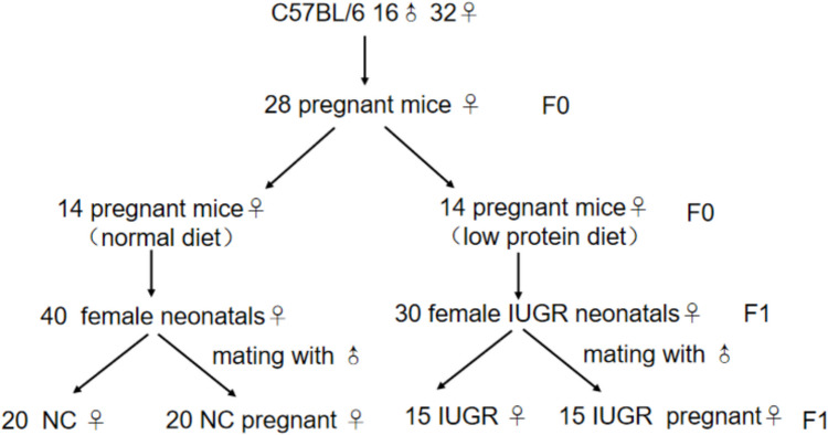
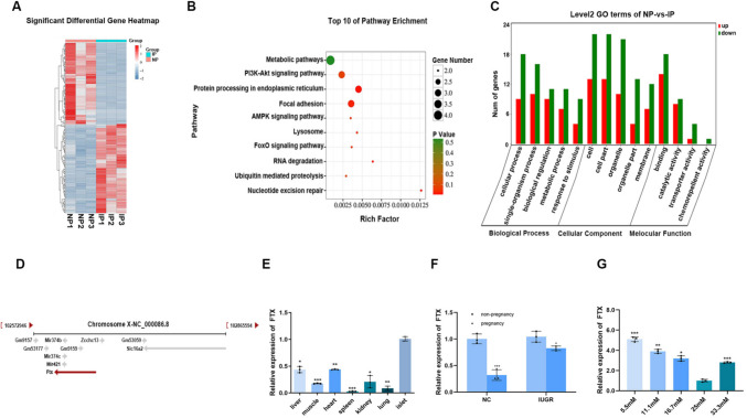
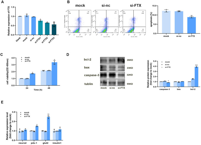
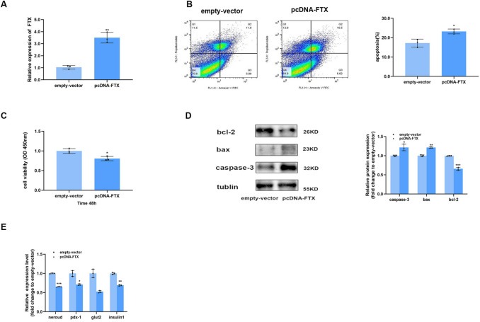
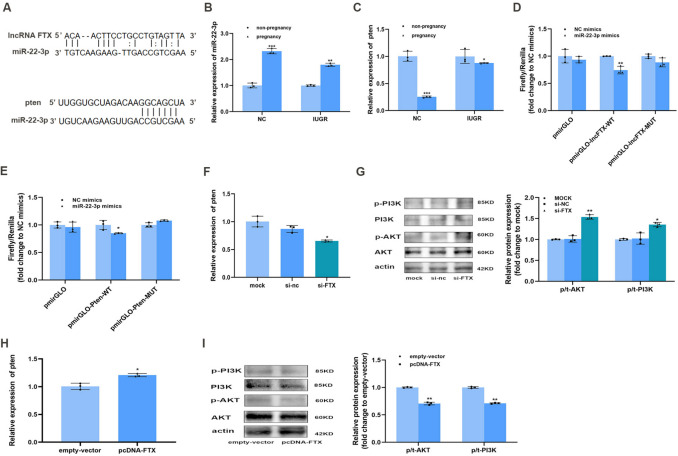
Glucose metabolism during pregnancy in adult females born with intrauterine growth restriction (IUGR) remains inadequately understood. This study aims to investigate how LncRNA FTX regulates islet function during pregnancy in F1 female mice born with IUGR (F1 IUGR pregnant mice). A pregnant mouse model was established using F1 female mice born with IUGR (F1 IUGR pregnant mouse model). Intraperitoneal glucose tolerance test (IPGTT), immunohistochemistry (IHC) staining, quantitative real-time PCR (qPCR) were performed in both F1 IUGR and normal mice during pregnancy and non-pregnancy periods. RNA-sequencing was conducted on islets from F1 IUGR and normal pregnant mice. Insulin-related gene expression analysis, cell proliferation, and apoptosis assessment were performed in TC6 cells following FTX knockdown or overexpression. A luciferase reporter assay was conducted to validate the molecular interactions. F1 IUGR pregnant mice exhibited a smaller increase in insulin-staining area and lower upregulation of insulin-related gene expression levels compared to normal pregnant mice. There were 1,007 differentially expressed lncRNAs between F1 IUGR and normal pregnant islets; among these, FTX was down-regulated during pregnancy, although its downregulation in F1 IUGR pregnant mice was less pronounced than in normal pregnant mice. FTX was closely related to cell proliferation activity, apoptosis, insulin-related transcription factor expression. The pten/PI3K/AKT pathway was also regulated by FTX. Luciferase reporter assay confirmed FTX acted as a competing endogenous RNA (CeRNA) to target pten by sponging miR-22-3p. LncRNA FTX regulates islet function during pregnancy in F1 mice born with IUGR via the miR-22-3p/pten axis.

Keywords: Diabetes; IUGR (intrauterine growth retardation); Islet function; LncRNA FTX; Low protein diet; Pregnancy.


Conflict of interest statement

Declarations. Conflict of interest: The authors declare that they have no competing interests.

Figures

Fig. 1
Fig. 1
Model of normal and IUGR pregnant mice
Fig. 2
Fig. 2
Islet proliferation occurred during pregnancy, but weaker in IUGR mice. (A) The appearance and birthweight of normal control and IUGR mice. (B) The weight of normal control and IUGR mice during the growth process. (C) Intraperitoneal glucose tolerance test of normal control and IUGR mice at 4 weeks. (D) The weight of NC, NC pregnancy, IUGR, IUGR pregnancy (gestational day12). (E) Blood glucose and (F) serum insulin levels of NC, NC pregnancy, IUGR, IUGR pregnancy (gestational day12). (G) Pancreas weight of NC, NC pregnancy, IUGR, IUGR pregnancy (gestational day15). (H) Pancreatic immunohistochemistry for insulin of NC, NC pregnancy, IUGR, IUGR pregnancy (gestational day15), Scale-bar = 50 µm. (I) Expression levels of genes associated with insulin synthesis and secretion of NC, NC pregnancy, IUGR, IUGR pregnancy (gestational day15), compaired with non-pregnant. NC, Normal Control mice; NP, Normal Control pregnant mice; IUGR, Intrauterine Growth Restriction; IP, Intrauterine Growth Restriction pregnant mice. *p < 0.05, **p < 0.01, ***p < 0.001, ****p < 0.0001
Fig. 3
Fig. 3
LncRNA FTX was insufficiently down-regulated in IUGR pregnant mice. The (A) heatmap of differentially expressed lncRNAs between IUGR pregnant mice and normal pregnant mice. The (B) KEGG enrichment and (C) GO analysis on target mRNA of differentially expressed LncRNA. (D) The localization of lncRNA FTX in chromosome X. (E) Expression levels of lncRNA FTX in different tissues of 8-week-old normal control mice, compared with islets. (F) LncRNA FTX expression level in islets of NC, NC pregnancy, IUGR, IUGR pregnancy, compared with non-pregnancy. (G) The expression level of LncRNA FTX in TC6 cells under different glucose concentrations, compared with 25 mM. NC, Normal Control mice; NP, Normal Control pregnant mice; IUGR, Intrauterine Growth Restriction; IP, Intrauterine Growth Restriction pregnant mice. *p < 0.05, **p < 0.01, ***p < 0.001
Fig. 4
Fig. 4
Down-regulation of LncRNA FTX promoted cell proliferation and the expression of genes related to insulin synthesis and secretion, and inhibited cell apoptosis. (A) Interference efficiency of three LncRNA FTX interfering sequences in TC6 cells, compared with the control group. (B) The representative graph of apoptosis ratio of TC6 cells after downregulation of lncRNA FTX, compared with the mock group. (C) Proliferation activity of TC6 cells after FTX intervention 24 h and 48 h later, compared with the mock group. (D) Expression of apoptosis-related proteins in TC6 cells after downregulation of lncRNA FTX, compared with the mock group. The expression of insulin-related genes after lncRNA FTX was down-regulated in TC6 cells at (E) nucleic acid level, compared with the mock group. *p < 0.05, **p < 0.01, ***p < 0.001
Fig. 5
Fig. 5
Overexpression of lncRNA FTX inhibited cell proliferation and the expression of insulin-related genes, and promoted apoptosis. (A) The overexpression efficiency of pcDNA-FTX in TC6 cells, compared with the empty-vector group. (B) The representative graph of apoptosis ratio of TC6 cells after the overexpression of lncRNA FTX, compared with the empty-vecor group. (C) The proliferation activity of TC6 cells after FTX was over-expressed, compared with the empty-vecor group. (D) The expression of apoptosis-related protein after the overexpression of lncRNA FTX in TC6 cells, compared with the empty-vecor group. The expression of insulin-related genes at (E) nucleic acid level after the overexpression of FTX in TC6 cells, compared with the empty- vector group. *p < 0.05, **p < 0.01, ***p < 0.001
Fig. 6
Fig. 6
FTX was a sponge of miR- 22 - 3p which regulated pten through PI3 K/AKT signaling pathway. (A) StarBase V2.0 predicted that miR- 22 - 3p was a target of lncRNA FTX and pten was a target of miR- 22 - 3p. The levels of miR- 22 − 3p (B) and pten (C) in NC, NC pregnancy, IUGR, IUGR pregnancy, compared with non-pregnancy. (D) Dual luciferase report assay in WT- lncRNA FTX or MUT-lncRNA FTX co-transfected with miR-NC or miR- 22 - 3p mimics. (E) Dual luciferase assay in WT-pten or MUT-pten co-transfected with miR-NC or miR- 22 - 3p mimics, compared with NC mimics.The mRNA expression level of (F) pten and the protein expression levels of (G) p-PI3 K, PI3 K, p-AKT and AKT after down-regulation of lncRNA FTX in TC6 cells, compared with the mock group. The mRNA expression level of (H) pten and the protein expression levels of (I) p-PI3 K, PI3 K, p-AKT and AKT after the overexpression of lncRNA FTX in TC6 cells, compared with the empty-vector group. NC, Normal Control mice; IUGR, Intrauterine Growth Restriction.*p < 0.05, **p < 0.01, *p < 0.001

References

    1. Hryciw DH. Early life nutrition and the development of offspring metabolic health. Int J Mol Sci. Jul 22 2022;23(15). 10.3390/ijms23158096 - PMC - PubMed
    1. He X, Xie Z, Dong Q, Chen P, Li W, Wang T. Dynamic p53 protein expression and phosphorylation in the kidneys of rats that experienced intrauterine growth restriction. Ren Fail. 2015;37(5):896–902. 10.3109/0886022x.2015.1015428. - PubMed
    1. Boehmer BH, Limesand SW, Rozance PJ. The impact of IUGR on pancreatic islet development and β-cell function. J Endocrinol. 2017;235(2):R63-r76. 10.1530/joe-17-0076. - PMC - PubMed
    1. Golden TN, Simmons RA. Immune dysfunction in developmental programming of type 2 diabetes mellitus. Nat Rev Endocrinol. 2021;17(4):235–45. 10.1038/s41574-020-00464-z. - PMC - PubMed
    1. Rashid CS, Bansal A, Simmons RA. Oxidative stress, intrauterine growth restriction, and developmental programming of type 2 diabetes. Physiology (Bethesda). 2018;33(5):348–59. 10.1152/physiol.00023.2018. - PMC - PubMed

LinkOut - more resources