[Preprint]. 2025 May 2:2025.05.01.25326605.
doi: 10.1101/2025.05.01.25326605.

Charcot-Marie-Tooth disease type 1E: Clinical Natural History and Molecular Impact of PMP22 Variants

Affiliations

Charcot-Marie-Tooth disease type 1E: Clinical Natural History and Molecular Impact of PMP22 Variants

Kailee S Ward et al. medRxiv. .

Update in

  • Charcot-Marie-Tooth disease type 1E: clinical natural history and molecular impact of PMP22 variants.
    Ward KS, Ptak CP, Pashkova N, Grider T, Peterson TA, Pareyson D, Pisciotta C, Saveri P, Moroni I, Laura M, Burns J, Menezes MP, Cornett K, Finkel R, Mukherjee-Clavin B, Sumner CJ, Greene M, Abdul Hamid O, Herrmann D, Sadjadi R, Walk D, Züchner S, Reilly MM, Scherer SS; Inherited Neuropathy Consortium; Piper RC, Shy ME. Ward KS, et al. Brain. 2025 Jun 9:awaf219. doi: 10.1093/brain/awaf219. Online ahead of print. Brain. 2025. PMID: 40488457

Abstract

Charcot-Marie-Tooth disease type 1E (CMT1E) is a rare, autosomal dominant peripheral neuropathy caused by missense variants, deletions, and truncations within the peripheral myelin protein-22 (PMP22) gene. CMT1E phenotypes vary depending on the specific variant, ranging from mild to severe, and there is little natural history and phenotypic progression data on individuals with CMT1E. Patients with CMT1E were evaluated during initial and follow-up visits at sites within the Inherited Neuropathy Consortium. Clinical characteristics were obtained from history, neurological exams, and nerve conduction studies. Clinical outcome measures were used to quantify baseline and longitudinal changes, including the Rasch-modified CMT Examination Score version 2 (CMTESv2-R) and the CMT Pediatric Scale (CMTPedS). The trafficking of PMP22 variants in transfected cells was correlated to disease severity. Twenty-four, presumed disease-causing PMP22 variants were identified in 50 individuals from 35 families, including 19 missense variants, three in-frame deletions, and two truncations. Twenty-nine patients presented with delayed walking during childhood. At their baseline evaluation, the mean CMTESv2-R in 46 patients was 16 ± 7.72 (out of 32), and the mean CMTPedS from 17 patients was 28 ± 6.35 (out of 44). Six individuals presented with hearing loss, eleven with scoliosis, three with hip dysplasia, and one with both scoliosis and hip dysplasia. Twenty variants were localized within in transmembrane domains; 31 of 35 individuals with these variants had moderate to severe phenotypes. Three variants were found in the extracellular domain and were associated with milder phenotypes. Reduced expression of PMP22 at the cell surface, and the location of missense variants within in the transmembrane domain correlated with disease severity. Pathogenic PMP22 variants located within the transmembrane regions usually cause a moderate to severe clinical phenotype, beginning in early childhood, and have impaired trafficking to the plasma membrane.

Keywords: Charcot-Marie-Tooth type 1E (CMT1E); PMP22; natural history.


Conflict of interest statement

Competing interests The authors declare no competing interests.

Figures

Figure 1.
Figure 1.. Genotype and structure descriptions of CMT1E patient PMP22 gene variants included in this study.
(A) The protein schematic depicts the full sequence and transmembrane topology of human PMP22 with amino acid positions of single point mutations and deletions colored. Patient variants are listed by protein region. (B) The predicted 3D folded structure of PMP22 illustrates the four transmembrane helical bundle. Locations of patient variants are denoted by their alpha carbon only (cyan) and labelled.
Figure 2.
Figure 2.. Phenotype evaluation of patients with PMP22 sequence variants using clinical outcome measures.
(A) Rasch modified CMT exam score version 2 (CMTESv2-R) measuring PMP22 sequence variant disease severity for individual patients are plotted by age, labelled by patient variant, and colored by protein region. (B) For patients under age 20, the CMT Pediatric Scale (CMTPedS) was performed to measure the patient’s muscle strength, balance and coordination, and upper and lower extremity function. These scores are plotted by age at evaluation, labeled by patient variant, and colored by protein region. (C) The distribution of PMP22 sequence variants across transmembrane helices and extracellular/intracellular regions for patients who completed the CMTESv2-R. (D) The distribution of PMP22 sequence variants across transmembrane helices for patients who completed the CMTPedS.
Figure 3.
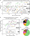
Figure 3.. Longitudinal progression of CMT1E patient symptoms.
(A) Rasch modified CMT exam score version 2 (CMTESv2-R) measuring CMT1E disease severity for individual patients are plotted by age and colored by achievement of walking milestone (variants are labeled in Fig. 2A). Whether a patient met, exhibited a delay in, or never met their walking milestones is inherently tied to the severity of CMT1E symptom progression. Dashed curves follow the age-dependent trajectory of CMTESv2-R progression by a defined equation. Curves are labeled 0 through 10 to indicate their age-adjusted CMTESv2-R value with 0 signifying late CMTESv2-R progression and 10 signifying early CMTESv2-R progression. Fitting of individual patient CMTES-R to the same equation was used to obtain a patient’s age-adjusted CMTESv2-R value (Supplementary Fig. 4). Labels denoting the variant for individual patients can be found in the longitudinal CMT1E patient data plotted in Fig. 2A. (B) The standard response mean was calculated for CMTESv2-R and grouped by the patient’s follow-up age. For each CMT1E patient, (C) CMTESv2-R baseline values or (D) age-adjusted CMTESv2-R values were grouped by walking milestones (Met, Delayed, and Never Met). There is no significant difference in CMTESv2-R baseline values between walking milestone groups (p>0.05). In contrast, the age-adjusted CMTESv2-R values are significantly different between walking milestone groups (***=p<0.0002; **=p<0.002; *=p<0.05).
Figure 4.
Figure 4.. Cell surface trafficking of PMP22 variants.
(A) Confocal microscope images of HEK293 cells co-transfected with PMP22-GFP (green) and mTAGBFP-KDEL (depicted in false color red) as marker of Endoplasmic Reticulum (ER). Green arrows highlight PMP22-GFP localized to the plasma membrane. Large black openings in the cells are nucleus. Wild type and S131C PMP22-GFP localized in ER and plasma membrane, while S72L variant PMP22-GFP mostly found in ER. Scale bar: 10 μm. (B) PMP22-mycexo expression vector scheme (top panel). The plasmid also expresses a nuclear-localized blue fluorescent protein via an internal ribosome entry site (IRES) within the same mRNA. This allows the expression level of PMP22 to be normalized via the level of BFP. The PMP22 expression plasmids were stably integrated via piggyBac transposase and selection with puromycin. Low panel - confocal microscope image of a stable PMP22-mycexo HEK293 cell line with immunofluorescent cell surface labeling as used for flow cytometry experiments. Wild type PMP22-mycexo can be found localized to the cell surface (in red) but not G150D mutant protein. (C) Cell surface localization of PMP22-mycexo variants analyzed after Flow cytometry surface immunostaining of HEK293 stable cell lines. Graph bars represent geometric mean of Alexa 568 fluorescence intensity for tested PMP22 variants normalized to the wild type. (D) PMP22-mycexo protein expression in the cell lines used for Flow cytometry experiments was tested by Western blotting with anti-myc antibodies. Equal amounts of cell lysates were loaded onto SDS-PAGE. Anti-b-actin antibody were used as a loading control. Full variant data values are available in Supplementary Table 2.
Figure 5:
Figure 5:. PMP22-mycexo variants can form a complex with PMP22-GFP.
(A) HEK293 stable cell lines with PMP22-mycexo variants were transfected with wild type PMP22-GFP. After cell lysis, the proteins were extracted and co-immunoprecipitated using nanobody GFP beads (depicted by the cartoon). Eluted with SDS-sample buffer proteins were immunoblotted with anti-myc antibody to detect bound PMP22-mycexo and with anti-GFP antibody to monitor recovered PMP22-GFP. A 5% equivalent of the input lysate was included in the immunoblots. (B) Immunoblotting of WT, unsolved PMP22 variant (p.T118M), negative control variant (p.A135T), and a patient variant (p.G150C). (C) Immunoblotting of PMP22 sequence variants in this cohort and TrJ variant (p.G150D).
Figure 6.
Figure 6.. Correlations between CMT1E symptom progression, PMP22 trafficking, and variant position relative to the membrane.
(A) Age-adjusted CMTESv2-R values, (B) relative expression-adjusted PMP22 cell surface localization, and (C) predicted depth in the membrane bilayer are mapped onto the PMP22 structure to depict the visual correlation between these variant parameters in 3-dimensional space (variant positions are labeled in Fig. 1B). The membrane depth of each variant position was derived from the membrane-embedded position of the PMP22 AlphaFold model as predicted by the PPM method. For variants, plots of the linear correlation for (D) CMT1E symptom progression and PMP22 cell surface localization, (E) CMT1E symptom progression and predicted depth in the membrane bilayer, and (F) PMP22 cell surface localization and predicted depth in the membrane bilayer. For each correlation plot, R2 are indicated for the linear fits (p<0.01 for each) and only variants with |standardized residuals| > 0.5 are labeled. Full variant data values are available in Supplementary Table 2.

Similar articles

  • Charcot-Marie-Tooth disease type 1E: clinical natural history and molecular impact of PMP22 variants.
    Ward KS, Ptak CP, Pashkova N, Grider T, Peterson TA, Pareyson D, Pisciotta C, Saveri P, Moroni I, Laura M, Burns J, Menezes MP, Cornett K, Finkel R, Mukherjee-Clavin B, Sumner CJ, Greene M, Abdul Hamid O, Herrmann D, Sadjadi R, Walk D, Züchner S, Reilly MM, Scherer SS; Inherited Neuropathy Consortium; Piper RC, Shy ME. Ward KS, et al. Brain. 2025 Jun 9:awaf219. doi: 10.1093/brain/awaf219. Online ahead of print. Brain. 2025. PMID: 40488457
  • Autosomal Dominant TRPV4 Disorders.
    McCray BA, Schindler A, Hoover-Fong JE, Sumner CJ. McCray BA, et al. 2014 May 15 [updated 2020 Sep 17]. In: Adam MP, Feldman J, Mirzaa GM, Pagon RA, Wallace SE, Amemiya A, editors. GeneReviews® [Internet]. Seattle (WA): University of Washington, Seattle; 1993–2025. 2014 May 15 [updated 2020 Sep 17]. In: Adam MP, Feldman J, Mirzaa GM, Pagon RA, Wallace SE, Amemiya A, editors. GeneReviews® [Internet]. Seattle (WA): University of Washington, Seattle; 1993–2025. PMID: 24830047 Free Books & Documents. Review.
  • Prescription of Controlled Substances: Benefits and Risks.
    Preuss CV, Kalava A, King KC. Preuss CV, et al. 2025 Jul 6. In: StatPearls [Internet]. Treasure Island (FL): StatPearls Publishing; 2025 Jan–. 2025 Jul 6. In: StatPearls [Internet]. Treasure Island (FL): StatPearls Publishing; 2025 Jan–. PMID: 30726003 Free Books & Documents.
  • Clinical Characteristics of Charcot-Marie-Tooth Disease Type 4J.
    Sadjadi R, Picher-Martel V, Morrow JM, Thedens D, DiCamillo PA, McCray BA, Pareyson D, Herrmann DN, Reilly MM, Li J, Castro D, Shy ME; Inherited Neuropathy Consortium. Sadjadi R, et al. Neurology. 2024 Sep 10;103(5):e209763. doi: 10.1212/WNL.0000000000209763. Epub 2024 Aug 12. Neurology. 2024. PMID: 39133880
  • The clinical effectiveness and cost-effectiveness of enzyme replacement therapy for Gaucher's disease: a systematic review.
    Connock M, Burls A, Frew E, Fry-Smith A, Juarez-Garcia A, McCabe C, Wailoo A, Abrams K, Cooper N, Sutton A, O'Hagan A, Moore D. Connock M, et al. Health Technol Assess. 2006 Jul;10(24):iii-iv, ix-136. doi: 10.3310/hta10240. Health Technol Assess. 2006. PMID: 16796930

References

    1. Skre H. Genetic and clinical aspects of Charcot-Marie-Tooth’s disease. Clinical genetics 1974;6:98–118. - PubMed
    1. Lupski JR, de Oca-Luna RM, Slaugenhaupt S, et al. DNA duplication associated with Charcot-Marie-Tooth disease type 1A. Cell 1991;66:219–232. - PubMed
    1. Raeymaekers P, Timmerman V, Nelis E, et al. Duplication in chromosome 17p11.2 in Charcot-Marie-Tooth neuropathy type 1a (CMT 1a). The HMSN Collaborative Research Group. Neuromuscular disorders : NMD 1991;1:93–97. - PubMed
    1. Chance PF, Alderson MK, Leppig KA, et al. DNA deletion associated with hereditary neuropathy with liability to pressure palsies. Cell 1993;72:143–151. - PubMed
    1. Li J, Parker B, Martyn C, Natarajan C, Guo J. The PMP22 gene and its related diseases. Mol Neurobiol 2013;47:673–698. - PMC - PubMed

Publication types

LinkOut - more resources