

Morphometric analysis of the abducens nerve in the petroclival region
- PMID: 40343054
- PMCID: PMC12058666
- DOI: 10.3389/fsurg.2025.1574047
Morphometric analysis of the abducens nerve in the petroclival region
Abstract
Background: The abducens nerve (AN), our sixth cranial nerve, is responsible for the innervation of the lateral rectus muscle of the eye. The abducens nerve is a vulnerable structure at the skull base with its long intracranial course and complex topographic relationships. The AN anatomy in the petroclival region, where the nerve passes from the posterior to the middle cranial fossa, is of great interest for neurosurgical procedures. Despite detailed studies of its anatomy from the past 150 years, there is a need for more recent data on macroscopical and microscopical aspects of the AN in well defined populations.
Methods: We investigated macroscopical variations and the number of nerve fibers of the AN in the petroclival region in German body donors.
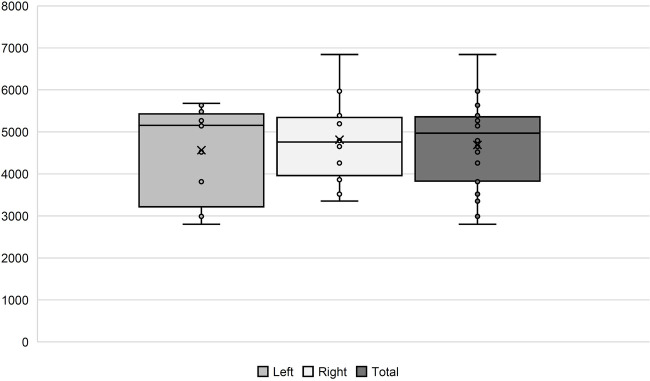
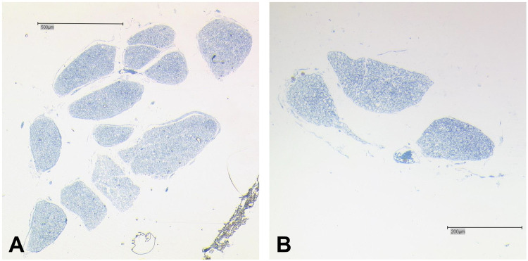
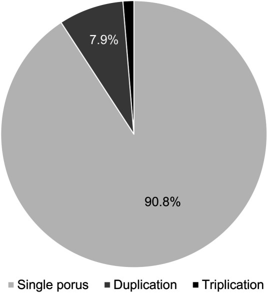
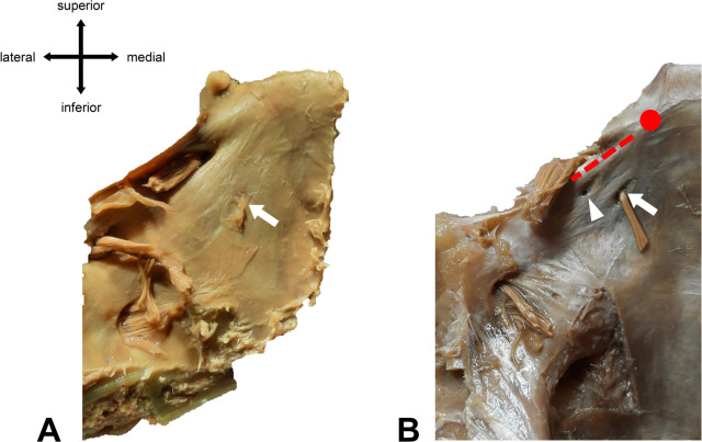
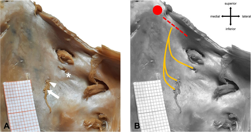
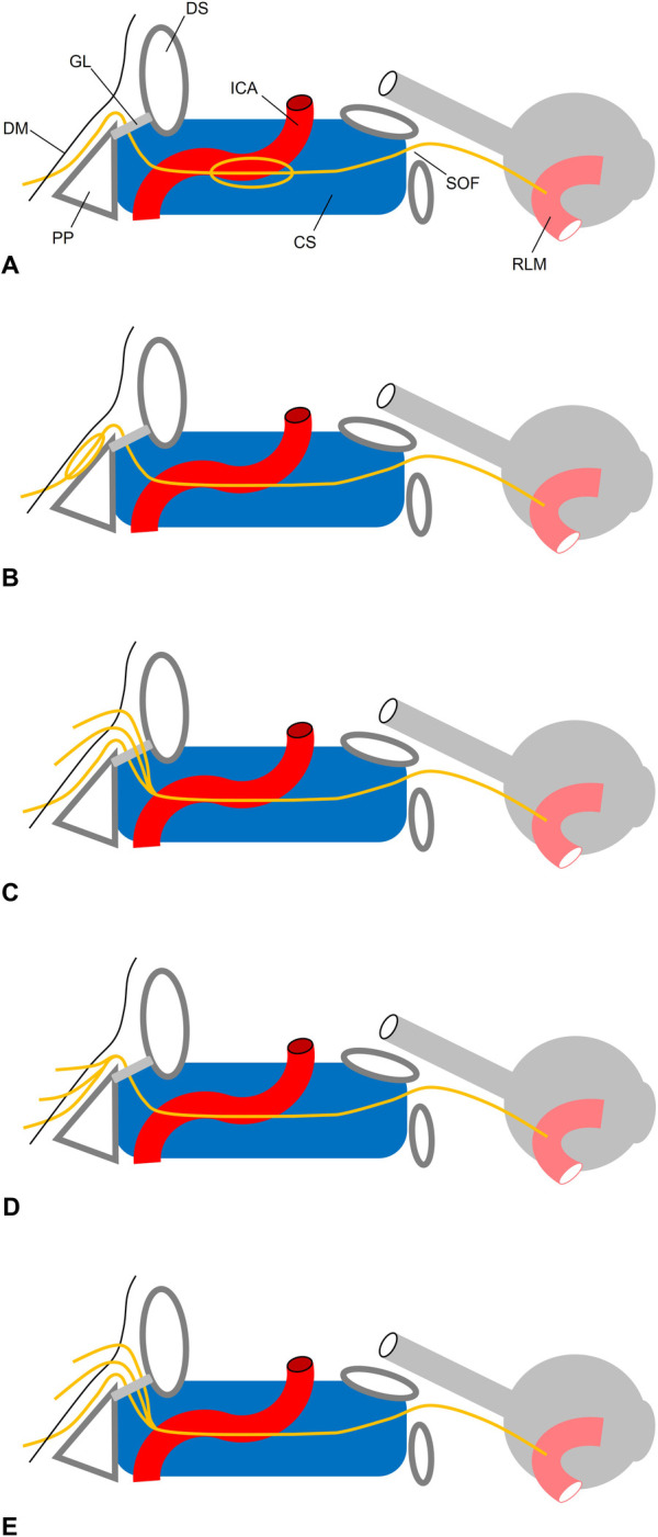
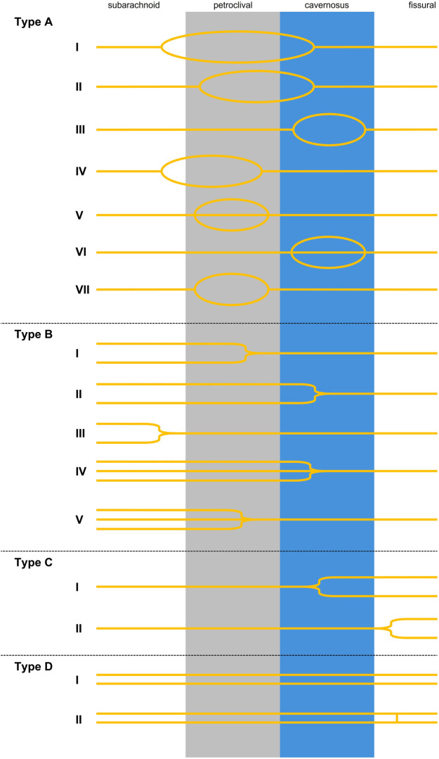
Results: In our histological samples (n = 24) we counted 4688 (+/-1,041) nerve fibers per AN. There was no correlation between sex, age and body side regarding the number of nerve fibers. In our macroscopic examination (n = 76 skull base sides), we found six duplications (four left-sided, two right-sided; 7.9%) and one triplication (right-sided; 1.3%) of the AN in the petroclival region. The AN triplication was further examined: Three nerve bundles pierce the dura mater separately and united before passing under the petrosphenoidal ligament (of Gruber).
Conclusion: Variations of the AN in the petroclival region are not a rare phenomenon but occur very frequently. Consequently, we have developed a new classification system for AN variations. This knowledge might help neurosurgeons, as it prepares them to be aware of such variations and adapt their surgical approaches accordingly.
Keywords: anatomical variation; cranial nerve; dura mater; neurosurgery; skull base.
© 2025 Bach, Schmeisser and Schumann.
Conflict of interest statement
The author(s) declared that they were an editorial board member of Frontiers, at the time of submission. This had no impact on the peer review process and the final decision. The authors declare that the research was conducted in the absence of any commercial or financial relationships that could be construed as a potential conflict of interest.
Figures
Similar articles
-
Bilateral duplication of the abducens nerve - case study.Folia Med Cracov. 2019;59(4):13-20. doi: 10.24425/fmc.2019.131376. Folia Med Cracov. 2019. PMID: 31904746
-
Duplication of the abducens nerve at the petroclival region: an anatomic study.Neurosurgery. 2003 Mar;52(3):645-52; discussion 651-2. doi: 10.1227/01.neu.0000048186.18741.3c. Neurosurgery. 2003. PMID: 12590690
-
Meningovenous structures of the petroclival region: clinical importance for surgery and intravascular surgery.Neurosurgery. 2002 Apr;50(4):829-36; discussion 836-7. doi: 10.1097/00006123-200204000-00027. Neurosurgery. 2002. PMID: 11904035
-
Surgical anatomy of the petrous apex and petroclival region.Adv Tech Stand Neurosurg. 2007;32:91-146. doi: 10.1007/978-3-211-47423-5_5. Adv Tech Stand Neurosurg. 2007. PMID: 17907476 Review.
-
Bilateral Extensive Ossification of the Posterior Petroclinoid Ligament: An Anatomical Case Report and Literature Review.J Neurol Surg A Cent Eur Neurosurg. 2019 Mar;80(2):122-126. doi: 10.1055/s-0038-1666782. Epub 2018 Jul 30. J Neurol Surg A Cent Eur Neurosurg. 2019. PMID: 30060260 Review.
References
-
- Limmroth V. Erkrankungen der hirnnerven. In: Limmroth V, Diener HC, editors. Neurologie für Praktiker. Darmstadt: Steinkopff-Verlag; (2006). p. 401–17. 10.1007/3-7985-1621-9_14 - DOI
LinkOut - more resources
Full Text Sources
Miscellaneous