. 2025 Apr 8:32:101743.
doi: 10.1016/j.mtbio.2025.101743. eCollection 2025 Jun.

Polymer oxidation: A strategy for the controlled degradation of injectable cryogels

Affiliations

Polymer oxidation: A strategy for the controlled degradation of injectable cryogels

Alexandra Nukovic et al. Mater Today Bio. .

Abstract

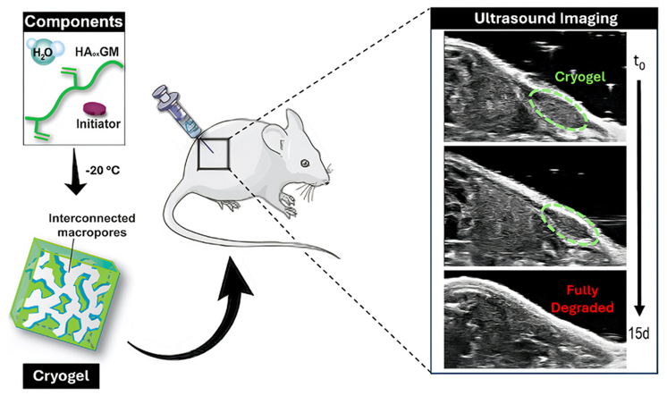
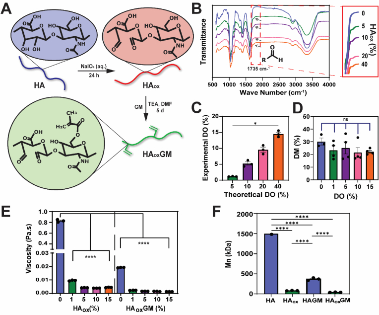
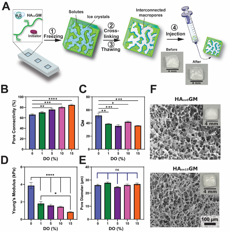
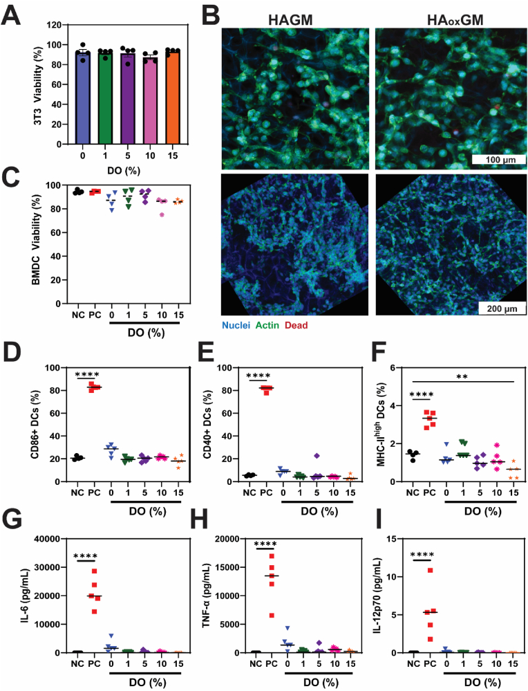
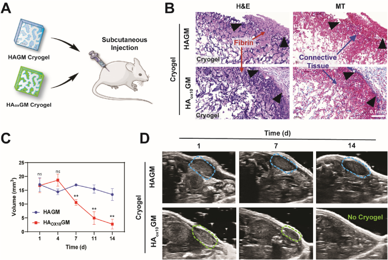
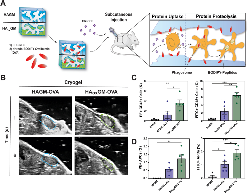
Cryogels, an advanced subclass of hydrogels, are widely used in biomedical applications such as tissue engineering, drug delivery, and immunotherapy. Biopolymers, like hyaluronic acid (HA), are key building blocks for cryogel fabrication due to their intrinsic biological properties, biocompatibility, and biodegradability. HA undergoes biodegradation through hydrolysis, enzymatic degradation, and oxidation, but becomes less susceptible to degradation once chemically modified. This modification is necessary for producing HA-based cryogels with unique properties, including an open macroporous network, mechanical resilience, shape memory, and syringe injectability. Endowing cryogels with resorbable features is essential for meeting regulatory requirements and improving treatment outcomes. To this end, HA was oxidized with sodium periodate (HAox) and chemically modified with glycidyl methacrylate (HAoxGM) to create HAoxGM cryogels with controlled degradation. Oxidation of HA increased the susceptibility of the polymer backbone to breakdown through various mechanisms, including oxidative cleavage and alkaline hydrolysis. Compared to their poorly degradable counterparts, HAoxGM cryogels retained their advantageous properties despite reduced compressive strength. HAoxGM cryogels were highly cytocompatible, biocompatible, and tunable in degradation. When injected subcutaneously into mice, the HAoxGM cryogels were biocompatible and resorbed within two weeks. To validate the beneficial effect of controlled biodegradation in a relevant in vivo setting, we demonstrated that the degradation of HAoxGM cryogels accelerates ovalbumin release and enhances its uptake and response by immune cells in mice. This versatile oxidation strategy can be applied to a wide range of polymers, allowing better control over cryogel degradation, and advancing their potential for biomedical applications and clinical translation.

Keywords: Biocompatibility; Cryogel; Degradation; Hydrolysis; Oxidation.


Conflict of interest statement

The authors declare that they have no known competing financial interests or personal relationships that could have appeared to influence the work reported in this paper.

Figures

Image 1
Graphical abstract
Fig. 1
Fig. 1
Synthesis and characterization of HAoxGM. A) Overview of the chemical synthesis of HAoxGM. HA is oxidized with NaIO4 and subsequently methacrylated with GM. B) ATR-FTIR of HAox with varying DOs (DO = 0–40 %). The characteristic aldehyde peaks are observed at 1725 cm−1 (shown in the inset). C) Comparison of experimental and theoretical DO for HAox. D) DM of HAoxGM quantified using 1H NMR spectroscopy. E) Viscosity measurements of HAox and HAoxGM in diH2O (0.5 % w/v) at shear rate of 10 s−1. F) Number average molecular weight (Mn) of HA, HAox (DO = 1 %), HAGM, and HAoxGM (DO = 1 %). Data are presented as mean ± SEM (n = 3–4). Statistical analysis was performed using one-way ANOVA and Tukey's post hoc test: ns (not significant) ≥ 0.05, ∗p < 0.05, ∗∗∗∗p < 0.0001.
Fig. 2
Fig. 2
Fabrication and physical properties of HAoxGM cryogels. A) Schematic depicting the process of cryogelation and syringe injectability: HAoxGM is first dissolved in diH2O and mixed with a redox initiator system, frozen at −20 °C in a precooled mold for 20 h, and then thawed at RT, resulting in a macroporous and interconnected network suitable for syringe injection. B–E) Comparative analysis of pore connectivity, swelling ratio (QM), Young's modulus, and pore diameter of HAoxGM (DO = 1–15 %) cryogels vs. unoxidized HAGM cryogels. F) Scanning electron microscopy images of HAGM and HAoxGM (DO = 15 %) cryogels. Data are presented as mean ± SEM (n = 3–4). Statistical analysis was performed using one-way ANOVA and Tukey's post hoc test: ns ≥ 0.05, ∗p < 0.05, ∗∗p < 0.01, ∗∗∗p < 0.001, and ∗∗∗∗p < 0.0001.
Fig. 3
Fig. 3
HAoxGM cryogels exhibit enhanced degradation across various conditions. A) Mass loss rate (%/d) and corresponding degradation kinetics of HAoxGM (DO = 0–15 %) cryogels incubated in PBS (pH 7.4) at 37 °C across various time points (0–63 d). B) Mass loss rate (%/d) and corresponding degradation kinetics of HAoxGM (i.e., DO = 5 %) cryogels incubated in different buffer solutions (pH 3–8.5) at 37 °C across various time points (0–24 d). C) Visual representation of the degradation of HAGM (non-oxidized: DO = 0 %) and HAoxGM (DO = 15 %) cryogels incubated in a sodium bicarbonate solution (pH 8.5) at 37 °C after 28 d. D) Degradation kinetics of HAoxGM (DO = 0–15 %) cryogels incubated in a buffer solution (pH 5.35) supplemented with hyaluronidase (HYAL) at 37 °C across various time points (0–14 d). Data are presented as mean ± SEM (n = 3–5). Statistical analysis was performed using one-way ANOVA: ns ≥ 0.05, ∗∗p < 0.01, ∗∗∗p < 0.001, and ∗∗∗∗p < 0.0001.
Fig. 4
Fig. 4
HAoxGM cryogels are cytocompatible and do not trigger DC activation. A) Cell viability (%) of NIH 3T3 cells after 24 h of culture within HAoxGM (DO = 0–15 %) cryogels. B) Confocal microscopy images of NIH 3T3 cells within HAGM and HAoxGM (DO 15 %), stained with DAPI (nuclear stain), cytoskeleton ActiStainTM, and FarRed viability stain. C–F) Fractions of live, CD86+, CD40+, and MHC-II+ BMDCs after 24 h of culture in various conditions: negative control (NC: cryogel-free RPMI media), positive control (PC: LPS-supplemented RPMI media), and HAoxGM (DO = 0–15 %) cryogels. G–I) Concentrations (pg/mL) of secreted proinflammatory cytokines: IL-6, TNF-α, and IL-12p70. Data are presented as mean ± SEM (n = 4–5). Statistical analysis was performed using one-way ANOVA and Tukey's post hoc test: ∗∗p < 0.01 and ∗∗∗∗p < 0.0001.
Fig. 5
Fig. 5
HAoxGM cryogels are biocompatible and biodegradable in vivo. A) Cryogels are injected subcutaneously into mouse flanks. B) H&E and MT staining of explanted HAGM and HAoxGM (DO 10 %) cryogels after 7 d. The black arrows indicate the boundary between the cryogel and the host tissue. C–D) Volume measurements of cryogels in mouse flanks over 14 d (C), calculated from 3D ultrasound imaging (D). Data are presented as mean ± SEM (n = 4–5). Statistical analysis was performed using a Welch's t-test to compare each timepoint: ns ≥ 0.05, ∗∗p < 0.01.
Fig. 6
Fig. 6
Degradable cryogels enhance OVA delivery to infiltrating immune cells. A) pHrodo-red and BODIPY labeled-OVA were conjugated to HAGM/HAoxGM cryogels using EDC/NHS chemistry and subcutaneously injected with mGM-CSF into the mouse flank. B) Ultrasound images taken 1 and 6 d after injection of OVA-loaded HAGM and HAoxGM cryogels. C) Fractions of PE+ (pHrodo-red) and FITC+ (BODIPY) CD45+ cells infiltrating the cryogels. D) Fractions of infiltrated PE+ (pHrodo-red) and FITC+ (BODIPY) MHC-II+ APCs. Data are presented as mean ± SEM (n = 4–5). Statistical analysis was performed using one-way ANOVA and Tukey's post hoc test: ∗p < 0.1, ∗∗p < 0.01, ∗∗∗p < 0.001, ∗∗∗∗p < 0.0001. (For interpretation of the references to colour in this figure legend, the reader is referred to the Web version of this article.)

Similar articles

References

    1. Savina I.N., Zoughaib M., Yergeshov A.A. Design and assessment of biodegradable macroporous cryogels as advanced tissue engineering and drug carrying materials. Gels. 2021;7(3) doi: 10.3390/gels7030079. - DOI - PMC - PubMed
    1. Deshayes S., Kasko A.M. Polymeric biomaterials with engineered degradation. J. Polym. Sci. Part Polym. Chem. 2013;51(17):3531–3566. doi: 10.1002/pola.26765. - DOI
    1. Yang D., Xiao J., Wang B., Li L., Kong X., Liao J. The immune reaction and degradation fate of scaffold in cartilage/bone tissue engineering. Mater. Sci. Eng. C. 2019;104 doi: 10.1016/j.msec.2019.109927. - DOI - PubMed
    1. Ulery B.D., Nair L.S., Laurencin C.T. Biomedical applications of biodegradable polymers. J. Polym. Sci., Part B: Polym. Phys. 2011;49(12):832–864. doi: 10.1002/polb.22259. - DOI - PMC - PubMed
    1. Hemshekhar M., Thushara R.M., Chandranayaka S., Sherman L.S., Kemparaju K., Girish K.S. Emerging roles of hyaluronic acid bioscaffolds in tissue engineering and regenerative medicine. Int. J. Biol. Macromol. 2016;86:917–928. doi: 10.1016/j.ijbiomac.2016.02.032. - DOI - PubMed

LinkOut - more resources