Review
. 2025 May 4:20:5763-5777.
doi: 10.2147/IJN.S518655. eCollection 2025.

Macrophage-Derived Extracellular Vesicles: A Novel Therapeutic Alternative for Diabetic Wound

Affiliations
Review

Macrophage-Derived Extracellular Vesicles: A Novel Therapeutic Alternative for Diabetic Wound

Shaoyan Shi et al. Int J Nanomedicine. .

Abstract

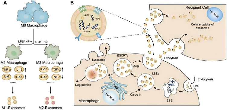
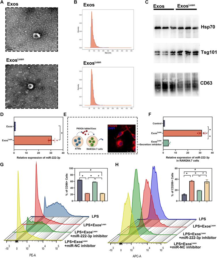
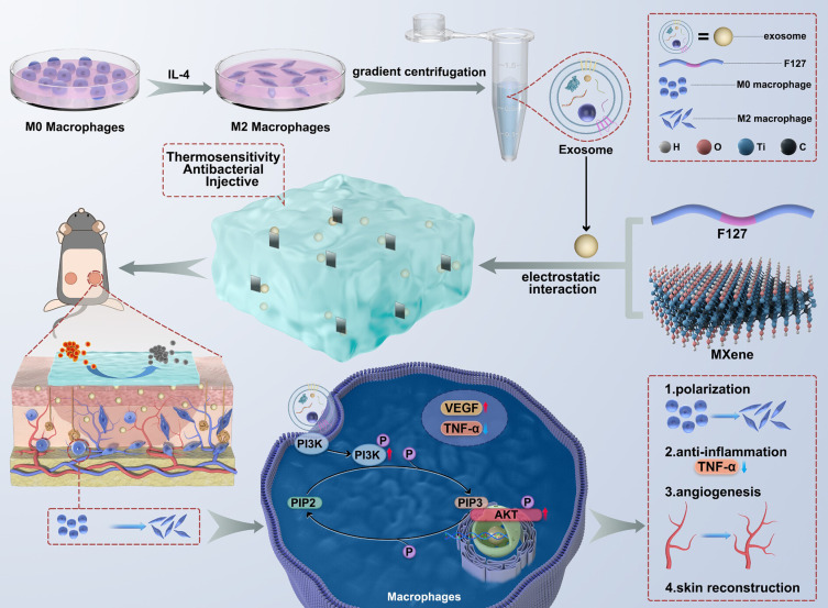
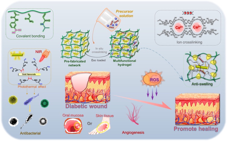
Diabetic wounds represent a significant clinical and economic challenge owing to their chronicity and susceptibility to complications. Dysregulated macrophage function is a key factor in delayed wound healing. Recent studies have emphasized the therapeutic potential of macrophage-derived extracellular vesicles (MDEVs), which are enriched with bioactive molecules such as proteins, lipids, and nucleic acids that mirror the state of their parent cells. MDEVs influence immune modulation, angiogenesis, extracellular matrix remodeling, and intercellular communication. In this review, we summarize and discuss the biological properties and therapeutic mechanisms of MDEVs in diabetic wound healing, highlighting strategies to enhance their efficacy through bioengineering and advanced delivery systems. We also explore the integration of MDEVs into innovative wound care technologies. Addressing current limitations and advancing clinical translation of MDEVs could advance diabetic wound management, offering a precise, effective, and versatile therapeutic option.

Keywords: diabetic wound; extracellular vesicle; macrophage; nanomedicine; therapy.


Conflict of interest statement

The author(s) report no conflicts of interest in this work.

Figures

Figure 1
Figure 1
Macrophage Polarization and Small EVs Formation. (A) Macrophages (Mφs) can be classified into two major subtypes, M1 and M2, based on their responses to distinct environmental signals. M1 Mφs are typically activated by IFN-γ or LPS, leading to the production of proinflammatory cytokines like TNF-α, IL-1β, IL-6, IL-12, and IL-23, which amplify inflammatory and cytotoxic reactions. In contrast, M2 Mφs, stimulated by IL-4 or IL-10, secrete small EVs that not only dampen proinflammatory responses but also enhance anti-inflammatory factors like IL-10 and TGF-β, contributing to the resolution of inflammation and tissue repair. (B) The process of small EVs biogenesis begins with the invagination of the macrophage plasma membrane, forming endocytic vesicles. These vesicles then merge to create early sorting endosomes, which mature into late sorting endosomes, or multivesicular bodies. Subsequently, multivesicular bodies fuse with the plasma membrane, releasing their cargo as small EVs into the extracellular milieu. Reproduced from Ye J, Liu X. Macrophage-derived small extracellular vesicles in multiple diseases: biogenesis, function, and therapeutic applications. Front Cell Dev Biol. 2022;10:913110. Copyright © 2022 Ye and Liu. This is an open-access article distributed under the terms of the Creative Commons Attribution License (CC BY).
Figure 2
Figure 2
(A) Representative TEM image of exosomes. (B) Size distribution of exosomes confirmed by NTA. (C) Western blot analysis demonstrating the presence of exosomal markers HSP70, TSG101, and CD63 in both Exos and ExosLean. (D) MiR-222-3p levels in exosomes verified through qRT-PCR, with statistical significance (*P < 0.05, paired t-test, n = 6). (E) Exosomes labeled with PKH26 were incubated with RAW264.7 macrophages. (F) Impact of an EV secretion inhibitor on miR delivery via exosomes, analyzed using repeated-measures ANOVA (*P < 0.05, n = 6). LPS-stimulated RAW264.7 cells were treated with ExosLean, ExosLean combined with a miR-222-3p inhibitor, or ExosLean plus a miR-NC inhibitor. (G) Flow cytometry analysis quantifying CD86+ cells (*P < 0.05, #P < 0.05, paired t-test, n = 3). (H) Flow cytometry analysis quantifying CD206+ cells (*P < 0.05, #P < 0.05, paired t-test, n = 3). A-H Reproduced from Xia W, Liu Y, Jiang X, et al. Lean adipose tissue macrophage derived exosome confers immunoregulation to improve wound healing in diabetes. J Nanobiotechnology. 2023;21:128. Copyright © 2023, The Author(s).This article is licensed under a Creative Commons Attribution 4.0 International License.
Figure 3
Figure 3
Schematic representation of the FM-Exo hydrogel, designed with multifunctional properties to enhance diabetic wound healing and facilitate skin reconstruction. Reproduced rom Jiang X, Ma J, Xue K, et al. Highly bioactive MXene-M2-exosome nanocomposites promote angiogenic diabetic wound repair through reconstructing high glucose-derived immune inhibition. ACS Nano. 2024;18:4269–4286. Copyright 2024, American Chemical Society.
Figure 4
Figure 4
Schematic illustration of the hydrogel system integrating functionalized AuNRs and M2-Exos designed to enhance granulation tissue formation and facilitate diabetic wound healing. Reproduced from Li W, Wu S, Ren L, et al. Development of an antiswelling hydrogel system incorporating M2-exosomes and photothermal effect for diabetic wound healing. ACS Nano. 2023;17:22106–22120. Copyright 2023, American Chemical Society.
Figure 5
Figure 5
Bidirectional communication between wound-edge macrophages (WE mϕ) and resident keratinocytes is essential for effective wound closure. (A) Design of a Krt14-promoter-driven tetraspanin plasmid linked via the IRES element to a “don’t eat me” CD47 sequence, incorporating GFP and RFP reporters. (B) Confocal images illustrating coexpression of RFP (red) and GFP (green) in murine keratinocytes. Scale bar, 20 μm. (C) Flow cytometry of murine Exoκ captured on pan-CD magnetic beads, showing dual GFP and RFP positivity in “don’t eat me” Exoκ-GFP-CD47-RFP. (D) Experimental setup schematic. (E) Digital images of excisional stented punch wounds (6 mm) at day 0 and day 12 post-wounding in C57BL/6 mice treated with either TNTκ-GFP or TNTκ-GFP-CD47-RFP. (F) Wound area quantification via digital planimetry after treatment with TNTκ-GFP or TNTκ-GFP-CD47-RFP. Scale bar, 2 mm (n = 20). (G) TEWL measurements in C57BL/6 mice at day 12 post-wounding following treatment with either TNTκ-GFP or TNTκ-GFP-CD47-RFP, with gray dots representing normal skin (n = 16). (H) H&E staining of wounds in C57BL/6 mice at day 12 post-wounding, showing either TNTκ-GFP or TNTκ-GFP-CD47-RFP treatment. Scale bar, 1000 μm. Blue arrowheads indicate complete reepithelialization; red arrowheads indicate the wound edge (WE). (I) Morphometric analysis of epithelial gap, wound length, and granulation tissue area in C57BL/6 mice at day 12 post-wounding (n = 6). (J) Coimmunofluorescence staining of F4/80 (red) with iNOS (green) and DAPI in WE granulation tissue at day 12 post-wounding, following either TNTκ-GFP or TNTκ-GFP-CD47-RFP treatment. (K and L) Quantification of F4/80 and iNOS intensity in WE tissue, with each dot representing a quantified ROI, and blue/red dots indicating the mean per mouse (n = 4–5, with at least 5 ROI per mouse). (M) Heat map showing expression of proinflammatory markers COX2, MCP-1, and MIP-1α in WE granulation tissue at day 12 post-wounding in C57BL/6 mice, following treatment with TNTκ-GFP or TNTκ-GFP-CD47-RFP. Data in panels (C, F, G, I, K, and L) are presented as mean ± SEM and analyzed using a two-tailed unpaired Student’s t-test. Reproduced from Sharma A, Srivastava R, Gnyawali SC, et al. Mitochondrial bioenergetics of functional wound closure is dependent on macrophage-keratinocyte exosomal crosstalk. ACS Nano. 2024;18:30405–30420. Copyright © 2024 The Authors. Published by American Chemical Society. This publication is licensed under CC-BY 4.0.

Similar articles

References

    1. Sen CK. Human wound and its burden: updated 2020 compendium of estimates. Adv Wound Care. 2021;10:281–292. doi:10.1089/wound.2021.0026 - DOI - PMC - PubMed
    1. Malone-Povolny MJ, Maloney SE, Schoenfisch MH. Nitric oxide therapy for diabetic wound healing. Adv Healthc Mater. 2019;8:e1801210. doi:10.1002/adhm.201801210 - DOI - PMC - PubMed
    1. Louiselle AE, Niemiec SM, Zgheib C, Liechty KW. Macrophage polarization and diabetic wound healing. Transl Res. 2021;236:109–116. doi:10.1016/j.trsl.2021.05.006 - DOI - PubMed
    1. Sharifiaghdam M, Shaabani E, Faridi-Majidi R, De Smedt SC, Braeckmans K, Fraire JC. Macrophages as a therapeutic target to promote diabetic wound healing. Mol Ther. 2022;30:2891–2908. doi:10.1016/j.ymthe.2022.07.016 - DOI - PMC - PubMed
    1. Liu W, Yu M, Xie D, et al. Melatonin-stimulated MSC-derived exosomes improve diabetic wound healing through regulating macrophage M1 and M2 polarization by targeting the PTEN/AKT pathway. Stem Cell Res Ther. 2020;11:259. doi:10.1186/s13287-020-01756-x - DOI - PMC - PubMed

LinkOut - more resources