Review
. 2025 Apr 24:4:1571516.
doi: 10.3389/frtra.2025.1571516. eCollection 2025.

Regulated cell death and DAMPs as biomarkers and therapeutic targets in normothermic perfusion of transplant organs. Part 1: their emergence from injuries to the donor organ

Affiliations
Review

Regulated cell death and DAMPs as biomarkers and therapeutic targets in normothermic perfusion of transplant organs. Part 1: their emergence from injuries to the donor organ

Walter G Land et al. Front Transplant. .

Abstract

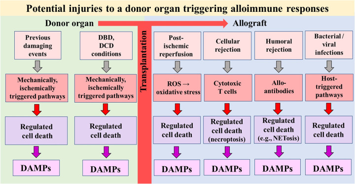
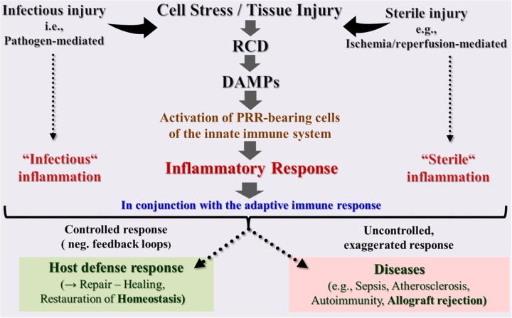
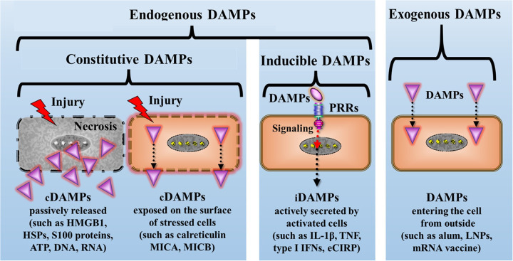
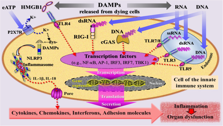
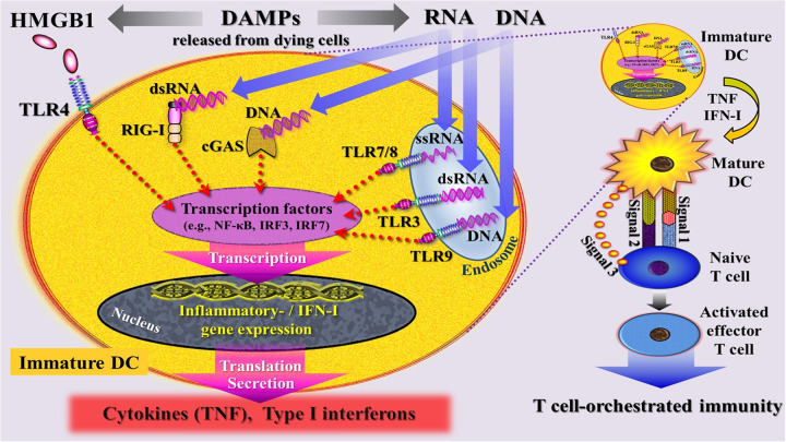
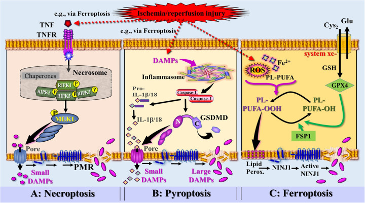
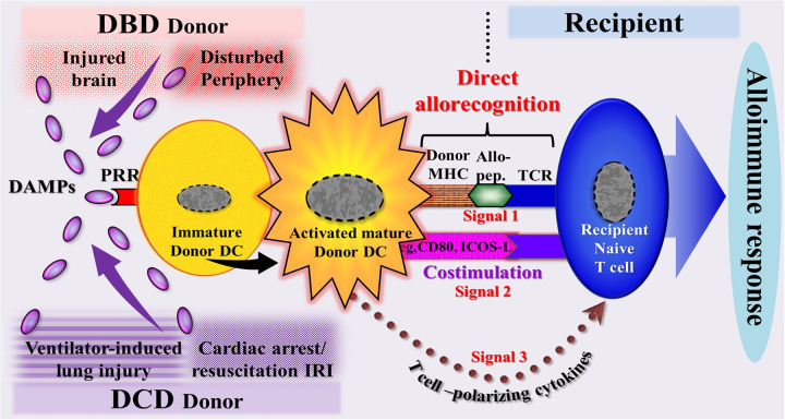
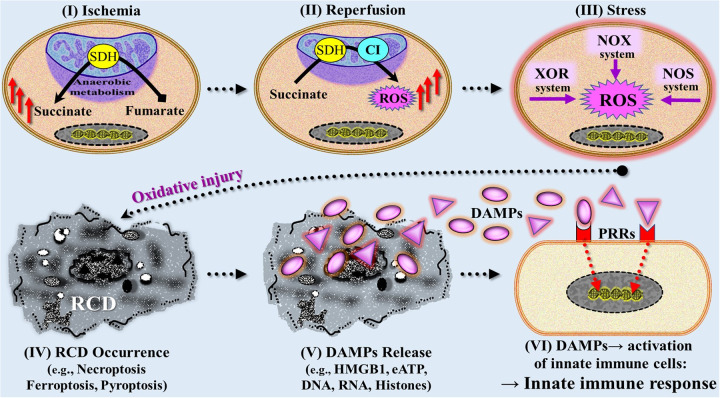
This Part 1 of a bipartite review commences with a succinct exposition of innate alloimmunity in light of the danger/injury hypothesis in Immunology. The model posits that an alloimmune response, along with the presentation of alloantigens, is driven by DAMPs released from various forms of regulated cell death (RCD) induced by any severe injury to the donor or the donor organ, respectively. To provide a strong foundation for this review, which examines RCD and DAMPs as biomarkers and therapeutic targets in normothermic regional perfusion (NRP) and normothermic machine perfusion (NMP) to improve outcomes in organ transplantation, key insights are presented on the nature, classification, and functions of DAMPs, as well as the signaling mechanisms of RCD pathways, including ferroptosis, necroptosis, pyroptosis, and NETosis. Subsequently, a comprehensive discussion is provided on major periods of injuries to the donor or donor organs that are associated with the induction of RCD and DAMPs and precede the onset of the innate alloimmune response in recipients. These periods of injury to donor organs include conditions associated with donation after brain death (DBD) and donation after circulatory death (DCD). Particular emphasis in this discussion is placed on the different origins of RCD-associated DAMPs in DBD and DCD and the different routes they use within the circulatory system to reach potential allografts. The review ends by addressing another particularly critical period of injury to donor organs: their postischemic reperfusion following implantation into the recipient-a decisive factor in determining transplantation outcome. Here, the discussion focuses on mechanisms of ischemia-induced oxidative injury that causes RCD and generates DAMPs, which initiate a robust innate alloimmune response.

Keywords: DAMPs; donation after brain death; donation after circulatory death; injuries to donor organs; innate alloimmunity; normothermic machine perfusion; normothermic regional perfusion; regulated cell death.


Conflict of interest statement

The authors declare that the research was conducted in the absence of any commercial or financial relationships that could be construed as a potential conflict of interest.

Figures

Figure 1
Figure 1
Simplified schematic diagram of a model illustrating potential injuries that can affect a donor organ before and after transplantation into a recipient. The injuries can induce types of regulated cell death that serve as sources of DAMPs. In turn, DAMPs—via activation of pattern recognition receptor-bearing cells of the donor's and recipient's innate immune system—drive alloimmune responses (not shown). DBD, donation after brain death; DCD, donation after circulatory death; ROS, reactive oxygen species.
Figure 2
Figure 2
Schematic overview of the innate immune system as a highly sensitive organ of perception. This conserved first-line defense system, composed of somatic cells bearing pattern recognition receptors (PRRs), senses any cell stress or tissue injury and triggers either infectious or sterile inflammatory responses to maintain homeostasis. However, uncontrolled dysregulation of this system results in pathologies and diseases. DAMPs, damage-associated molecular patterns; PRRs, pattern recognition receptors; RCD, regulated cell death.
Figure 3
Figure 3
Simplified schematic diagram of the principal classification of DAMPs: A primary framework for categorizing DAMPs involves dividing them into four major groups. They are broadly classified as endogenous or exogenous DAMPs, with exogenous DAMPs representing molecules originating outside the host. Endogenous DAMPs are further subdivided into constitutive DAMPs (cDAMPs, either passively released or cell surface-exposed), and inducible DAMPs (iDAMPs) that are secreted by DAMP-activated innate immune cells. alum, aluminum hydroxide; eCIRP, extracellular cold-inducible RNA-binding protein; HMGB1, high mobility group protein B1; HSPs, heat shock proteins; IFNs, interferons; IL-1β, interleukin-1beta; LNPs, lipid nanoparticles; MICA, MICB, MHC class I chain-related protein A and B; mRNA, messenger RNA; PRRs, pattern recognition receptors; TNF, tumor necrosis factor.
Figure 4
Figure 4
Simplified schematic diagram of a narrative model depicting injury induced, DAMP-triggered, pattern recognition receptor (PRR)-mediated trajectories that promote an inflammatory response associated with organ dysfunction. DAMPs [exemplified by high mobility group protein B1 (HMGB1), RNA, DNA] released by cells dying from regulated cell death are sensed by PRRs that are located at the plasma and endosomal membrane (TLRs) and in the cytosol (RIG-I, cGAS) of an innate immune cell. PRR-mediated signaling pathways (adaptor molecules not shown) lead—via transcriptional and translational processes (details not specified)—to secretion of inflammatory mediators that drive inflammation. The DAMP extracellular ATP (eATP)—via activation of the purinergic P2X7 receptor (P2X7R), induces dyshomeostatic DAMPs (dysDAMPs) that are sensed by NLRP3 receptor to contribute to the assembly of the inflammasome, which is associated with production of IL-1β and IL-18. These cytokines are released via transmembrane pores. AP-1, activating protein-1; cGAS, cyclic GMP-AMP synthase; dsRNA, double-stranded RNA; IL, interleukin; IRF3/7, interferon regulatory factor 3/7; NLRP3, nucleotide oligomerization domain (NOD)-like receptor protein 3; RIG-I, retinoic acid inducible gene I; ssRNA, single-stranded RNA; TBK1, TANK-binding kinase 1; TLRs, Toll-like receptors.
Figure 5
Figure 5
Simplified schematic diagram of a narrative model depicting injury induced, DAMP-triggered, pattern recognition receptor (PRR)-mediated trajectories that promote T cell-orchestrated immunity through activation of immature dendritic cells (DC) into mature DCs. DAMPs [exemplified by high mobility group protein B1 (HMGB1), RNA, DNA] released by cells dying from regulated cell death are sensed by PRRs that are located at the plasma and endosomal membrane (TLRs) and in the cytosol (RIG-I, cGAS) of an immature DC. These DAMP- triggered signaling pathways are believed to intersect at the level of interferon regulatory factors (IRFs) and nuclear factor kappa-light-chain-enhancer of activated B cells (NF- κB), leading to transcriptional changes that drive—via gene expression—the maturation of DCs and promote the subsequent secretion of cytokines and type I interferons. signal 1: upregulation of peptide/MHC molecules; signal 2: upregulation of costimulatory molecules; signal 3: secretion of T cell-polarizing cytokines. cGAS, cyclic GMP-AMP synthase; DC, dendritic cell; dsRNA, double-stranded RNA; HMGB1, high mobility group protein B1; IFN-I, type I interferons; IRF7, interferon regulatory factor 7; RIG-I, retinoic acid inducible gene I; ssRNA, single-stranded RNA; TLRs, Toll-like receptors; TNF, tumor necrosis factor.
Figure 6
Figure 6
Simplified and rough schematic diagram illustrating a model of DAMPs released from injury-induced RCD types and controlled by ninurin-1 (NINJ1)-dependent plasma membrane rupture (PRM) (injury exemplified by ischemia/reperfusion injury). (A) Death receptor signaling [here exemplified by tumor necrosis factor receptor (TNFR) bound to TNF secreted by innate immune cells activated by DAMPs, e.g., released from ferroptosis] leads to the formation of the necrosome, which activates the receptor-interacting protein kinase 3 (RIPK3). RIPK3 phosphorylates the molecule MLKL, which forms pores to instigate a PMR-associated necroptotic cell death, which is (partially) dependent on NINJ1. (B) Perception of DAMPs (e.g., released from ferroptosis) triggers the canonical inflammasome pathway and the activation of the inflammatory caspase-1. Caspase-1 is capable to cleave pro-IL-1β and pro-IL-18 into the mature cytokines. Activation and assembly of the inflammasome results in activation of Gasdermin D (GSDMD), whereby the caspase-1-cleaved N-terminal of GSDMD oligomerizes in membranes to proceed to final pores that release small DAMPs including the iDAMPs IL-1β and IL-18. GSDMD pores also drive the pyroptotic cell death associated with PMR that requires NINJ1 activation. (C) Accumulation of ischemia/reperfusion injury-induced reactive oxygen species (ROS) and iron (Fe2+) generate phospholipid hydroperoxides (PL-PUFA- OOH) leading to induction of ferroptosis. Ferroptosis suppressor protein 1 (FSP1) and glutathionperoxidase 4 (GPX4)—requiring the cofactor glutathione (GSH)—counterbalance the ferroptotic pathway by reducing PL-PUFA-OOH into lipid alcohols (PL-PUFA-OH). Phospholipid peroxides cause plasma membrane lipid peroxidation associated with permeabilization and the formation of NINJ1, which progresses into large NINJ1 oligomers that execute PMR. Source: Ramos et al. Ref. 69; C, C-terminal domain of GSDMD; Cyto-c, cytochrome c; Glu, glutamine; Lipid Perox., lipid peroxidation; MLKL, mixed lineage kinase domain like pseudokinase; N, N-terminal domain of GSDMD.
Figure 7
Figure 7
Schematic diagram of a model illustrating the DAMP-triggered, donor dendritic cell (DC)-mediated direct allorecognition process resulting in an alloimmune response of the recipient. This scenario reflects the immunogenicity of a potential allograft, as demonstrated by a powerful immune response of the recipient to the deceased donor's alloantigens. In organs from DBD donors, DAMPs originate (1) from injury-induced RCD occurring during brain death conditions in the cerebrum and migrating to the periphery (intracerebrally generated → circulating DAMPs) and (2) from the periphery (DAMPs emitted primarily in the periphery during pathophysiological events). In organs from DCD donors, DAMPs are supposed to originate mainly from injury-induced RCD occurring during lung ventilation and cardiac arrest→successful resuscitation -induced ischemia/reperfusion injury. Allo-pep., allogeneic peptide; DBD, donation after brain death; DCD, donation after circulatory death; ICOS-L, inducible costimulator-ligand; IRI, ischemia reperfusion injury; MHC, major histocompatibility complex; PRR, pattern recognition receptor; TCR, T cell receptor.
Figure 8
Figure 8
Simplified and rough schematic diagram illustrating a model with six stages mechanistically involved in ischemia reperfusion injury-induced innate immune responses. (I) During initial (warm) ischemic tissue condition, the succinate dehydrogenase (SDH) operates in reverse, reducing fumarate to succinate that accumulates within mitochondria. (II) On subsequent reperfusion, the accumulated succinate is rapidly oxidized by SDH causing generation of reactive oxygen species (ROS) as a result of reverse electron transport through mitochondrial complex I (CI). (III) Following initial reperfusion, other ROS-producing mechanisms contribute to increased generation of ROS including the extramitochondrial xanthine oxidasoreductase (XOR) system, NADPH oxidase (NOX) system, and nitric oxide synthase (NOS) system. (IV) Severe ROS-mediated oxidative injury leads to various types of regulated cell death (RCD) such as ferroptosis, necroptosis, and pyroptosis, which are associated with (V) release of large amounts of DAMPs (compare Figure 6). (VI) DAMPs interact with pattern recognition receptors (PRRs) on/in multiple cells of the innate immune system, thereby triggering an innate immune response (compare Figures 4, 5). eATP, extracellular ATP; HMGB1, high mobility group protein B1.

Similar articles

References

    1. Land W. Postischemic reperfusion injury to allografts—a case for ‘innate immunity’? Eur Surg Res. (2002) 34(1–2):160–9. 10.1159/000048904 - DOI - PubMed
    1. Land WG. Injury to allografts: innate immune pathways to acute and chronic rejection. Saudi J Kidney Dis Transpl. (2005) 16:520–39. Available online at: https://pubmed.ncbi.nlm.nih.gov/18202507/. - PubMed
    1. Land WG. Innate Alloimmunity: Part 2: Innate Immunity and Allograft Rejection. Lengerich: Baskent University, Ankara, Pabst Science Publishers; (2011). p. 760. Available online at: https://www.pabst-publishers.com/shop-checkout/suche/suchedetailergb?tt_...
    1. Land W, Schneeberger H, Schleibner S, Illner W-D, Abendroth D, Rutili G, et al. The beneficial effect of human recombinant superoxide dismutase on acute and chronic rejection events of cadaveric renal transplants. Transplantation. (1994) 57(2):211–7.. 10.1097/00007890-199401001-00010 - DOI - PubMed
    1. Matzinger P. Tolerance, danger, and the extended family. Annu Rev Immunol. (1994) 12(1):991–1045. 10.1146/annurev.iy.12.040194.005015 - DOI - PubMed

LinkOut - more resources