

Surgical Outcomes of Single-Stage Correction Using Cervical Pedicle Screw Fixation Rather Than Lateral Mass Fixation in NF1-Associated Pediatric Cervical Kyphosis: A Retrospective Study with a Minimum 2-Year Follow-Up
- PMID: 40343228
- PMCID: PMC12055176
- DOI: 10.2106/JBJS.OA.24.00252
Surgical Outcomes of Single-Stage Correction Using Cervical Pedicle Screw Fixation Rather Than Lateral Mass Fixation in NF1-Associated Pediatric Cervical Kyphosis: A Retrospective Study with a Minimum 2-Year Follow-Up
Abstract
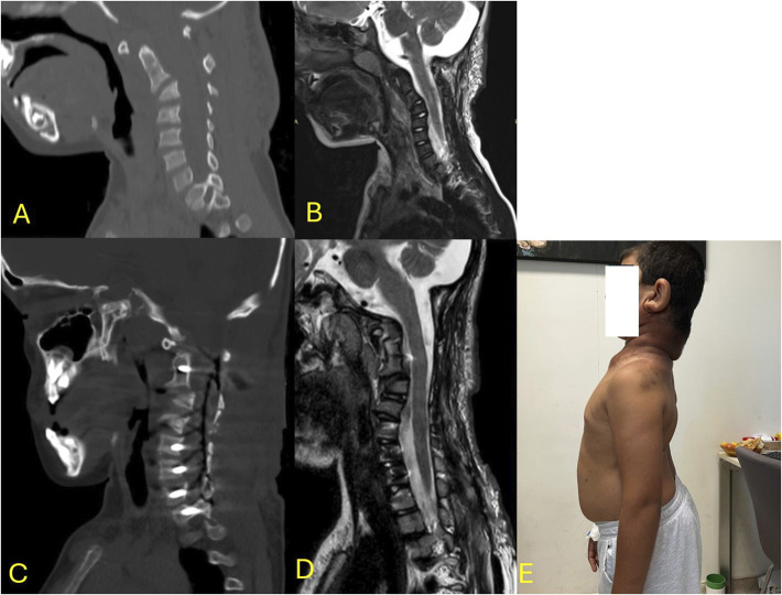
Background: Neurofibromatosis type 1 (NF-1) can cause severe kyphosis in the cervical vertebrae. There is no consensus on the optimal surgical treatment for this rare condition, although long-segment fixation and combined approaches are generally preferred. To our knowledge, this study is the first to report the clinical outcomes of patients with NF-related cervical kyphosis who underwent stand-alone posterior pedicle fixation surgery.
Methods: The outcomes of 14 patients who underwent surgery using the pedicle screw were retrospectively examined between 2015 and 2022. Only patients with at least 2 years of follow-up were included. For each patient, the following parameters were recorded and evaluated at 1 month postoperatively and at the end of the follow-up period: cervical lordosis (CL), local kyphosis angle (LKA), T1 slope, cervical sagittal vertical axis, visual analog score for neck pain, modified Japanese Orthopedic Association score, and Neck Disability Index. Complications, surgical duration, blood loss, levels of instrumentation, and length of hospital stay were also recorded.
Results: In terms of radiographic parameters, all patients achieved lordosis, with the cervical LKA improving from an average of 76.7° preoperatively to an average of 20.4° in the early postoperative period. At the 2-year follow-up, the postoperative CL significantly improved compared with preoperative values (p < 0.001) with only approximately 4° correction loss. Moreover, by the end of the follow-up, all postoperative symptoms showed improvement compared with the preoperative symptoms. The average surgical duration was 211.86 ± 49.83 min. During the follow-up, junctional kyphosis was observed in 4 patients all of whom required revision surgery. C5 palsy was detected in 3 patients. Infection-related complications occurred in 6 patients, with wound infection in only 1 patient.
Conclusion: Cervical pedicle screw fixation is an effective treatment for NF-1-related cervical kyphosis. Although this technique is considered difficult and dangerous to apply by several spine surgeons, it exerts a positive effect on clinical improvement and provides optimal correction.
Level of evidence: Therapeutic Level IV. See Instructions for Authors for a complete description of levels of evidence.
Copyright © 2025 The Authors. Published by The Journal of Bone and Joint Surgery, Incorporated. All rights reserved.
Conflict of interest statement
Disclosure: The Disclosure of Potential Conflicts of Interest forms are provided with the online version of the article (http://links.lww.com/JBJSOA/A820).
Figures


References
-
- Hirbe AC, Gutmann DH. Neurofibromatosis type 1: a multidisciplinary approach to care. Lancet Neurol. 2014;13(8):834-43. - PubMed
-
- Akbarnia BA, Gabriel KR, Beckman E, Chalk D. Prevalence of scoliosis in neurofibromatosis. Spine (Phila Pa 1976). 1992;17(8 Suppl):S244-248. - PubMed
-
- Kaspiris A, Savvidou OD, Vasiliadis ES, Hadjimichael AC, Melissaridou D, Iliopoulou-Kosmadaki S, Iliopoulos ID, Papadimitriou E, Chronopoulos E. Current aspects on the pathophysiology of bone metabolic defects during progression of scoliosis in neurofibromatosis type 1. J Clin Med. 2022;11(2):444. - PMC - PubMed
LinkOut - more resources
Full Text Sources
Research Materials
Miscellaneous