. 2025 Apr 11;28(5):112416.
doi: 10.1016/j.isci.2025.112416. eCollection 2025 May 16.

Multifaceted profiling of virus-specific CD8 T cells reveals distinct immune signatures against cytomegalovirus infection states during pregnancy

Affiliations

Multifaceted profiling of virus-specific CD8 T cells reveals distinct immune signatures against cytomegalovirus infection states during pregnancy

Ayumi Taguchi et al. iScience. .

Abstract

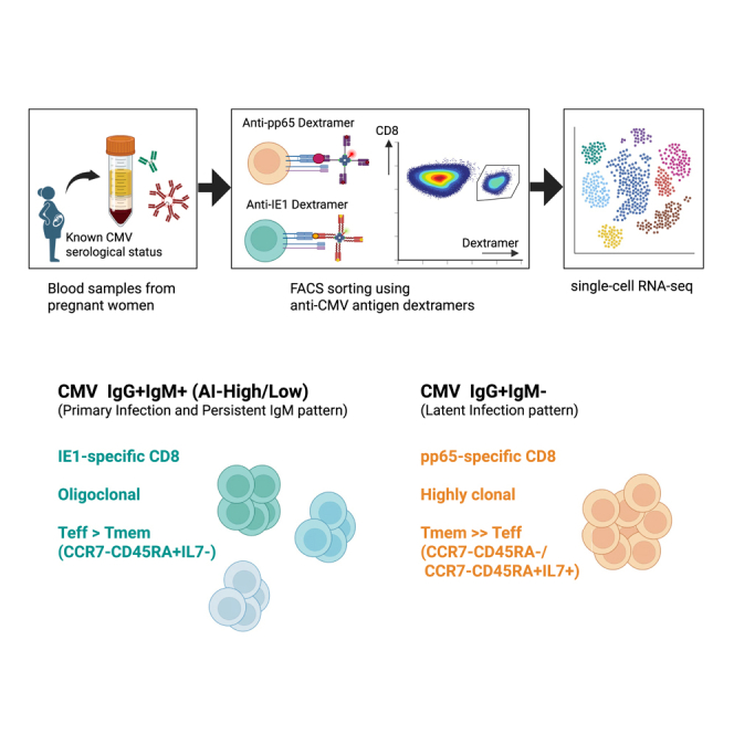
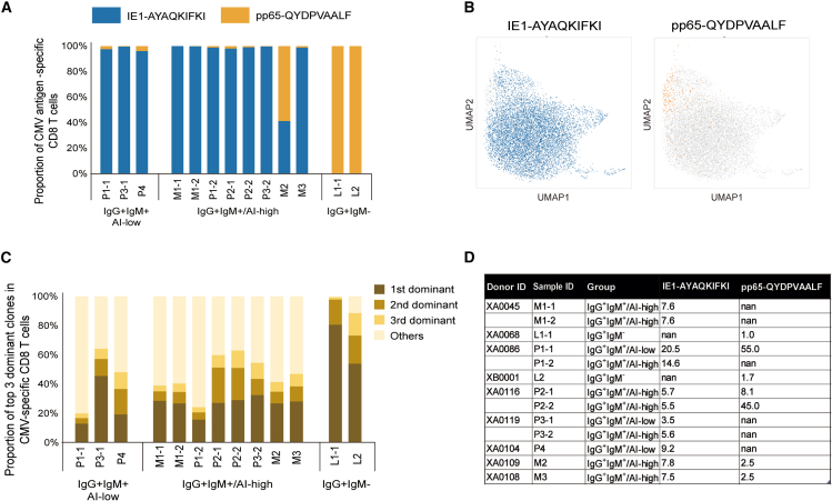
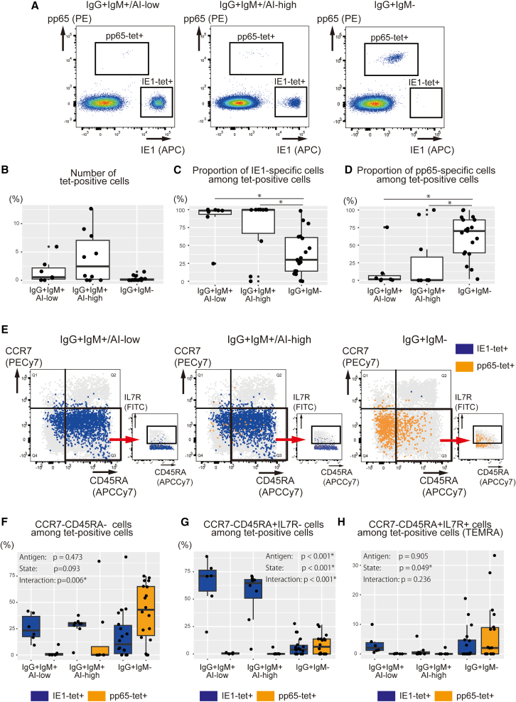
Anti-cytomegalovirus (CMV) serological testing, including the IgG avidity index (AI), is used to assess CMV infection phases during pregnancy. However, little is known about anti-CMV cellular immunity during pregnancy, particularly its relation to serological diagnosis. Herein, using MHC-dextramer single-cell RNA sequencing and flow cytometry, we characterized IE1 and pp65 CMV-antigen specific CD8 T cells from pregnant women with different anti-CMV serological patterns, including IgG+IgM+/AI-low, IgG+IgM+/AI-high, and IgG+IgM-. In IgG+IgM+/AI-low and IgG+IgM+/AI-high specimens, CMV-specific T cells consisted largely of effectors, with a minor but characteristic proportion of memory T cells, including HLA-DR-positive memory precursors and granzyme K-high memory cells reactive to IE1. Conversely, IgG+IgM- cases had a distinctive expansion of pp65-specific terminally differentiated T effector memory with a signature of convergent clonal selection. Our findings revealed that different CMV infection phases have characteristic patterns of CD8 cell phenotype and antigen recognition, potentially offering a new approach for assessing congenital infection risk.

Keywords: biological sciences; immunology; pregnancy.


Conflict of interest statement

The authors declare no competing interests.

Figures

None
Graphical abstract
Figure 1
Figure 1
Kinetics of CMV IgM decay vary widely among individuals (A) Summary of anti-CMV antibody status in pregnant donors and their neonatal outcomes. Results from CMV serological tests using enzyme immunoassay (EIA) in early pregnancy (<16 weeks) and their corresponding neonatal outcome are summarized (N = 3122). Donors were classified into the following four groups: IgG+IgM+/AI-low group, CMV IgG-positive and IgM-positive with low IgG avidity index (AI); IgG+IgM+/AI-high group, IgG-positive and IgM-positive with high AI; IgG+IgM group, CMV IgG-positive and CMV IgM-negative; and IgG group, CMV IgG-negative. Asterisk indicates a case in which serological data were converted to IgG and IgM positive in the late pregnancy. (B) Temporal changes in IgM titers among donors who tested CMV IgG+ and IgM+ within 16 weeks of conception. Donors who had multiple test results during pregnancy were included (N = 95). The threshold of positivity was set at IgM titer 0.8. “Day 0” refers to the date of the first serological testing, and time (days) indicates time after first serological testing. Cases with negative conversion are marked in blue, and those without negative conversion are marked in orange. (C) Changes in CMV IgM titers between two consecutive pregnancies. Left, IgG-positive, IgM-negative (IgG+ IgM-) donors (N = 125); right, IgG-positive, IgM-positive (IgG+ IgM+) donors (N = 27) in their first pregnancy in the cohort (P1). The measurements at the time of first pregnancy (P1) are marked with ●, and those at the time of second pregnancy (P2) are marked with ♦. “Time” indicates the time (year) from the date of the initial examination at first pregnancy (P1) to the time of the initial examination at second pregnancy (P2). Cases with positive IgM at second pregnancy are marked in orange, and cases with negative IgM are marked in blue. (D) Summary of serological tests in the second pregnancy (P2) based on serological status in the first pregnancy (P1).
Figure 2
Figure 2
scRNA-seq analysis of CMV-specific T cells in pregnant women (A) UMAP projections of the single cell transcriptome of CMV-specific CD8 T cells captured by HLA-A∗24:02 AYAQKIFKI (IE1) or HLA-A∗24:02 QYDPVAALF (pp65) MHC-dextramers. CMV-specific CD8 T cells were divided into four clusters: C0, Teff; C1, Tmem; C2, Tsle; and C3, Tpro. (B) Violin plots showing the expression level of characteristic marker genes in each cluster. Y axis is presented on a log scale. (C) Heatmap representation of DEGs in each cluster. The top 10 DEGs in each cluster are shown. (D) Proportion of T cell subsets in individual samples according to serological groups.
Figure 3
Figure 3
Functional compositions of CMV-specific memory by serological groups (A) UMAP projection of the single cell transcriptome of the CMV-specific memory fraction. CD8 T cells in C1 (Tmem) were divided into four clusters: C1.0, GZMKhigh Tem; C1.1, Tmp; C1.2 term-Tem; C1.3, Tcm. (B) Violin plots showing the expression levels of characteristic marker genes in each cluster. Y axis is presented on a log scale. (C) Heatmap representation of DEGs in each cluster. The top 10 DEGs in each cluster are shown. (D) Proportion of memory T cell subsets in individual samples according to serological groups.
Figure 4
Figure 4
Target antigen and clonality of CMV-specific CD8 T cells (A) Bar graph showing the proportion of IE1-and pp65-specific CD8 T cells determined by MHC-dextramers in individual samples according to serological groups. (B) UMAP projections of the single cell transcriptome of the IE1-AYAQKIFKI-, or pp65-QYDPAVAALF-specific CD8 T cells. (C) Proportions of the top 3 largest clones in CMV-specific T cells. (D) Inverse Simpson index (ISI) values of pp65-and IE-specific clones.
Figure 5
Figure 5
Longitudinal changes in CMV-specific clones (A) Bar graphs showing the longitudinal changes in the percentages of the five largest clones for donors XA0045, XA0086, XA0116, and XA0119. (B) UMAP projections illustrating the longitudinal changes in the single cell transcriptome of CMV-specific clones in these donors. AI, IgG avidity index.
Figure 6
Figure 6
TCR repertoire analysis of CMV-specific T cells (A and B) TCR Vα-Jα, and Vβ-Jβ gene segment usage and Vα-Jα, Vβ -Jβ pairings in IE1-specific T cell clonotypes (A) and pp65-specific T cell clonotypes (B). In the visualization, the contribution of each QYDPVAALF clone was normalized by multiplying #AYA/#QYD to ensures that the same color represents the same fraction across respective T cell population. #AYA: total number of IE1-AYAQKIFKI clones, #QYD: total number of pp65-QYDPAVAALF clones. (C and D) Examples of similar T clonotypes specific to IE1 across donors. Sequencing logos of CDR3α and β from all clonotypes encoded by TRAV27-TRAJ19 and TRBV30-TRBJ1-1 with 11 amino acid-long CDR3α and β in the dataset (C). Clonal distribution across donors of the top 20 clonotypes in this class (D). Group A, IgG+IgM+/AI-low; group B, IgG+IgM+/AI-high; and group C, IgG+IgM.
Figure 7
Figure 7
Surface phenotypes of CMV-specific CD8 T cells differ by target antigens and infection states (A) Detection of CMV-specific CD8 T cells by flow cytometry. Representative FACS plots gated on 7AAD CD8 cells stained with APC-conjugated HLA-A∗24:02 AYAQKIFKI (IE1) and PE-conjugated HLA-A∗24:02 QYDPVAALF (pp65) tetramers: left panel, IgG+IgM+/AI-low group; middle panel, IgG+IgM+/AI-high group; and right panel, IgG+IgM group. (B) Number of tetramer-positive cells by CMV serological status. The total number of HLA-A∗24:02 IE1-AYAQKIFKI-specific and pp65-QYDPAVAALF-specific cells was represented as tetramer-positive cells. (C) Proportion of IE1-AYAQKIFKI-specific cells among tetramer-positive cells. (D) Proportion of pp65-QYDPAVAALF-specific cells among tetramer-positive cells. (E–H) Surface phenotypes of CMV-specific CD8 T cells at different phases of infection. Representative FACS plots gated on 7AAD CD8 cells: left panel, IgG+IgM+/AI-low group; middle panel, IgG+IgM+/AI-high group; and right panel, IgG+IgM group (E). Proportion of CD45RA CCR7- effector memory T cells (F), proportion of CD45RA+ CCR7- IL7R effector T cells (G), and proportion of CD45RA+ CCR7- IL7R+ TEMRA cells (H) among tetramer-positive CD8 T cells. The Kruskal-Wallis test with Holm correction was used in (B–D), and a two-way ANOVA was used in (F–H). The boxes represent the interquartile range (IQR) of the data, with the horizontal line inside each box indicating the median and whiskers extend to the most extreme data points within 1.5 times the IQR from Q1 and Q3 (B–D, G − H). Antigen: Whether the antigen (IE1 or pp65) affected the change in the proportion of each fraction; State: Whether infectious status (IgG+IgM+/AI-low, IgG+IgM+/AI-high, or IgG+IgM) affected the change in the percentage of each fraction; Interaction: Whether “antigen” and “state” interact with each other in the proportion of each fraction (F–H). Asterisks indicate statistical significance (p < 0.05). ng tetramer-positive cells.

Similar articles

References

    1. Cannon M.J., Schmid D.S., Hyde T.B. Review of Cytomegalovirus Seroprevalence and Demographic Characteristics Associated with Infection. Rev. Med. Virol. 2010;20:202–213. doi: 10.1002/rmv.655. - DOI - PubMed
    1. Drew W.L. Cytomegalovirus Infection in Patients with AIDS. Clin. Infect. Dis. 1992;14:608–615. doi: 10.1093/clinids/14.2.608-a. - DOI - PubMed
    1. Kotton C.N. CMV: Prevention, Diagnosis and Therapy. Am. J. Transplant. 2013;13:24–40. doi: 10.1111/ajt.12006. quiz 40. - DOI - PubMed
    1. Rawlinson W.D., Boppana S.B., Fowler K.B., Kimberlin D.W., Lazzarotto T., Alain S., Daly K., Doutré S., Gibson L., Giles M.L., et al. Congenital Cytomegalovirus Infection in Pregnancy and the Neonate: Consensus Recommendations for Prevention, Diagnosis, and Therapy. Lancet Infect. Dis. 2017;17:e177–e188. doi: 10.1016/S1473-3099(17)30143-3. - DOI - PubMed
    1. Fowler K.B., Boppana S.B. Congenital Cytomegalovirus (CMV) Infection and Hearing Deficit. J. Clin. Virol. 2006;35:226–231. doi: 10.1016/j.jcv.2005.09.016. - DOI - PubMed

LinkOut - more resources