Review
. 2025 May 8;15(19):14966-15016.
doi: 10.1039/d5ra01427a. eCollection 2025 May 6.

Protein-based nanoparticles for antimicrobial and cancer therapy: implications for public health

Affiliations
Review

Protein-based nanoparticles for antimicrobial and cancer therapy: implications for public health

Ikhazuagbe Hilary Ifijen et al. RSC Adv. .

Abstract

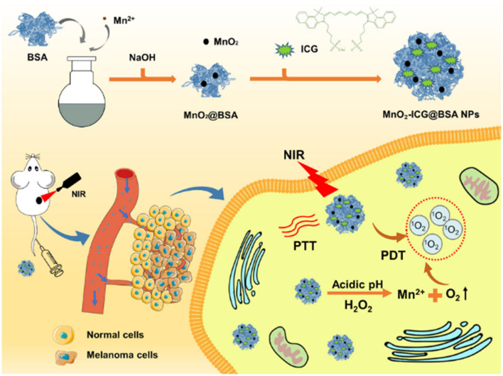
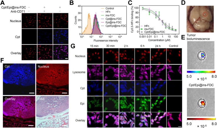
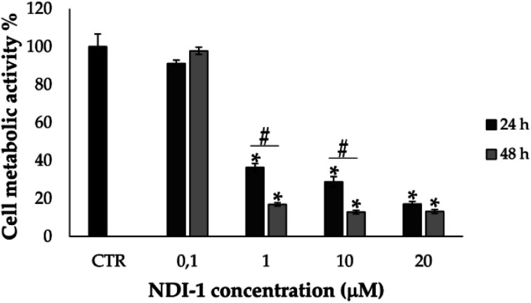
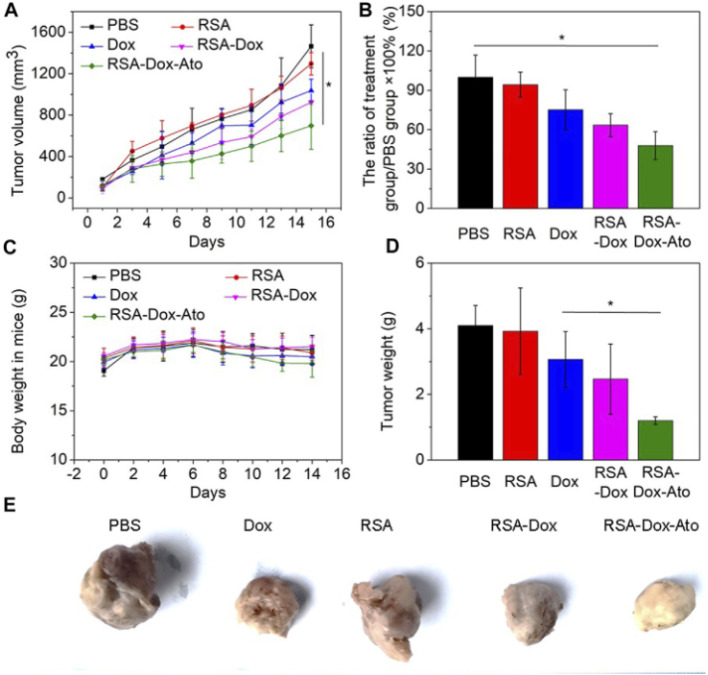
This review discusses the growing potential of protein-based nanoparticles (PBNPs) in antimicrobial and cancer therapies, emphasizing their mechanisms of action, applications, and future prospects. In antimicrobial therapy, PBNPs exhibit several mechanisms of action, including disruption of microbial membranes, enhanced antibiotic delivery, immune modulation, and biofilm disruption. Protein nanoparticles like albumin, lactoferrin, gelatin, and peptide-based variants enhance the efficacy of antibiotics, offering targeted approaches to combat multidrug-resistant pathogens. Their ability to improve drug localization and enhance microbial eradication represents a significant advancement in infectious disease management. In cancer therapy, PBNPs facilitate targeted drug delivery, controlled release, tumor microenvironment modulation, and photothermal and photodynamic therapies. Nanoparticles such as Abraxane® and engineered ferritin nanocages are at the forefront of cancer treatment, enhancing the precision and effectiveness of chemotherapy while minimizing adverse effects. Additionally, silk fibroin nanoparticles are being explored for their biodegradability and targeting capabilities. Despite their promise, challenges remain, including the scalability of production, long-term safety concerns, regulatory approval processes, and environmental impact. Addressing these issues through rigorous research and innovation is crucial for integrating PBNPs into mainstream therapeutic practices. PBNPs offer transformative solutions in both antimicrobial and cancer therapies, with significant implications for improving public health outcomes globally.


Conflict of interest statement

On behalf of all authors, the corresponding author states that there is no conflict of interest.

Figures

Fig. 1
Fig. 1. Peptide-induced morphological changes in E. coli cells. All peptides were treated at minimum inhibitory concentrations (MICs).
Fig. 2
Fig. 2. Schematic mode of action of the designed peptides in bacterial membranes. Red-dotted lines indicate electrostatic interaction between cationic amino acids and anionic head group of lipids. Hydrophobic interaction between hydrophobic amino acids and fatty acids of lipids. Hydrophobic interaction between hydrophobic amino acids and fatty acids of lipids.
Fig. 3
Fig. 3. Permeabilizing effect of AgNP–AMP/AP conjugates on Escherichia coli ML-35p outer and inner membranes. The effects of the conjugates are compared to those of the corresponding antimicrobial peptides and proteins (AMPs and APs) and gelatin-only coated silver nanoparticles (AgNPs) alone. The optical density (OD) increase correlates to the hydrolysis of the chromogenic markers by bacterial enzymes. Permeabilization of the outer membrane gives the nitrocefin marker the access to periplasmic β-lactamase, while inner membrane permeabilization gives the o-nitrophenyl-β-d-galactoside marker (ONPG) the access to cytoplasmic β-galactosidase. Bacterial membranes are impenetrable to the markers under normal conditions; hence the dynamics of their degradation allow assessing the scale and velocity of membrane damage inflicted by the tested substances. The concentration of antimicrobials used was equal to 4 × MIC (minimal inhibitory concentration); typical curves are shown.
Fig. 4
Fig. 4. (a) Results of the viability experiments carried out with and without AuNPs and AuNPs + HSA at three different illumination times, along with representative infrared thermographic images of (b) E. coli and (c) E. coli + NPs samples after 20 min of laser irradiation. The values represent the mean of three independent triplicate experiments with standard error.
Fig. 5
Fig. 5. Schematic representation of the interaction between HSA and AuNPs, and the effect on E. coli bacteria with and without 532 nm laser irradiation.
Scheme 1
Scheme 1. Schematic illustration of using CP/HA/RH-NPs in the treatment of UC in mice. (1) Schematic illustration of the passage of orally administered CP/HA/RH-NPs through GIT. CP could protect CP/HA/RH-NPs to pass through stomach and small intestine, and further release HA/RH-NPs into colonic lumen due to its degradation. (2) Schematic illustration of enhancing the effects of RH in repairing intestinal damage by adjusting ZO-1 and Claudin-1 expression in the UC mice model by colonic epithelial cell target. (3) Schematic illustration of targeting macrophage could effectively promote RH's anti-inflammatory effect through the TLR4/MyD88/NF-κB pathway in in vivo anti-UC therapeutic efficacy.
Fig. 6
Fig. 6. Representative photos of bacteria colonies counting on Mueller–Hinton agar plates after 18 h incubation at 37 °C and exposed to our topical formulations with 1.1% and 1.5% (w/v) gelatin with 0.7% (w/v) PVA and 20 μg per mL GO–Ag NPs, the commercial treatments, and the positive control (n = 15).
Fig. 7
Fig. 7. Bacterial growth for the 1.1% and 1.5% (w/v) gelatin with 0.7% (w/v) PVA and 20 μg per mL GO–Ag NPs treatment, the commercial treatments, and the positive control (n = 15). *, p < 0.05.
Fig. 8
Fig. 8. Inhibition dynamics of metabolic activity in E. coli ML-35p by AgNP–AMP/AP conjugates. The metabolic inhibitory effects of AgNP–AMP/AP conjugates are compared with those of free antimicrobial peptides and proteins (AMPs/APs), gelatin-only coated silver nanoparticles (AgNPs), and ionic silver (AgNO3). Actively metabolizing bacterial cells reduce the redox-sensitive marker resazurin to its fluorescent product, resorufin. An increase in fluorescence intensity reflects active bacterial metabolism, while a decline indicates metabolic inhibition. The antimicrobials were tested at a concentration of 4× the minimal inhibitory concentration (MIC). Representative response curves illustrate the rapid and potent metabolic suppression induced by the conjugates and their components.
Fig. 9
Fig. 9. SEM analysis of bacterial cell damage and EDX spectra of metal content in the test samples. P. aeruginosa with different treatments (A–H) and S. aureus with different treatments (I–P). P. aeruginosa and S. aureus cells with untreated (A & I), D2A21 treated (B & J), D2A10 treated (C & K), D4E1 treated (D & L), positive control (heat treatment in boiling water bath for 30 min) (E & M), nanocomposites D2A21–AgNPs (F & N), D2A10–AgNPs (G & O), D4E1–AgNPs (H & P). The red coloured arrows indicating the damage of cell wall and white coloured arrows indicating the presence of silver.
Fig. 10
Fig. 10. (a) Cytotoxicity of the I3K/PNIPAM hydrogel against NIH 3T3 cells as determined by the MTT assay. (b) The cumulative release of FITC-G(IIKK)3I-NH2 as a function of time.
Fig. 11
Fig. 11. Interactions within the tumor microenvironment (TME), comprising tumor cells and three major classes of stromal cells—angiogenic vascular cells, infiltrating immune cells, and tumor fibrosis-related cells—significantly influence cancer progression. This figure illustrates the peptide-assembled nanoparticles designed to target tumor cells and stromal cells for enhanced cancer therapy.
Fig. 12
Fig. 12. (A) Molecular structures of the RGD peptide LKR. The red represents hydrophobic areas, and the blue represents hydrophilic areas. (B) Self-assembly behavior and acid-responsive morphological transformation of LKR. After the targeted nanoparticles reached the tumor site, the slightly acidic tumor environment triggered the rupture of the spherical nanoparticles, resulting in the release of the encapsulated antitumor drug and achieving a combined antitumor effect.
Fig. 13
Fig. 13. Illustration of tumor microenvironment-responsive, oxygen self-sufficient oil droplet nanoparticles designed for enhanced photothermal and photodynamic combination therapy, effectively addressing hypoxia in tumor treatment.
Scheme 2
Scheme 2. A preparation and application schematic illustrating the PTT–PDT of melanoma mediated by MnO2-ICG@BSA under NIR irradiation. NIR, near-infrared; PTT–PDT, photothermal–photodynamic combination therapy.
Fig. 14
Fig. 14. Enhanced antitumor efficacy and survival through combination therapy using Nano-PI and anti–PD-1 in a mouse model of metastatic breast cancer. (A) Schematic representation of the experimental design for treatment of MMTV-PyMT mice with Nano-PI (nanoparticle-loaded PI3K inhibitor), anti–PD-1, or their combination. Mice received intraperitoneal (IP) anti–PD-1 and intravenous (IV) Nano-PI or control treatments starting on day 105 after birth. Lungs were collected at the end of treatment to evaluate tumor burden. (B) Tumor burden progression over time in individual mice treated with vehicle, Nano-P (nanoparticles without drug), anti–PD-1 alone, Nano-PI, or combinations thereof. The combination of Nano-PI + anti–PD-1 showed the most significant inhibition of tumor growth. (C) Representative hematoxylin and eosin (H&E)-stained lung tissue sections and gross lung images showing metastatic tumor nodules across treatment groups. Quantification of metastatic nodules per lung reveals significantly reduced tumor burden in the Nano-PI + anti–PD-1 group. (D) Kaplan–Meier survival curves of treated mice indicate improved survival in the Nano-PI + anti–PD-1 group compared to monotherapies and control. (E) Diagram showing the experimental timeline for immune profiling of Nano-PI + anti–PD-1–treated PyMT tumor-bearing mice. Tumors were inoculated subcutaneously on day 132, and immune organs (lymph nodes, spleen, bone marrow, blood, and lungs) were collected on day 170. (F) Quantification of immune cell populations from flow cytometry in various tissues of treated mice. The combination therapy group exhibited enhanced T-cell infiltration and activation compared to other groups. (G) Schematic of another treatment model and timeline for validation using IV injection of Nano-PI in late-stage tumor-bearing MMTV-PyMT mice. (H) Tumor burden progression in mice treated with vehicle, PTX/PI (paclitaxel and PI3K inhibitor combination), or Nano-PI + anti–PD-1. (I) Representative H&E lung sections and gross lung images show reduced metastasis in Nano-PI + anti–PD-1–treated mice. Bar graphs quantify metastatic nodules. (J) Kaplan–Meier survival curve from validation experiment confirming extended survival in the combination treatment group. Statistical significance is indicated as follows: p < 0.05, p < 0.01, and p < 0.001. Data are presented as mean ± SEM.
Fig. 15
Fig. 15. Viability of HL60 cells treated with free ellipticine/doxorubicin or with HFt-W4 loaded with ellipticine or doxorubicin (mean ± SD, n = 4). When conducting experiments with HFt-W4 loaded with ellipticine or doxorubicin, the specific concentration of the encapsulated drug was taken into account.
Fig. 16
Fig. 16. Tumor-targeting and cellular internalization of Cpt/Epi@ins-FDC. (A) Cpt/Epi@ins-FDC binds to CD71 receptors on tumor cells. Confocal laser scanning microscopy (CLSM) images of U87MG cells treated with Cpt/Epi@ins-FDC in the presence or absence of anti-CD71 monoclonal antibodies (mAbs). Red: propidium iodide (PI) (λex/λem = 535 nm/615 nm); blue: Cpt (λex/λem = 365 nm/500 nm). Scale bar: 20 μm. (B) Flow cytometry histograms showing the interaction of U87MG cells with different formulations after 2 hours of incubation. (C) Competitive binding assay comparing the binding efficiency of Cpt/Epi@ins-FDC, ins-FDC, and HFn. (D) Digital image of the brain from a U87MG tumor xenograft mouse model. (E) In vivo near-infrared fluorescence (NIRF) imaging demonstrating specific tumor accumulation of Cpt/Epi@ins-FDC following intravenous injection (λex/λem = 365 nm/500 nm). (F) Fluorescence staining using Cpt/Epi@ins-FDC and H&E staining of paraffin-embedded lung slices from HepG2 lung metastasis mouse models. Red: PI (λex/λem = 535 nm/615 nm); blue: Cpt (λex/λem = 365 nm/500 nm). Scale bar: 200 μm. (G) CLSM visualization of Cpt/Epi@ins-FDC internalization by U87MG cells and the sequential release of Cpt and Epi at specified time points. Purple: Cy5-labeled lysosomal-associated membrane protein 1 (LAMP1) (λex/λem = 650 nm/700 nm); red: PI (λex/λem = 535 nm/615 nm); blue: Cpt (λex/λem = 365 nm/500 nm); green: Epi (λex/λem = 485 nm/575 nm). Scale bar: 20 μm.
Fig. 17
Fig. 17. Cell metabolic activity % of U373 cells treated with increasing doses of NDI-1 for 24 and 48 h. Untreated cells at 24 hours were considered the control (CTR), representing 100% of metabolic activity. Multifactor ANOVA, mean values ± least significant difference (LSD), n = 4. *p < 0.01 with respect to CTR-; #p < 0.05.
Scheme 3
Scheme 3. Schematics demonstration the RSA-Dox-Ato alleviating tumor hypoxia and improving the antitumor efficacy. (A) Scheme illustrating the synthesis of RSA-Dox-Ato NPs; (B) the mechanism of RSA-Dox-Ato NPs alleviate tumor hypoxia in vivo.
Fig. 18
Fig. 18. The behavior of RSA-Dox-Ato NPs post-i.v. injection in vivo. (A) The tumor volume growth curves of 4T1 tumor-bearing mice injected with nanodrugs for 14 days. The data were represented as the mean ± SD (n = 5, pp < 0.05, ppp < 0.01); (B) the tumor growth inhibition ratio of each group; (C) the body weight curve of 4T1 tumor-bearing mice upon the period of treatment; (D) the tumor weight curve of 4T1 tumor-bearing mice upon the period of treatment; (E) representative photos of tumor-bearing mice from different treatment groups at the end of the study.

Similar articles

References

    1. Bratti V. Wilson B. Fazelzad R. Pabani A. Zurn S. Johnson S. et al., Scoping review protocol on the impact of antimicrobial resistance on cancer management and outcomes. BMJ Open. 2023;13:e068122. doi: 10.1136/bmjopen-2022-068122. - DOI - PMC - PubMed
    1. Egwonor L. I. Awoyemi R. F. Owolabi B. Aworinde O. R. Kajola R. O. Hazeez A. et al., Cutting-edge developments in zinc oxide nanoparticles: Synthesis and applications for enhanced antimicrobial and UV protection in healthcare solutions. RSC Adv. 2024;14:20992–21034. doi: 10.1039/D4RA02452D. - DOI - PMC - PubMed
    1. Ifijen I. H. Christopher A. T. Lekan O. K. Aworinde O. R. Faderin E. Obembe O. F. et al., Advancements in tantalum-based nanoparticles for integrated imaging and photothermal therapy in cancer management. RSC Adv. 2024;14:33681–33740. doi: 10.1039/D4RA05732E. - DOI - PMC - PubMed
    1. Di Pietro M. Filardo S. Sessa R. Editorial for the Special Issue “Antibacterial Activity of Drug-Resistant Strains”. Int. J. Mol. Sci. 2024;25:1878. doi: 10.3390/ijms25031878. - DOI - PMC - PubMed
    1. Maliki M., Omorogbe S. O., et al., Incisive review on magnetic iron oxide nanoparticles and their use in the treatment of bacterial infections, in TMS 2023 152nd Annual Meeting & Exhibition Supplemental Proceedings, Cham, Springer, 2023, 10.1007/978-3-031-22524-6_44 - DOI

LinkOut - more resources