[Preprint]. 2025 May 2:rs.3.rs-6422592.
doi: 10.21203/rs.3.rs-6422592/v1.

Cryo-EM reveals remodeling of a tandem riboswitch at 2.9 Å resolution

Affiliations

Cryo-EM reveals remodeling of a tandem riboswitch at 2.9 Å resolution

Nathan Jespersen et al. Res Sq. .

Abstract

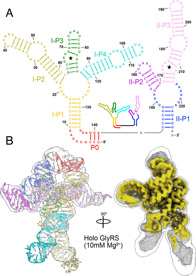
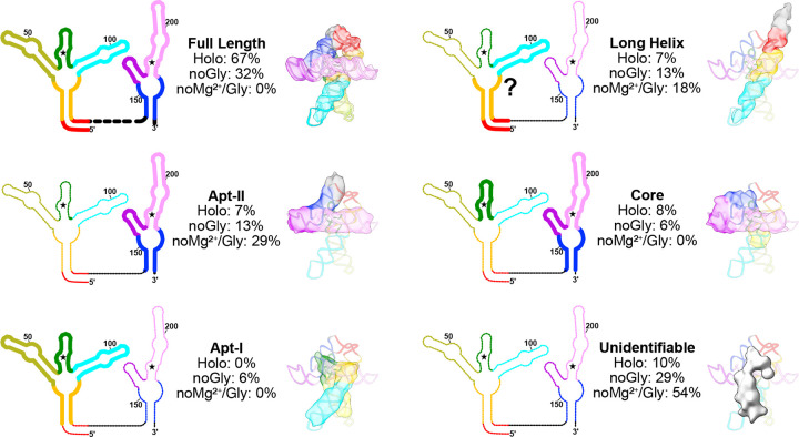
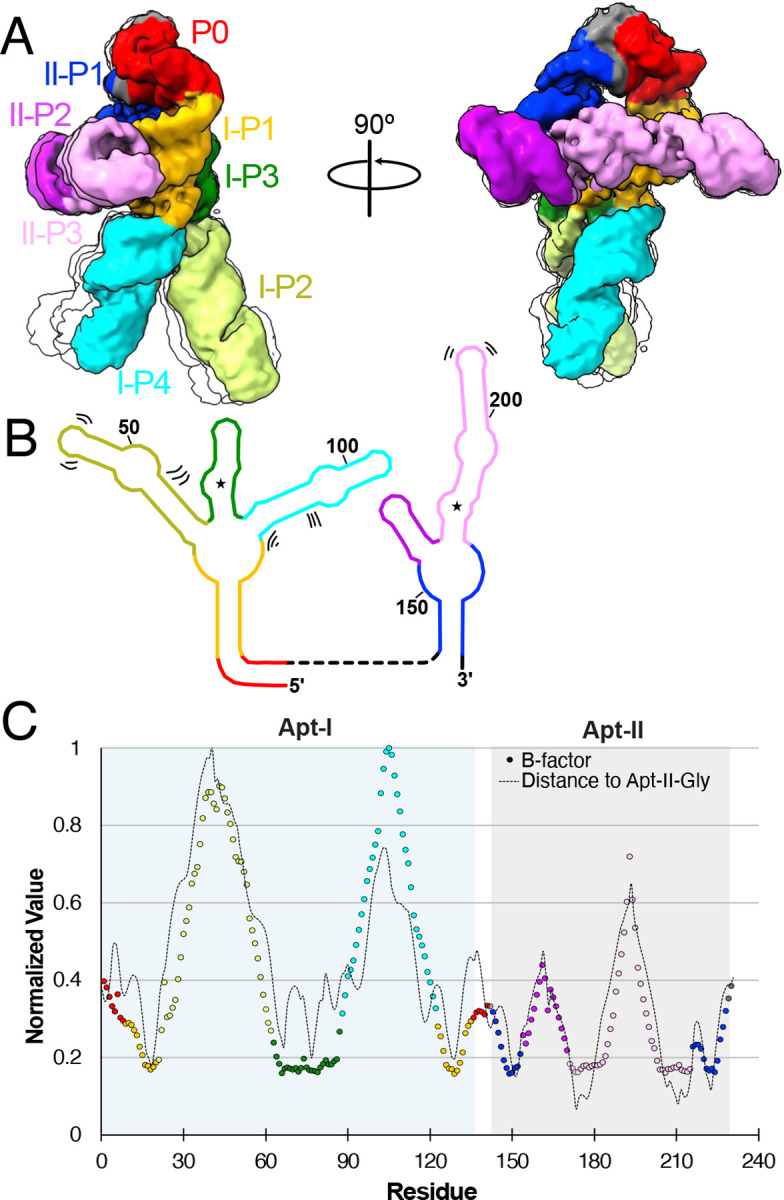
Riboswitches are non-coding RNA sequences that control cellular processes through ligand binding. Conformational heterogeneity is fundamental to riboswitch functionality, yet this same attribute makes structural characterization of these mRNA elements challenging. Here, we use a combination of molecular dynamics and cryo-electron microscopy to expound the flexible nature of the glycine riboswitch tandem aptamers and characterize diMerent structural populations. We find that Mg2+ partially stabilizes the fully folded state, resulting in one-third of the particles adopting a unique "walking man" conformation, consisting of a rigidified core and two dynamic helices, and two-thirds adopting distinct, partially folded states. Glycine interactions double the relative population of fully folded particles by stabilizing a conserved inter-aptamer Hoogsteen base pair, enabling our capture of a 2.9 Å structure for this RNA-only system. The population data show that glycine and Mg2+ operate synergistically: glycine enhances Mg2+ occupancy, while Mg2+ drives glycine specificity. Our findings indicate that cryo-electron microscopy oMers a promising avenue to characterize RNA folding ensembles.


Conflict of interest statement

Additional Declarations: There is NO Competing Interest.

Figures

Extended Data Fig. 1:
Extended Data Fig. 1:. Cryo-EM data processing scheme.
Data collection and processing procedures for the three glycine riboswitch datasets. From left to right: holo (2mM glycine, 10mM Mg2+), apo (no glycine, 10mM Mg2+), and neither glycine nor Mg2+.
Extended Data Fig. 2:
Extended Data Fig. 2:. Overall and local resolution estimation.
a Fourier Shell Correlation (FSC) plot generate in cryoSPARC for the holo (left) and apo (right) glycine riboswitch structures. b Surface representations of the respective maps colored based on local resolution. c Expanded view of the boxed region in b, showing the density and model fit in the core region.
Extended Data Fig. 3:
Extended Data Fig. 3:. The P0 region of the glycine riboswitch adopts RNA suites consistent with a kink-turn conformation.
a Overlay of residues 4–8 in the holo glycine riboswitch structure with a transparent density map. Colors correspond to base identity. Suites for each sugar-to-sugar set of torsion angles are included. b Chemical view of each nucleotide and suite in the kink-turn.
Extended Data Fig. 4:
Extended Data Fig. 4:. The glycine riboswitch core is conserved in both sequence and structure.
a Normalized occupancy of each residue within the V. cholerae glycine riboswitch across all identified homologs. b Normalized conservation for each residue within the V. cholerae glycine riboswitch across all identified homologs. Normalized values were calculated with gaps included. Values from a,b were calculated using 11,626 glycine riboswitch sequences identified, and alignments made, in Torgerson et al. (2020). c RMSD calculated in ChimeraX using the F. nucleatum glycine riboswitch (PDB code: 3P49) and the V. cholerae structure solved here. Regions colored in black are absent in F. nucleatum.
Extended Data Fig. 5:
Extended Data Fig. 5:. Apt-II is stabilized by an additional G-C pair proximal to the glycine binding site.
Residues within 4 Å of the bound glycine are shown for both Apt-I (top) and Apt-II (bottom). Glycines are colored with carbons in grey and magnesium ions proximal to the binding site are shown and colored dark green. Nucleotides are colored according to base identity. Binding sites are largely identical, with the exception of the A70-U80 (Apt-I) to G177-C209 (Apt-II) change.
Extended Data Fig. 6:
Extended Data Fig. 6:. Glycines remain stable in our proposed configurations for Apt-I and Apt-II.
From unbiased and metadynamics simulations trajectories, several thousand frames representing the initial state conformations (Fig. 6) are extracted, and approximately one hundred conformations are depicted for glycine in each aptamer. Glycines maintain constant distance from the Mg2+ ions, with the carboxyl group facing to towards the Mg+2 ions and the amino group orientated towards the phosphate group of A68 and A175 in Apt-I and Apt-II, respectively, indicating our proposed glycine configurations are most stable.
Extended Data Fig. 7:
Extended Data Fig. 7:. Glycine conformations in metastable states seen in vicinity of modeled state.
a Free energy surfaces with respect to the Mg2+-glycine distance and glycine orientation for Apt-I (consistent with Fig. 6b). Five minima over the free energy surface, including a minimum representing the initial state of glycine modeled from the cryo-EM map, denoted as state I, are marked. b Conformation of glycine in minimum I, along with the neighboring residues, Mg2+ ions and cryo-EM-derived, low-pass filtered glycine density, are shown from two perspectives. c Similarly, glycine conformations in four additional minima are illustrated.
Fig. 1:
Fig. 1:. Overall walking man fold for the holo glycine riboswitch.
a Secondary structural representation of the tandem glycine riboswitch aptamers, colored to highlight structural elements. Stars denote glycine binding pockets. A schematic representation is included as an inset. b Cryo-EM density of the holo glycine riboswitch structure, with the model colored consistent with panel a. A triple overlay of raw (mesh), sharpened (gold), and gaussian filtered (silhouette) maps is shown on the right to demonstrate how diMerent regions of the model were fit and refined.
Fig. 2:
Fig. 2:. The full glycine riboswitch fold is stabilized by three main regions of inter-aptamer contacts.
a CryoEM map and model colored to highlight residues derived from Apt-I (light green), Apt-II (pink), or linker regions (grey). b Regions of Inter-aptamer contact. Residues of one aptamer that approach within 2.5 Å of the other aptamer were considered as a contact, leading to three regions involved in stabilization of the fully folded conformation. This includes two sets of A-minor motifs (1,2), and one Hoogsteen base pair (3). CryoEM density for contact regions is extracted to demonstrate fit. A schematic representation of these contact regions is included (right) to emphasize regions involved in stabilization of the full fold. Model and schematic representation are colored to emphasize structural elements.
Fig. 3:
Fig. 3:. Populations of glycine riboswitch folds are highly responsive to solution conditions.
Six general classes of structures are show as both a schematic representation and with a representative transparent map overlaid with a potential structural element from the model. Fractional populations within the holo, no glycine, and no glycine/Mg2+ sample are noted for each class. A question mark is used to emphasize the uncertainty inherent in defining which specific residues are involved in the structure.
Fig. 4:
Fig. 4:. The glycine riboswitch P2 and P4 regions are highly dynamic.
a Four representative models derived from 3D classification of the final holo particle pool, limited to 5 Å resolution. One structure is colored based on structural elements, while three others are shown as simple transparent silhouettes. b Schematic representation of secondary structure, with bending and shifting motions denoted by sets of two or three lines. c Normalized B-factor (colored circles) and distance of Apt-II glycine (dashed line) per residue for the holo glycine riboswitch cryoEM structure. Circles are colored consistent with structural elements in a,b. Apt-I and Apt-II regions are shaded in blue and grey, respectively.
Fig. 5:
Fig. 5:. Glycine and magnesium synergistically stabilize inter-aptamer glycine riboswitch contacts.
a Transparent cryoEM map of the holo glycine riboswitch, overlaid with a gaussian filtered diMerence map of the holo glycine riboswitch minus the apo glycine riboswitch, demonstrating structural changes are localized to the glycine binding loci. b Schematic representation of the glycine riboswitch secondary structure, colored to emphasize diMerent structural elements. c Enlarged view of regions altered in the presence of glycine, with residues colored consistent with panel b and overlaid with a silhouette view of the diMerence map. Glycine ligands are colored in grey and magnesium ions are shown in lime green. Note the direct path of stabilization between the two glycine residues, connected via the conserved inter-aptamer Hoogsteen base pair (here, U77-A206). Three key regions are further enlarged, with modified density in the Apt-I (1), and Apt-II (2) binding pockets, demonstrating a cooperative interaction between magnesium and glycine in these regions. Movement of U80 and U81 (3) may capture movement required to allow glycine access to the tight binding pocket.
Fig. 6:
Fig. 6:. Glycine binding orientation analysis using all-atom explicit solvent MD simulations.
a Glycine orientation is defined as the angle between the z-components of the vector passing from amino N to carboxyl C of glycine and the z-axis. To facilitate the angles calculations, the whole riboswitch structure is aligned to z-axis in the same manner as shown in the figure. Initial angles for Apt-I and Apt-II glycines are mentioned on subset panels. b Free energy surfaces with respect to two variables: i) distance between the Mg2+ and the center of mass of glycine, and ii) glycine orientation as described in panel a. Surfaces on both left panels for Apt-I and Apt-II glycines are obtained by running four 1 μs long equilibrium (unbiased) simulations, while on the right are derived after performing three 2 μs long metadynamics simulations, where the above described angle variables for both glycines were sampled to reorient glycines in all possible orientations. The starting position of the glycines with respect to both variables are marked with the * symbol on all panels.

Similar articles

References

    1. Simensen V. et al. Experimental determination of Escherichia coli biomass composition for constraint-based metabolic modeling. PLoS One 17, (2022). - PMC - PubMed
    1. Schneider B. et al. When will RNA get its AlphaFold moment? Nucleic Acids Res 51, 9522–9532 (2023). - PMC - PubMed
    1. Berman H. M. et al. The Protein Data Bank. Nucleic Acids Res 28, 235 (2000). - PMC - PubMed
    1. Hershkovitz E., Sapiro G., Tannenbaum A. & Williams L. D. Statistical Analysis of RNA Backbone. IEEE/ACM transactions on computational biology and bioinformatics / IEEE, ACM 3, 33 (2006). - PMC - PubMed
    1. Woodson S. A. Compact intermediates in RNA folding: Annual Reviews in Biophysics. Annu Rev Biophys 39, 61 (2010). - PMC - PubMed

Publication types

LinkOut - more resources