Case Reports
. 2025 Apr 11:12:139-146.
doi: 10.2176/jns-nmc.2024-0269. eCollection 2025.

Favorable Response to Conventional Chemoradiotherapy in Radiation-induced Glioma Harboring Coamplification of PDGFRA, KIT, and KDR: A Case Report and Literature Review

Affiliations
Case Reports

Favorable Response to Conventional Chemoradiotherapy in Radiation-induced Glioma Harboring Coamplification of PDGFRA, KIT, and KDR: A Case Report and Literature Review

Daisuke Tanikawa et al. NMC Case Rep J. .

Abstract

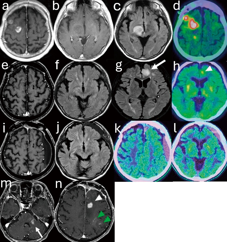
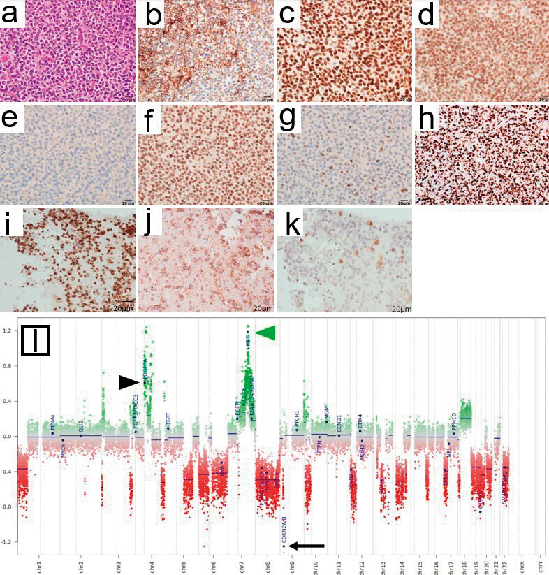
One of the most serious complications of cranial radiotherapy is the development of radiation-induced glioma, which is estimated to occur in 1%-4% of patients who have received cranial irradiation and has a worse prognosis than sporadic glioblastoma. Although comprehensive genetic analysis has recently uncovered the molecular characteristics of radiation-induced glioma, the full picture remains unclear due to its rarity. A 45-year-old man presented with generalized seizures caused by multiple brain tumors involving the right frontal lobe, thalamus, and brainstem. The patient had a history of whole-brain radiotherapy for recurrent Burkitt's lymphoma at the age of 12. He underwent craniotomy, and the histological diagnosis revealed a high-grade glioma with isocitrate dehydrogenase-wildtype, which was presumed to be a radiation-induced glioma that developed 33 years after whole-brain irradiation. Next-generation sequencing identified a CDKN2A/B deletion, as well as coamplification of several receptor tyrosine kinases-encoding genes, including PDGFRA, KIT, and KDR, all of which are located at 4q12. Amplification of this region is broadly observed across cancers and is associated with poor prognosis in sporadic glioblastoma. Nevertheless, the patient received chemoradiotherapy with temozolomide, followed by temozolomide maintenance therapy, resulting in a complete response of all lesions. Although radiation-induced gliomas are generally difficult to treat, our patient unexpectedly responded well to conventional chemoradiotherapy despite the coamplification of multiple receptor tyrosine kinases-encoding genes, which is typically suggestive of an aggressive phenotype. Our case indicates that some radiation-induced gliomas may have distinct molecular characteristics influencing the therapeutic response, which differ from those of sporadic glioblastomas.

Keywords: 4q12 amplification; next-generation sequencing; radiation-induced glioma; radiotherapy.


Conflict of interest statement

All authors have no conflict of interest.

Figures

Fig. 1
Fig. 1
MRI. T1-weighted gadolinium-enhanced MRI shows multiple enhancing tumors in the right frontal lobe (a) and a non-enhancing tumor involving the right thalamus and right midbrain (b), which appears hyperintense on FLAIR (c). 11C-methionine PET before starting CRT shows strong uptake in the residual right frontal lobe tumor (d). MRI, three months after the completion of CRT, shows complete response in all lesions on T1-weighted gadolinium-enhancement (e) and FLAIR (f). MRI, 12 months after completion of CRT, shows a hyperintense lesion on FLAIR, suggesting recurrence in the left frontal lobe (white arrow) (g), with central uptake on 11C-methionine PET (white arrowhead) (h). MRI, 22 months after the diagnosis of RIG, shows that the primary sites in the right frontal lobe, right thalamus, and midbrain maintain complete response on T1-weighted gadolinium-enhancement (i) and FLAIR (j). 11C-methionine PET shows no significant uptake in previously treated sites (k, l). T1-weighted gadolinium-enhanced MRI 28 months after the diagnosis of RIG shows multiple enhancing tumor lesions in the left cerebellar peduncle (white arrow) (m), left frontal lobe (white arrowhead) (n), and left parietal lobe (green arrowhead) (n), which are suggestive of recurrence. CRT: chemoradiotherapy; FLAIR: fluid-attenuated inversion recovery; MRI: magnetic resonance imaging; PET: positron emission tomography; RIG: radiation-induced glioma; T1: type 1
Fig. 2
Fig. 2
Pathological features of the right frontal lobe tumor at the initial operation. H&E staining shows diffuse infiltrative proliferation of neoplastic cells with scant cytoplasm (a). The neoplasm shows immunoreactivity for GFAP (b), OLIG2 (c), and SOX10 (d). Furthermore, those neoplastic cells show no expression of IDH1 R132H (e) and preserve the nuclear expression of ATRX (f). A few neoplastic cells exhibit weak p53 nuclear expression (g). The Ki-67 labeling index is approximately 70% (h). Tumor cells were diffusely and strongly immunopositive for PDGFRA (i) and c-MET (j), with lost MTAP expression (k). The copy-number profile derived from DNA methylation array data shows significant genome-wide copy-number alterations, including CDKN2A/B homozygous deletion (black arrow) and PDGFRA amplification (black arrowhead) (l), which are often observed in RIG. MET amplification is also observed (green arrowhead). ATRX: alpha-thalassemia/mental retardation syndrome X-linked; DNA: deoxyribonucleic acid; GFAP, glial fibrillary acidic protein; H&E: hematoxylin-eosin; IDH1: isocitrate dehydrogenase 1; OLIG2, oligodendrocyte transcription factor 2; RIG: radiation-induced glioma

Similar articles

References

    1. Leary JB, Anderson-Mellies A, Green AL. Population-based analysis of radiation-induced gliomas after cranial radiotherapy for childhood cancers. Neurooncol Adv. 2022;4(1):vdac159. doi: 10.1093/noajnl/vdac159 - DOI - PMC - PubMed
    1. Trkova K, Sumerauer D, Bubenikova A, et al. Clinical and molecular study of radiation-induced gliomas. Sci Rep. 2024;14(1):3118. doi: 10.1038/s41598-024-53434-0 - DOI - PMC - PubMed
    1. Paulino AC, Mai WY, Chintagumpala M, et al. Radiation-induced malignant gliomas: is there a role for reirradiation? Int J Radiat Oncol Biol Phys. 2008;71(5):1381-7. doi: 10.1016/j.ijrobp.2007.12.018 - DOI - PubMed
    1. Neglia JP, Robison LL, Stovall M, et al. New primary neoplasms of the central nervous system in survivors of childhood cancer: a report from the Childhood Cancer Survivor Study. J Natl Cancer Inst. 2006;98(21):1528-37. doi: 10.1093/jnci/djj411 - DOI - PubMed
    1. DeSisto J, Lucas Jr JT, Xu K, et al. Comprehensive molecular characterization of pediatric radiation-induced high-grade glioma. Nat Commun. 2021;12(1):5531. doi: 10.1038/s41467-021-25709-x - DOI - PMC - PubMed

Publication types